Histopathology-validated lesion detection rates of clinically significant prostate cancer with mpMRI, [ 68 Ga]PSMA-11-PET and [ 11 C]Acetate-PET
- PMID: 37615497
- PMCID: PMC10566593
- DOI: 10.1097/MNM.0000000000001743
Histopathology-validated lesion detection rates of clinically significant prostate cancer with mpMRI, [ 68 Ga]PSMA-11-PET and [ 11 C]Acetate-PET
Abstract
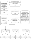
Objective: PET/CT and multiparametric MRI (mpMRI) are important diagnostic tools in clinically significant prostate cancer (csPC). The aim of this study was to compare csPC detection rates with [ 68 Ga]PSMA-11-PET (PSMA)-PET, [ 11 C]Acetate (ACE)-PET, and mpMRI with histopathology as reference, to identify the most suitable imaging modalities for subsequent hybrid imaging. An additional aim was to compare inter-reader variability to assess reproducibility.
Methods: During 2016-2019, all study participants were examined with PSMA-PET/mpMRI and ACE-PET/CT prior to radical prostatectomy. PSMA-PET, ACE-PET and mpMRI were evaluated separately by two observers, and were compared with histopathology-defined csPC. Statistical analyses included two-sided McNemar test and index of specific agreement.
Results: Fifty-five study participants were included, with 130 histopathological intraprostatic lesions >0.05 cc. Of these, 32% (42/130) were classified as csPC with ISUP grade ≥2 and volume >0.5 cc. PSMA-PET and mpMRI showed no difference in performance ( P = 0.48), with mean csPC detection rate of 70% (29.5/42) and 74% (31/42), respectively, while with ACE-PET the mean csPC detection rate was 37% (15.5/42). Interobserver agreement was higher with PSMA-PET compared to mpMRI [79% (26/33) vs 67% (24/38)]. Including all detected lesions from each pair of observers, the detection rate increased to 90% (38/42) with mpMRI, and 79% (33/42) with PSMA-PET.
Conclusion: PSMA-PET and mpMRI showed high csPC detection rates and superior performance compared to ACE-PET. The interobserver agreement indicates higher reproducibility with PSMA-PET. The combined result of all observers in both PSMA-PET and mpMRI showed the highest detection rate, suggesting an added value of a hybrid imaging approach.
Copyright © 2023 The Author(s). Published by Wolters Kluwer Health, Inc.
Conflict of interest statement
There are no conflicts of interest.
Figures


References
-
- Sung H, Ferlay J, Siegel RL, Laversanne M, Soerjomataram I, Jemal A, et al. Global Cancer Statistics 2020: GLOBOCAN estimates of incidence and mortality worldwide for 36 cancers in 185 countries. CA Cancer J Clin 2021; 71:209–249. - PubMed
-
- Fuchsjager M, Shukla-Dave A, Akin O, Barentsz JO, Hricak H. Prostate cancer imaging. Acta Radiol 2008; 49:107–120. - PubMed
-
- Turkbey B, Rosenkrantz AB, Haider MA, Padhani AR, Villeirs G, Macura KJ, et al. Prostate imaging reporting and data system version 2.1: 2019 update of prostate imaging reporting and data system version 2. Eur Urol 2019; 76:340–351. - PubMed
LinkOut - more resources
Full Text Sources
Miscellaneous

