. 2023 Sep 28:572:216363.
doi: 10.1016/j.canlet.2023.216363. Epub 2023 Aug 22.

Photodynamic augmentation of oncolytic virus therapy for central nervous system malignancies

Affiliations

Photodynamic augmentation of oncolytic virus therapy for central nervous system malignancies

Kazuhide Shimizu et al. Cancer Lett. .

Abstract

Oncolytic viruses (OVs) have emerged as a clinical therapeutic modality potentially effective for cancers that evade conventional therapies, including central nervous system malignancies. Rationally designed combinatorial strategies can augment the efficacy of OVs by boosting tumor-selective cytotoxicity and modulating the tumor microenvironment (TME). Photodynamic therapy (PDT) of cancer not only mediates direct neoplastic cell death but also primes the TME to sensitize the tumor to secondary therapies, allowing for the combination of two potentially synergistic therapies with broader targets. Here, we created G47Δ-KR, clinical oncolytic herpes simplex virus G47Δ that expresses photosensitizer protein KillerRed (KR). Optical properties and cytotoxic effects of G47Δ-KR infection followed by amber LED illumination (peak wavelength: 585-595 nm) were examined in human glioblastoma (GBM) and malignant meningioma (MM) models in vitro. G47Δ-KR infection of tumor cells mediated KR expression that was activated by LED and produced reactive oxygen species, leading to cell death that was more robust than G47Δ-KR without light. In vivo, we tested photodynamic-oncolytic virus (PD-OV) therapy employing intratumoral injection of G47Δ-KR followed by laser light tumor irradiation (wavelength: 585 nm) in GBM and MM xenografts. PD-OV therapy was feasible in these models and resulted in potent anti-tumor effects that were superior to G47Δ-KR alone (without laser light) or laser light alone. RNA sequencing analysis of post-treatment tumor samples revealed PD-OV therapy-induced increases in TME infiltration of variable immune cell types. This study thus demonstrated the proof-of-concept that G47Δ-KR enables PD-OV therapy for neuro-oncological malignancies and warrants further research to advance potential clinical translation.

Keywords: Glioblastoma; KillerRed; Malignant meningioma; Oncolytic herpes simplex virus; Photodynamic therapy.


Conflict of interest statement

Declaration of competing interest The authors declare the following financial interests/personal relationships which may be considered as potential competing interests: R.L.M. and S.D.R. are co-inventors on patents relating to oncolytic herpes simplex viruses, owned and managed by Georgetown University and Massachusetts General Hospital (MGH), which have received royalties from Amgen and ActiVec Inc. S.D.R. has received honoraria for expert panel participation from Replimune Inc. All the other authors have no competing interest to declare.

Figures

Fig. 1.
Fig. 1.
Characterizing the biological properties of KillerRed. A, Schematic presentation of light activation of KillerRed. B, The excitation and emission spectrum of KR in U87-KR displaying peaks at 570–590 nm (amber) and 620–640 nm (red), respectively. U87-WT, Wild-type U87 cells. C, Photobleaching induced by LED light exposure in U87-KR cells. D, Fluorometric intracellular ROS assay showing ROS production (shown by green) after LED illumination for 15 min. E, Quantitation of green fluorescence (indicative of ROS) in D. F and G, ATP-based cytotoxicity assay (F) and cell death assay (G) in U87-WT and U87-KR cells with and without light (3 h). *, P < 0.05; ****, P < 0.001 (Student t-test). Scale bar: 100 μm.
Fig. 2.
Fig. 2.
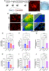
Characterizing photodynamic oncolytic virus therapy in glioblastoma and malignant meningioma cells in vitro. A, Schematic of the genomic structure of G47Δ-KR. KRM, KillerRed Membranous cDNA. B, Membrane localization of KillerRed was most notable in G47Δ-KR-infected Vero cells. C, X-gal staining of plaques in Vero cells (6-well plate). D, KillerRed expression (left) and the excitation and emission spectrum of KR in U87 and IOMM-Lee cells after infection with G47Δ-KR. E-G, ATP-based cytotoxicity assay (E), cell death assay (F) and ROS assay (G) in U87, comparing control, LED light (585–595 nm) alone (3 h), G47Δ-KR alone (MOI = 5), and PD-OV (G47Δ-KR followed by light). H-J, ATP-based cytotoxicity assay (H), cell death assay (I) and ROS assay (J) in IOMM-Lee, comparing control, light alone (2 h), G47Δ-KR alone (MOI = 0.2), and PD-OV (G47Δ-KR followed by light). *, P < 0.05; **, P < 0.01; ***, P < 0.005, ****, P < 0.001; ns, not significant (Student t-test). Scale bar, 100 μm.
Fig. 3.
Fig. 3.. Photodynamic oncolytic virus therapy in patient-derived GBM cells in vitro.
A, KillerRed expression (left) and the excitation and emission spectrum of KR in MGG4 cells after infection with G47Δ-KR. B, Photobleaching induced by LED light (585-595 nm) exposure in G47Δ-KR-infected MGG4 cells. C, ATP-based cytotoxicity assay in MGG4 cells, comparing control, light alone, G47Δ-KR alone (MOI = 5.0) and PD-OV (G47Δ-KR followed by light). *, P < 0.05; **, P < 0.01; ***, P < 0.005; ****, P < 0.001; ns, not significant (Student t-test). Scale bar, 100 μm.
Fig. 4.
Fig. 4.
G47Δ-KR can infect and spread in GBM and MM xenografts in vivo A, Subcutaneous U87 tumor in the right flank of a nude mouse (8.0 × 7.5 mm). B, Hematoxylin and eosin staining of U87 tumor, two days after intratumoral inoculation of G47Δ-KR. Necrotic area at the center (N) where the virus was inoculated. P, actively proliferating tumor cells in the tumor periphery. C, X-gal staining revealing the presence and distribution of G47Δ-KR infection within the U87 tumor. D, Fluorescent microscope detecting KillerRed expression in the area corresponding to the LacZ + area, depicted as rectangle in C. E, Hematoxylin and eosin staining of IOMM-Lee tumor, two days after intratumoral inoculation of G47Δ-KR. N, Necrotic area within the tumor. F, X-gal staining revealing the presence and distribution of G47Δ-KR infection within the IOMM-Lee tumor. G, Fluorescent microscope detecting KillerRed expression in the area corresponding to the necrotic area within IOMM-Lee meningioma.
Fig. 5.
Fig. 5.
Anti-tumor effects of photodynamic oncolytic virus therapy in GBM and MM xenograft models A, Experimental timeline for in vivo studies testing PD-OV therapy in U87 and IOMM-Lee xenograft models. Also shown is amber laser light treatment (585 nm, 100 mW). B, Laser light illuminating U87 tumors with and without G47Δ-KR inoculation two days prior. Top, Laser light group (tumors without G47Δ-KR). Bottom, PD-OV therapy group (tumors with G47Δ-KR). C, U87 tumor volume changes in four treatment arms: no treatment, laser light alone, G47Δ-KR alone (intratumoral), and PD-OV therapy (G47Δ-KR and laser light). D, U87 Tumor volume comparison between the four groups (Day 8 after treatment initiation). E, IOMM-Lee tumor volume changes in 4 treatment arms: PBS (intratumoral), laser light alone, G47Δ-KR alone (intratumoral), and PD-OV therapy (G47Δ-KR and laser light). F, IOMM-Lee tumor volume comparison between the four groups (Day 8 after treatment initiation). In C and E, black line indicates the mean. *, P < 0.05; **, P < 0.01; ****, P < 0.001; ns, not significant (Student t-test).
Fig. 6.
Fig. 6.
Transcriptomics and immunohistochemical analysis of GBM and MM xenografts receiving photodynamic oncolytic virus therapy A, Experimental schema for preparing tumor samples for RNA sequencing. B and C, Selected list of over-represented gene ontologies (GOs) based on differentially expressed (DE) genes in comparison of the PD-OV and OV (G47Δ-KR alone) groups. B, hg38; C, mm10. Plots depict the number of DE genes identified within each category, the percentage this represents in the GO category, and the p-value. Full list of GOs is given in Supplementary Fig. 6. D, Venn diagram depicting the overlap between the DE genes identified when OV is compared to PBS, and when PD-OV is compared to PBS. The number of GOs in which the DE genes are over-represented is shown below (blue). E, Heat map showing immune cell compositions in the tumor microenvironment, obtained by applying a deconvolution algorithm to the RNA-seq data, aligned to the mm10 genome. Colors depict the levels of each immune cell relative to those of other tumors, and can be compared between different tumors, but not different cell types. F, Left, Representative microscopic pictures of CD68 immunohistochemistry in the U87 model receiving different treatments (positive = brown). Scale bar: 100 μm. Right, Number of CD68+ cells per field. **, P < 0.01; ***, P < 0.005 (Student t-test).

References

    1. Russell SJ, Peng KW, Bell JC, Oncolytic virotherapy, Nat. Biotechnol 30 (2012) 658–670. - PMC - PubMed
    1. Kaufman HL, Kohlhapp FJ, Zloza A, Oncolytic viruses: a new class of immunotherapy drugs, Nat. Rev. Drug Discov 14 (2015) 642–662. - PMC - PubMed
    1. Shoaf ML, Desjardins A, Oncolytic viral therapy for malignant glioma and their application in clinical practice, Neurotherapeutics 19 (2022) 1818–1831. - PMC - PubMed
    1. Fudaba H, Wakimoto H, Oncolytic virus therapy for malignant gliomas: entering the new era, Expet Opin. Biol. Ther 23 (2023) 269–282. - PubMed
    1. Wen PY, Weller M, Lee EQ, Alexander BM, Barnholtz-Sloan JS, Barthel FP, Batchelor TT, Bindra RS, Chang SM, Chiocca EA, Cloughesy TF, DeGroot JF, Galanis E, Gilbert MR, Hegi ME, Horbinski C, Huang RY, Lassman AB, Le Rhun E, Lim M, Mehta MP, Mellinghoff IK, Minniti G, Nathanson D, Platten M, Preusser M, Roth P, Sanson M, Schiff D, Short SC, Taphoorn MJB, Tonn JC, Tsang J, Verhaak RGW, von Deimling A, Wick W, Zadeh G, Reardon DA, Aldape KD, van den Bent MJ, Glioblastoma in adults: a Society for Neuro-Oncology (SNO) and European Society of Neuro-Oncology (EANO) consensus review on current management and future directions, Neuro Oncol. 22 (2020) 1073–1113. - PMC - PubMed

Publication types