. 2023 Dec 1;22(12):1444-1453.
doi: 10.1158/1535-7163.MCT-22-0817.

SGN-B6A: A New Vedotin Antibody-Drug Conjugate Directed to Integrin Beta-6 for Multiple Carcinoma Indications

Affiliations

SGN-B6A: A New Vedotin Antibody-Drug Conjugate Directed to Integrin Beta-6 for Multiple Carcinoma Indications

Robert P Lyon et al. Mol Cancer Ther. .

Abstract

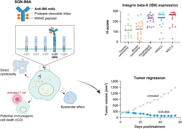
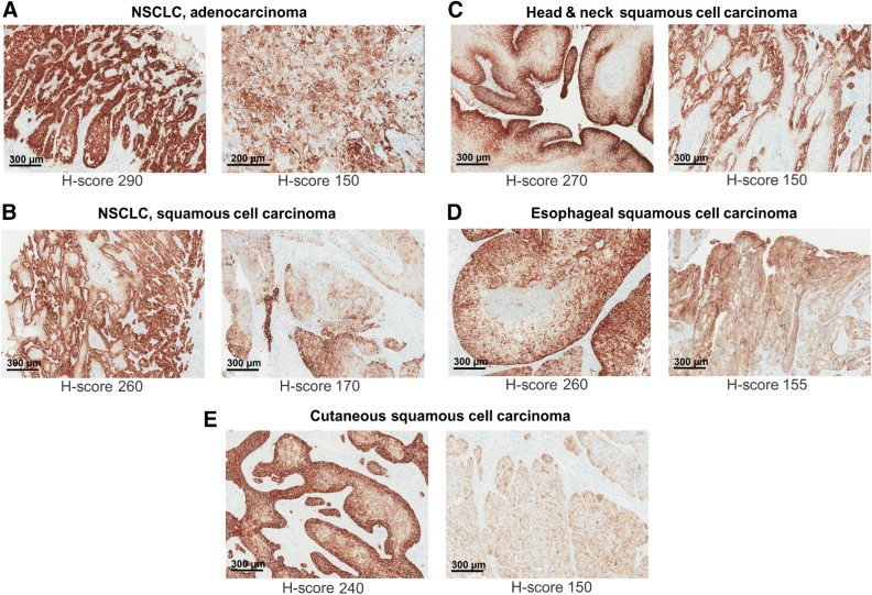
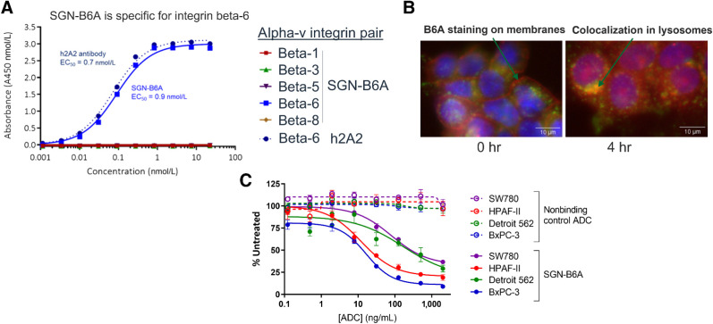
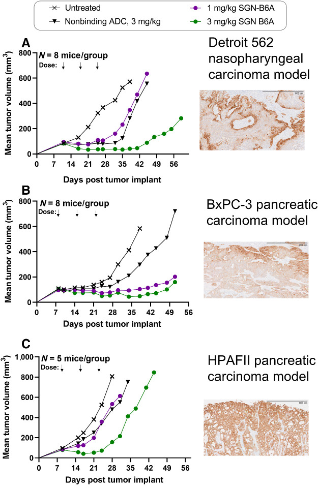
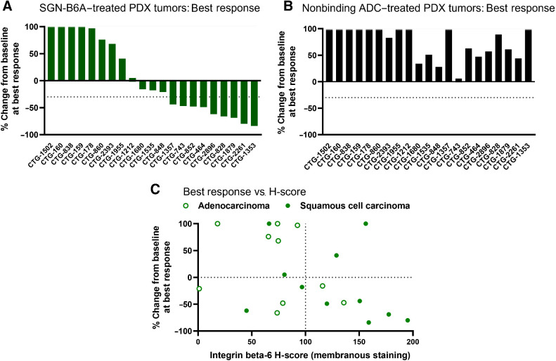
Integrin beta-6, a component of the heterodimeric adhesion receptor alpha-v/beta-6, is overexpressed in numerous solid tumors. Its expression has been shown by multiple investigators to be a negative prognostic indicator in diverse cancers including colorectal, non-small cell lung, gastric, and cervical. We developed SGN-B6A as an antibody-drug conjugate (ADC) directed to integrin beta-6 to deliver the clinically validated payload monomethyl auristatin E (MMAE) to cancer cells. The antibody component of SGN-B6A is specific for integrin beta-6 and does not bind other alpha-v family members. In preclinical studies, this ADC has demonstrated activity in vivo in models derived from non-small cell lung, pancreatic, pharyngeal, and bladder carcinomas spanning a range of antigen expression levels. In nonclinical toxicology studies in cynomolgus monkeys, doses of up to 5 mg/kg weekly for four doses or 6 mg/kg every 3 weeks for two doses were tolerated. Hematologic toxicities typical of MMAE ADCs were dose limiting, and no significant target-mediated toxicity was observed. A phase I first-in-human study is in progress to evaluate the safety and antitumor activity of SGN-B6A in a variety of solid tumors known to express integrin beta-6 (NCT04389632).


Figures

None
Graphical abstract
Figure 1. Expression of integrin beta-6 protein in cancer. Integrin beta-6 protein expression was confirmed by IHC using FFPE-sections from the indications shown. Representative H-scores of membranous staining are indicated below each image. H-score distributions are shown in Supplementary Fig. S3.
Figure 1.
Expression of integrin beta-6 protein in cancer. Integrin beta-6 protein expression was confirmed by IHC using FFPE-sections from the indications shown. Representative H-scores of membranous staining are indicated below each image. H-score distributions are shown in Supplementary Fig. S3.
Figure 2. In vitro characterization of SGN-B6A. A, SGN-B6A binds specifically to integrin beta-6. ELISA binding assay results with recombinant human alpha-v integrin dimers demonstrated that SGN-B6A bound specifically to integrin alpha-v/beta-6 (EC50 = 0.9 nmol/L) and not other alpha-v heterodimers. Binding of the parental unconjugated antibody h2A2 to integrin alpha-v/beta-6 is similar (EC50 = 0.7 nmol/L). B, SGN-B6A binds and internalizes into integrin beta-6 positive cancer cell lines. AlexaFluor 555 lysine-conjugated SGN-B6A (red) was visualized on BxPC-3 cells stained with LAMP1 lysosome marker (green), and Hoechst to label nuclear DNA (blue). At t = 0, SGN-B6A is primarily localized to the plasma membrane (left, green arrow). By 4 hours, SGN-B6A is largely absent from the plasma membrane and is colocalized inside the cells with the lysosomal marker (right, green arrow. C, SGN-B6A induces cytotoxic killing. Integrin beta-6 expressing cell lines were treated with SGN-B6A. Cytotoxicity was observed at concentrations in the range 20 to 300 ng/mL. Error bars represent standard deviation of triplicate samples.
Figure 2.
In vitro characterization of SGN-B6A. A, SGN-B6A binds specifically to integrin beta-6. ELISA binding assay results with recombinant human alpha-v integrin dimers demonstrated that SGN-B6A bound specifically to integrin alpha-v/beta-6 (EC50 = 0.9 nmol/L) and not other alpha-v heterodimers. Binding of the parental unconjugated antibody h2A2 to integrin alpha-v/beta-6 is similar (EC50 = 0.7 nmol/L). B, SGN-B6A binds and internalizes into integrin beta-6 positive cancer cell lines. AlexaFluor 555 lysine-conjugated SGN-B6A (red) was visualized on BxPC-3 cells stained with LAMP1 lysosome marker (green), and Hoechst to label nuclear DNA (blue). At t = 0, SGN-B6A is primarily localized to the plasma membrane (left, green arrow). By 4 hours, SGN-B6A is largely absent from the plasma membrane and is colocalized inside the cells with the lysosomal marker (right, green arrow. C, SGN-B6A induces cytotoxic killing. Integrin beta-6 expressing cell lines were treated with SGN-B6A. Cytotoxicity was observed at concentrations in the range 20 to 300 ng/mL. Error bars represent standard deviation of triplicate samples.
Figure 3. SGN-B6A shows antigen-selective antitumor activity in vivo. Mice engrafted with Detroit-562 (A), BxPC-3 (B), or HPAFII (C) tumor cell lines were treated with SGN-B6A at 1 or 3 mg/kg, a nonbinding control ADC at 3 mg/kg, or left untreated. Animals were dosed weekly for three doses. Immunologically selective antitumor activity was seen in the BxPC-3 model at ≥1 mg/kg and in the Detroit 562 and HPAFII models at 3 mg/kg. Staining by IHC confirming expression of ITGB6 is shown for each model at right. Tumor volume data for individual animals are shown in Supplementary Fig. S7.
Figure 3.
SGN-B6A shows antigen-selective antitumor activity in vivo. Mice engrafted with Detroit-562 (A), BxPC-3 (B), or HPAFII (C) tumor cell lines were treated with SGN-B6A at 1 or 3 mg/kg, a nonbinding control ADC at 3 mg/kg, or left untreated. Animals were dosed weekly for three doses. Immunologically selective antitumor activity was seen in the BxPC-3 model at ≥1 mg/kg and in the Detroit 562 and HPAFII models at 3 mg/kg. Staining by IHC confirming expression of ITGB6 is shown for each model at right. Tumor volume data for individual animals are shown in Supplementary Fig. S7.
Figure 4. SGN-B6A shows antitumor activity in PDX models of NSCLC. Mice (N = 3 per group) were engrafted with PDX models of NSCLC representing adenocarcinoma or squamous cell carcinoma histologies and dosed with 3 mg/kg SGN-B6A or nonbinding control ADC weekly for three doses. A, The waterfall plot of SGN-B6A activity depicts the percent change from baseline tumor volume at best response. This is calculated as 100 × (Tn – Ti) / Ti, where Ti is the initial tumor volume and Tn is the smallest volume observed after the final dose (or the first measurement after the final dose for growing tumors), with values over 100 (tumor size doubling) plotted as 100. SGN-B6A induced tumor volume reductions in 12 of 21 models, including reductions of > 30% (dashed line) in 9 of 21 (model designation indicated at the bottom). B, Corresponding waterfall plot for nonbinding control ADC given at the same dose and schedule, illustrating the impact of integrin beta-6 targeting by comparison with panel A. C, Best response to SGN-B6A for each model plotted against integrin beta-6 expression as assessed by IHC H-Score. Adenocarcinoma models are plotted as open circles, squamous cell models shown as filled circles. Tumor growth curves for all study groups are included in Supplementary Fig. S9 and additional model information in Supplementary Table S4.
Figure 4.
SGN-B6A shows antitumor activity in PDX models of NSCLC. Mice (N = 3 per group) were engrafted with PDX models of NSCLC representing adenocarcinoma or squamous cell carcinoma histologies and dosed with 3 mg/kg SGN-B6A or nonbinding control ADC weekly for three doses. A, The waterfall plot of SGN-B6A activity depicts the percent change from baseline tumor volume at best response. This is calculated as 100 × (Tn – Ti) / Ti, where Ti is the initial tumor volume and Tn is the smallest volume observed after the final dose (or the first measurement after the final dose for growing tumors), with values over 100 (tumor size doubling) plotted as 100. SGN-B6A induced tumor volume reductions in 12 of 21 models, including reductions of > 30% (dashed line) in 9 of 21 (model designation indicated at the bottom). B, Corresponding waterfall plot for nonbinding control ADC given at the same dose and schedule, illustrating the impact of integrin beta-6 targeting by comparison with panel A. C, Best response to SGN-B6A for each model plotted against integrin beta-6 expression as assessed by IHC H-Score. Adenocarcinoma models are plotted as open circles, squamous cell models shown as filled circles. Tumor growth curves for all study groups are included in Supplementary Fig. S9 and additional model information in Supplementary Table S4.
Figure 5. PK of SGN-B6A in cynomolgus monkeys. Cynomolgus monkeys received 4 weekly doses of SGN-B6A at 3, 4, or 5 mg/kg or 2 doses of SGN-B6A at 6 mg/kg every 3 weeks. Plasma exposure of SGN-B6A was approximately linear with dose and did not exhibit a PK profile suggestive of target-mediated drug disposition.
Figure 5.
PK of SGN-B6A in cynomolgus monkeys. Cynomolgus monkeys received 4 weekly doses of SGN-B6A at 3, 4, or 5 mg/kg or 2 doses of SGN-B6A at 6 mg/kg every 3 weeks. Plasma exposure of SGN-B6A was approximately linear with dose and did not exhibit a PK profile suggestive of target-mediated drug disposition.

Similar articles

Cited by

References

    1. Hamidi H, Ivaska J. Every step of the way: integrins in cancer progression and metastasis. Nat Rev Cancer 2018;18:533–48. - PMC - PubMed
    1. Desgrosellier JS, Cheresh DA. Integrins in cancer: biological implications and therapeutic opportunities. Nat Rev Cancer 2010;10:9–22. - PMC - PubMed
    1. Nemeth JA, Nakada MT, Trikha M, Lang Z, Gordon MS, Jayson GC, et al. . Alpha-v integrins as therapeutic targets in oncology. Cancer Invest 2007;25:632–46. - PubMed
    1. Bengs S, Becker E, Busenhart P, Spalinger MR, Raselli T, Kasper S, et al. . β6-integrin serves as a novel serum tumor marker for colorectal carcinoma. Int J Cancer 2019;145:678–85. - PubMed
    1. Elayadi AN, Samli KN, Prudkin L, Liu YH, Bian A, Xie XJ, et al. . A peptide selected by biopanning identifies the integrin alphavbeta6 as a prognostic biomarker for non–small cell lung cancer. Cancer Res 2007;67:5889–95. - PubMed

Associated data

Grants and funding