. 2023 Aug 24;14(1):5159.
doi: 10.1038/s41467-023-40961-z.

Immune stress suppresses innate immune signaling in preleukemic precursor B-cells to provoke leukemia in predisposed mice

Affiliations

Immune stress suppresses innate immune signaling in preleukemic precursor B-cells to provoke leukemia in predisposed mice

Marta Isidro-Hernández et al. Nat Commun. .

Abstract

The initial steps of B-cell acute lymphoblastic leukemia (B-ALL) development usually pass unnoticed in children. Several preclinical studies have shown that exposure to immune stressors triggers the transformation of preleukemic B cells to full-blown B-ALL, but how this takes place is still a longstanding and unsolved challenge. Here we show that dysregulation of innate immunity plays a driving role in the clonal evolution of pre-malignant Pax5+/- B-cell precursors toward leukemia. Transcriptional profiling reveals that Myd88 is downregulated in immune-stressed pre-malignant B-cell precursors and in leukemic cells. Genetic reduction of Myd88 expression leads to a significant increase in leukemia incidence in Pax5+/-Myd88+/- mice through an inflammation-dependent mechanism. Early induction of Myd88-independent Toll-like receptor 3 signaling results in a significant delay of leukemia development in Pax5+/- mice. Altogether, these findings identify a role for innate immunity dysregulation in leukemia, with important implications for understanding and therapeutic targeting of the preleukemic state in children.


Conflict of interest statement

O.B. reports personal fees from Takeda and Clinigen outside the submitted work. K.N. reports grants from Incyte outside the submitted work. No disclosures were reported by the other authors.

Figures

Fig. 1
Fig. 1. Genetic susceptibility to ALL shapes a specific gut microbiota in predisposed mice.
a Pairwise Jaccard distances (beta diversity) were computed for all samples. Distance metric was ordinated into 3D via Principal Coordinates Analysis (PCoA) and visualized via Emperor. Axes indicate percent of explained variance. b Pairwise Permanova tests with 999 permutations were applied to test for differences in beta diversity grouped by mouse genotype (WT n = 12, Sca1-BCR-ABLp190 n = 15, Sca1-Lmo2 n = 15 and Pax5+/− n = 10). Scatter dot plots visualize Jaccard distances between samples of the same genotype. Data are shown as Mean ± SD,“n = ” reports measured distances, “p“ is the p-value of the Permanova test. c Leukemia specific survival curve of Sca1-BCR-ABLp190 treated (n = 13) or untreated (n = 7) with antibiotic6s (p-value = 0.6514), Sca1-Lmo2 treated (n = 24) or untreated (n = 14) with antibiotics (p-value = 0.7935), and Sca1-ETV6-RUNX1 treated (n = 6) or untreated (n = 10) with antibiotics mice p-value > 0.9999), and Pax5+/− treated (n = 19) or untreated (n = 20) with antibiotics (p-value = 0.0002). None of them were exposed to common infections. P-values are from Log-rank (Mantel–Cox) test. Source data are provided as a Source Data file. Abx: treated with a cocktail of antibiotics for 8 weeks.
Fig. 2
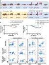
Fig. 2. Early exposure to infection promotes the development of B-cell precursor acute lymphoblastic leukemia in Pax5+/− mice.
a Experimental setup. Female mice of the “early exposure group” got pregnant already in a conventional facility (i.e., non-SPF conditions) where they are exposed to common infections during their lifespan, and the born pups were kept in these conditions for their lifetime. Female mice from the “delayed-exposure group” got pregnant under SPF conditions, and pups were kept in SPF until one month of age, when they were moved to the conventional facility. The mice were monitored periodically. The endpoint was established at 2 years of age (22–24 months) or when the mice showed signs of disease (clinical signs or presence of blasts in peripheral blood); the animals were sacrificed and the hematopoietic and non-hematopoietic tissues were analyzed. E: embryonic period; PN: postnatal period; SPF: specific pathogen free. b B-ALL specific survival curve of Pax5+/− animals born and kept into non-SPF conditions (red, n = 28) compared to WT control mice (black, n = 21) showing a significantly (log-rank P value 0.0151) shortened life span and compared to Pax5+/− animals always kept into SPF conditions (green, n = 20) showing a significantly (log-rank P value 0.0177) shortened life span (left panel), and B-ALL specific survival curve of Pax5+/− early exposure group (red, n = 28) compared to Pax5+/− delayed-exposure group (blue, n = 41) showing similar life span (log-rank P value 0.5491) and comparing Pax5+/− animals always kept into SPF conditions (green, n = 20) to Pax5+/− delayed-exposure group (blue, n = 41) showing a significantly (log-rank P value 0.0098) shortened life span (right panel). Source data are provided as a Source Data file. c Flow cytometric analysis of hematopoietic subsets in diseased Pax5+/− early exposure mice. Representative plots of cell subsets from the bone marrow and peripheral blood show accumulation of blast B cells in Pax5+/− early exposure mice (n = 7, age: 9–17 months) compared to control age-matched littermate WT mice (n = 4, age: 8–16 months). Pax5+/− early exposure mice show a similar immune-phenotype than Pax5+/− delayed-exposure mice (n = 9, age: 6–16 months).
Fig. 3
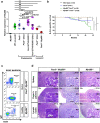
Fig. 3. Reduction in Myd88 levels significantly increases the development of leukemias in Pax5+/− mice upon exposure to natural infections.
a Q-PCR of Myd88 expression levels in preleukemic Pax5+/− and Sca1-ETV6-RUNX1 and leukemic Pax5+/− proB cells. Preleukemic samples: sorted BM proB (B220low IgM-) cells from 3 months-old of wild-type (n = 5), Pax5+/− (n = 3), Sca1-ETV6-RUNX1 (n = 5) and Pax5+/− (n = 3) and Sca1-ETV6-RUNX1 (n = 5) mice treated with Abx for 8 weeks were analyzed by Q-PCR to quantified the expression of Myd88. Leukemic samples: BM leukemic cells from Pax5+/− mice treated with Abx for 8 weeks (n = 3) and Myd88+/−;Pax5+/− mice (n = 3) were analyzed by Q-PCR to quantify the expression of Myd88. All the mice analyzed were exposed to infections (in the conventional facility). Total BM from Myd88−/− mice was used as a negative control. Error bars represent the mean and SD. For the significant differences, unpaired t test p-values (two-tailed) are indicated in each case. Only relevant comparisons are shown. p = 0.0378 when comparing Pax5+/−(Abx) vs Sca1-ETV6-RUNX1(Abx); p = 0.001 when comparing Pax5+/− vs Pax5+/−(Abx); p = 0.012 when comparing Sca1-ETV6-RUNX1 vs Sca1-ETV6-RUNX1-(Abx); p = 0.004 when comparing Pax5+/− vs Sca1-ETV6-RUNX1 and *** for p-value < 0.0001. b B-ALL-specific survival of Pax5+/− and Myd88+/−; Pax5+/− mice (Pax5+/−, blue line, n = 41), (Myd88+/−;Pax5+/− dark green line, n = 30), (WT, black line, n = 23), and (Myd88+/−;Pax5+/+, brown line, n = 26) all of them exposed to common infections. Log-rank (Mantel–Cox) test p-value > 0.9999 when comparing WT vs Myd88+/−:Pax5+/+ mice, p-value = 0.0117 when comparing Pax5+/− vs Myd88+/−;Pax5+/+ mice and p-value < 0.0001 when comparing Myd88+/−;Pax5+/− vs Myd88+/−;Pax5+/+ mice. c Flow cytometric analysis of bone marrow showing the accumulation of blast B cells (B220+ IgM+/−) in the leukemic Myd88+/−;Pax5+/− mice (X012 and W182) and compared with a healthy Myd88+/− mouse (X650), all of them exposed to common infections. d Haematoxylin and eosin staining of a tumor-bearing Myd88+/−;Pax5+/− mice showing infiltrating blast cells in the spleen, Peyer’s patches, and lymph nodes, compared with an age-matched Myd88+/− mouse. Loss of normal architecture due to leukemic cell infiltration can be seen. A representative example of 16 diseased mice is shown in the figure. Magnification and the corresponding scale bar are indicated in each case. Source data are provided as a Source Data file.
Fig. 4
Fig. 4. Pax5+/− mice injected with LPS develop B-ALL without infection exposure.
a B-ALL-specific survival of Pax5+/− mice injected with LPS (a Myd88-depenedent TLR ligand) (orange line, n = 10), Pax5+/− mice injected with poly(I:C) (a Myd88-indepenedent TLR ligand) (light blue line, n = 10), Pax5+/− mice injected with PBS (as control) (green line, n = 10), control wild type (WT) mice injected with LPS (red line, n = 10), WT mice injected with poly(I:C) (dark blue line, n = 10), and WT mice injected with PBS (black line, n = 10), all of them housed in an SPF facility (without exposure to common infections). Log-rank (Mantel–Cox) test p-value = 0,1464 when comparing Pax5+/− mice injected with LPS and Pax5+/− mice injected with PBS. b Flow cytometry representative illustration of the percentage of leukemic B cells (B220+IgM+ subsets) in PB, BM, spleen and LN from a diseased Pax5+/− mouse injected with LPS compared to an age-matched healthy WT mouse. c Haematoxylin and eosin staining of a tumor-bearing Pax5+/− mouse injected with LPS unexposed to common infections showing infiltrating blast cells in the spleen, liver, and lymph nodes and compared with a healthy WT mouse. Loss of normal architecture can be seen due to the infiltrating cells morphologically resembling lymphoblast. Magnification and the corresponding scale bar are indicated in each case. Source data are provided as a Source Data file.
Fig. 5
Fig. 5. Genetic impact of Myd88 downregulation in Pax5+/− infection-driven leukemia.
a GSEA showing that leukemic Pax5+/−;Myd88+/− cells from diseased mice present similar profiles for genesets previously identified in human B-ALL samples,,. The genesets shown are significantly enriched with a nominal p-value < 1%. Statistical analysis was done using Gene set enrichment analysis (GSEA) algorithm. b Whole Genome Sequencing in Pax5+/−;Myd88+/− B-ALLs. Oncoprint of somatic single nucleotide mutations and copy number alterations across 8 leukemia samples from Pax5+/−;Myd88+/− mice. Somatic alterations are clustered by gene. Tumor DNA was derived from whole leukemic BM or LN, while tail DNA of the respective mouse was used as reference germline material. Previously reported known human or mouse leukemia hotspot mutations are highlighted (red). Mean tumor variant allele fraction (VAF) for each single nucleotide mutation is shown on the dotplot on the right. c GSEA plots showing the 8 gene sets that are significantly enriched at nominal p-value < 1% in Pax5+/−;Myd88+/− proB cells from healthy mice. Enrichment analysis was done using the hallmarks collection from MSigDB. Statistical analysis was done using Gene set enrichment analysis (GSEA) algorithm.
Fig. 6
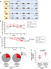
Fig. 6. Innate stimulation by poly(I:C) treatment delays B-ALL development in Pax5+/− mice exposed to infections.
a Study design. poly(I:C) treatment fully described in methods section. SPF: specific pathogen free facility; CF: conventional facility, where the mice area exposed to common infections. b Percentages of CD8+ T cells in peripheral blood of Pax5+/− and WT mice treated with poly(I:C) compared to untreated mice. An increase in CD8+ T cells can be observed in the blood of treated mice, only being statistically significant for the Pax5+/− mice (unpaired t test (two-tailed), p-value = 0.0087) (for WT mice the unpaired t test has a nonsignificant p-value = 0.5069). Each point represents the mean of the levels of the CD8+ T cells in all mice in each group for each of the different time points (n = 31 for Pax5+/− poly(I:C)-treated mice, n = 8 for Pax5+/− untreated mice, n = 14 for WT poly(I:C) treated mice and n = 23 for WT untreated mice). c B-ALL–specific survival of mice treated with poly(I:C) (Pax5+/−, pink dashed line, n = 31; WT, blue dashed line, n = 14) and nontreated mice (Pax5+/−, pink line, n = 35; WT, blue line, n = 23) following exposure to common infections. Log-rank (Mantel–Cox) test p = 0.7930 when comparing Pax5+/− mice treated with poly(I:C) versus nontreated Pax5+/− mice, p = 0.0034 when comparing Pax5+/− versus WT poly(I:C)-treated mice and p = 0.0154 when comparing Pax5+/− versus WT mice without treatment. d Absence of changes in B-ALL incidence in Pax5+/− mice treated with poly(I:C). The treatment of Pax5+/− mice with poly(I:C) did not reduce nor increase the rate of B-ALL development. Fisher exact test (two-tailed), p = 0.9999. 8 out 35 mice in the non-treated group developed the disease (22.86%) and 8 out of 31 mice in the poly(I:C)-treated group developed B-ALL (25.81%). e poly(I:C) treatment increases the latency of B-ALL development in Pax5+/− mice. Pax5+/− untreated mice (n = 8) were diagnosed with B-ALL between 6 and 15 months of age and the Pax5+/− mice treated with poly(I:C) (n = 8) were diagnosed between 10.5 and 20.8 months of age. Error bars represent the mean and SD. Statistic test Mann–Withney (two-tailed), p-value = 0.0019. Source data are provided as a Source Data file.
Fig. 7
Fig. 7. Suppression of innate immune signaling by immune stress triggers leukemia development from Pax5+/− preleukemic precursors.
Immune stress (e.g. infection exposure, antibiotic treatment, etc.) can affect both healthy (a) and Pax5+/− preleukemic carriers (b) alike, and in both of them it can elicit a partial downregulation of Myd88 expression in individual B cell progenitors. However, only in Pax5+/− preleukemic progenitors will this downregulation interfere with innate immune signaling in a way that allows the appearance of secondary mutations leading to the development of a full-blown leukemia. The incidence of B-ALL is directly proportional to the percentage of Pax5+/− preleukemic cells having a downregulation of Myd88 expression.

References

    1. Cobaleda C, Vicente-Duenas C, Ramirez-Orellana M, Sanchez-Garcia I. Revisiting the concept of childhood preleukemia. Trends Cancer. 2022;8:887–889. - PubMed
    1. Cobaleda C, Vicente-Duenas C, Sanchez-Garcia I. Infectious triggers and novel therapeutic opportunities in childhood B cell leukaemia. Nat. Rev. Immunol. 2021;21:570–581. - PubMed
    1. Martin-Lorenzo A, et al. Infection exposure is a causal factor in b-cell precursor acute lymphoblastic leukemia as a result of Pax5-inherited susceptibility. Cancer Discov. 2015;5:1328–1343. - PubMed
    1. Rodriguez-Hernandez G, et al. Infection exposure promotes ETV6-RUNX1 precursor B-cell leukemia via impaired H3K4 demethylases. Cancer Res. 2017;77:4365–4377. - PubMed
    1. Rodriguez-Hernandez G, et al. Infectious stimuli promote malignant B-cell acute lymphoblastic leukemia in the absence of AID. Nat. Commun. 2019;10:5563. - PMC - PubMed

Publication types

Substances