. 2023 Aug 24;14(1):5149.
doi: 10.1038/s41467-023-40786-w.

Molecular architecture and conservation of an immature human endogenous retrovirus

Affiliations

Molecular architecture and conservation of an immature human endogenous retrovirus

Anna-Sophia Krebs et al. Nat Commun. .

Abstract

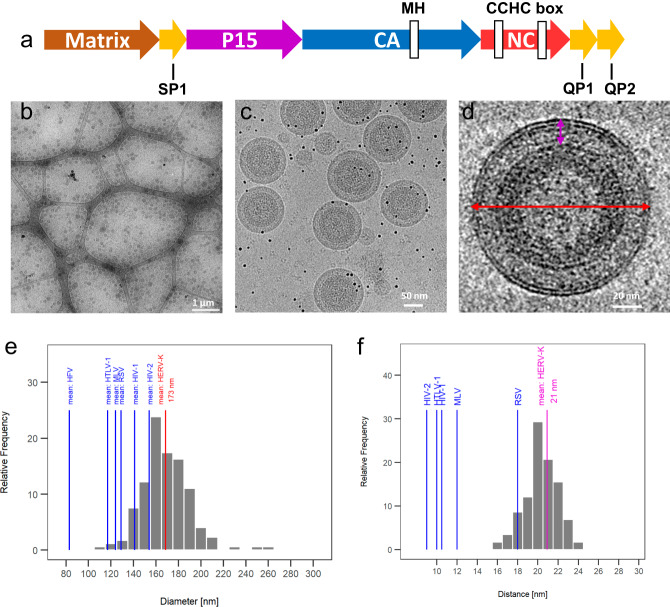
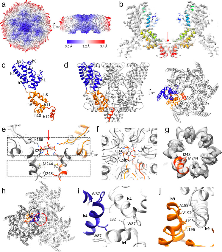
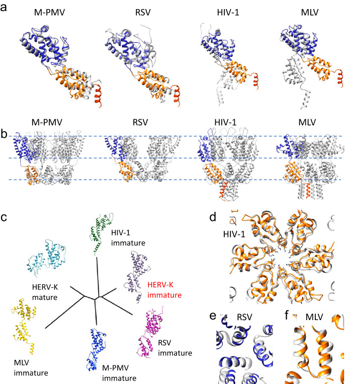
The human endogenous retrovirus K (HERV-K) is the most recently acquired endogenous retrovirus in the human genome and is activated and expressed in many cancers and amyotrophic lateral sclerosis. We present the immature HERV-K capsid structure at 3.2 Å resolution determined from native virus-like particles using cryo-electron tomography and subtomogram averaging. The structure shows a hexamer unit oligomerized through a 6-helix bundle, which is stabilized by a small molecule analogous to IP6 in immature HIV-1 capsid. The HERV-K immature lattice is assembled via highly conserved dimer and trimer interfaces, as detailed through all-atom molecular dynamics simulations and supported by mutational studies. A large conformational change mediated by the linker between the N-terminal and the C-terminal domains of CA occurs during HERV-K maturation. Comparison between HERV-K and other retroviral immature capsid structures reveals a highly conserved mechanism for the assembly and maturation of retroviruses across genera and evolutionary time.


Conflict of interest statement

The authors declare no competing interests.

Figures

Fig. 1
Fig. 1. CryoEM analysis of immature HERV-K virus-like particles (VLPs).
a Schematic representation of Gag domains. bd Representative cryoEM images of purified immature HERV-K VLPs at low (b), medium (c) and high (d) magnifications. e The mean (red line) and distribution (gray bars) of the diameter (red arrow in d) of HERV-K VLPs, compared with the mean diameters of other retroviruses (blue lines). f The mean (purple line) and distribution (gray bars) of the distance between lipid bilayer and capsid lattice (purple in d) of HERV-K VLPs, compared with the mean distance of other retroviruses (blue lines). The micrographs in (b) and (c, d) are representative of 20 and 124 measurements, respectively. Source data are provided as a Source Data file.
Fig. 2
Fig. 2. CryoET STA of immature HERV-K VLPs.
a CryoET STA map of immature HERV-K CA hexamer colored by local resolution (red to blue). b A slab of HERV-K CA hexamer map overlapped with the atomic model (rainbow colors blue to red, N-terminus to C-terminus. c The atomic model of the immature HERV-K CA monomer (NTD in blue, CTD in orange, h12 in red) with α-helices labeled. d The atomic model of the immature HERV-K hexamer viewed from the side and top with one monomer colored in blue and orange. e A central slice of the HERV-K hexamer density map, showing IP6 density with an IP6 fitted (red arrow) and two Lys rings (K166 and K240), in addition to the hydrophobic core of the 6HB (M244 and I248). f, g Density slabs at the top (f, top dashed box in e) and bottom (g, bottom dashed box in e) of the 6HB, with the protruding side chains shown as sticks. h The dimer (black circle) and trimer (red circle) interfaces between adjacent immature HERV-K hexamers. i A detailed view of the trimer interface involving helix 4 with the contributing side chains shown as sticks. j A detailed view of the dimer interface involving helix 9 with the involved side chains shown as sticks.
Fig. 3
Fig. 3. MD simulations and mutational validation of immature HERV-K CA intermolecular interactions.
ac Snapshots of the inter-hexamer trimer interface involving helix 4 with the contributing side chains shown as sticks. Residues in (b) and (c) show stable interactions. d A snapshot of the inter-hexamer dimer interface involving helix 9 with the contributing side chains. e, f Views of the 6HB with the contributing side chains of the hydrophobic core (M244 and I248) (e) and the K166 and K240 rings with the IP6 molecule (f). In each snapshot, protein is represented as cartoon (NTD in blue, CTD in orange). The amino acid side chains are colored based on the heavy-atom RMSF computed throughout the MD simulation. g Western-blot of VLP assembly carrying trimer interface mutants of HERV-K Gag. The Western-blot experiments, including independent infections, were repeated three times. Source data for (g) are provided as a Source Data file.
Fig. 4
Fig. 4. Comparison between immature and mature HERV-K CA.
a Overlay of the immature HERV-K structure (NTD blue, CTD orange, h12 red) with mature HERV-K (gray, PDB: 6SSM), aligned to the NTD (left), CTD (middle), and NTD/CTD separately (right). b, c Arrangement of HERV-K immature (b) and mature (c) lattices. NTD is colored in blue/cyan, CTD in orange/red. d, e Detailed views of the dimer (top) and trimer (bottom) interface in the immature (d) and mature (e) lattices.
Fig. 5
Fig. 5. Conservation among the immature retroviruses.
a Overlay of the immature CA monomer structures of HERV-K (NTD blue, CTD orange, h12 red) with those of other retroviruses (gray) aligned to the NTD, shown are M-PMV (PDB: 6HWI, beta-retrovirus), RSV (PDB: 5A9E, alpha-retrovirus), HIV-1 (PDB: 7ASL, lentivirus), MLV (PDB: 6HWW, gamma-retrovirus). b Hexamer structures of M-PMV, RSV, HIV-1 and MLV. c Structure based phylogenetic tree. df Conservation of immature CA intermolecular interfaces, compared with HIV-1 (hexamer interface, d), RSV (trimer interface, e) and MLV (dimer interface, f). HERV-K structure is colored in blue and orange, other retroviruses in gray.

Update of

References

    1. Lander, E., Linton, L., Birren, B. & Nusbaum, C. Initial sequencing and analysis of the human genome. Nature409, 860–921 (2001). - PubMed
    1. Garcia-Montojo, M., Doucet-O’Hare, T., Henderson, L. & Nath, A. Human endogenous retrovirus-K (HML-2): a comprehensive review. Crit. Rev. Microbiol.44, 715–738 (2018). - PMC - PubMed
    1. Herbst, H., Sauter, M. & Mueller-Lantzsch, N. Expression of human endogenous retrovirus K elements in germ cell and trophoblastic tumors. Am. J. Pathol.149, 1727–1735 (1996). - PMC - PubMed
    1. Büscher, K. et al. Expression of Human Endogenous Retrovirus K in Melanomas and Melanoma Cell Lines. Cancer Res.65, 4172–4180 (2005). - PubMed
    1. Kurth, R. & Bannert, N. Beneficial and detrimental effects of human endogenous retroviruses. Int J. Cancer126, 306–314 (2010). - PubMed

Publication types

Substances