. 2023 Aug 24;14(1):5157.
doi: 10.1038/s41467-023-40887-6.

Outer membrane vesicles from a mosquito commensal mediate targeted killing of Plasmodium parasites via the phosphatidylcholine scavenging pathway

Affiliations

Outer membrane vesicles from a mosquito commensal mediate targeted killing of Plasmodium parasites via the phosphatidylcholine scavenging pathway

Han Gao et al. Nat Commun. .

Abstract

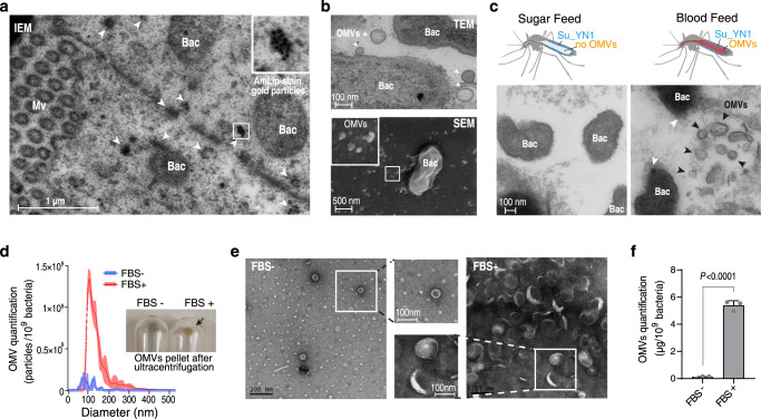
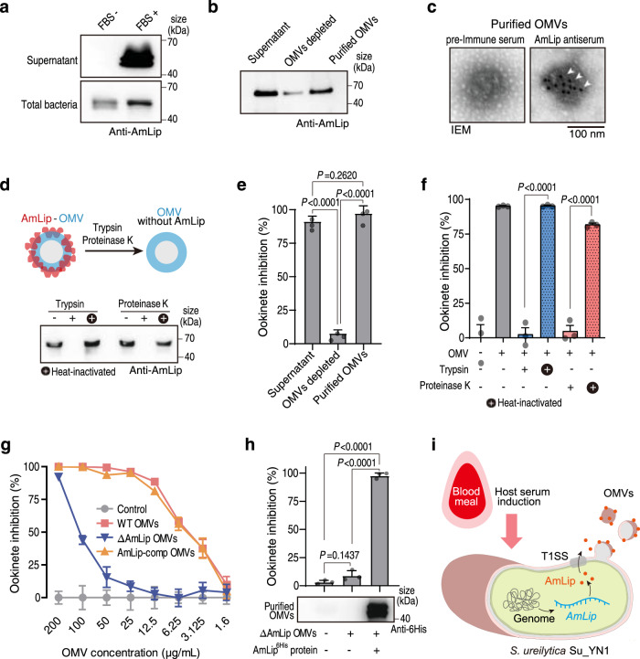
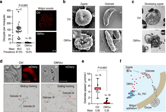
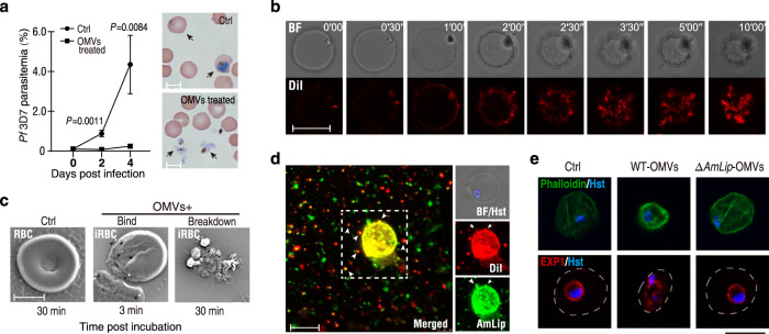
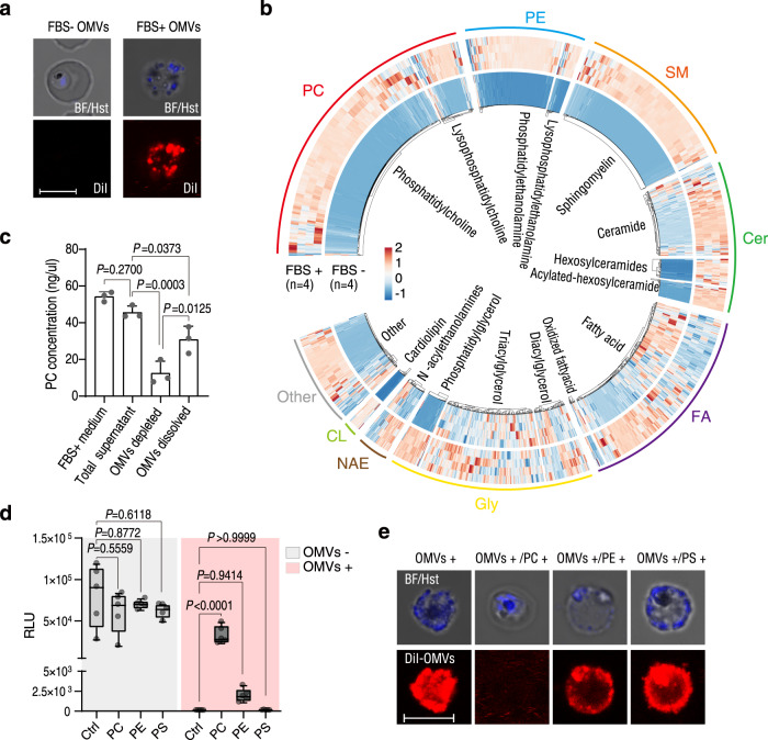
The gut microbiota is a crucial modulator of Plasmodium infection in mosquitoes, including the production of anti-Plasmodium effector proteins. But how the commensal-derived effectors are translocated into Plasmodium parasites remains obscure. Here we show that a natural Plasmodium blocking symbiotic bacterium Serratia ureilytica Su_YN1 delivers the effector lipase AmLip to Plasmodium parasites via outer membrane vesicles (OMVs). After a blood meal, host serum strongly induces Su_YN1 to release OMVs and the antimalarial effector protein AmLip into the mosquito gut. AmLip is first secreted into the extracellular space via the T1SS and then preferentially loaded on the OMVs that selectively target the malaria parasite, leading to targeted killing of the parasites. Notably, these serum-induced OMVs incorporate certain serum-derived lipids, such as phosphatidylcholine, which is critical for OMV uptake by Plasmodium via the phosphatidylcholine scavenging pathway. These findings reveal that this gut symbiotic bacterium evolved to deliver secreted effector molecules in the form of extracellular vesicles to selectively attack parasites and render mosquitoes refractory to Plasmodium infection. The discovery of the role of gut commensal-derived OMVs as carriers in cross-kingdom communication between mosquito microbiota and Plasmodium parasites offers a potential innovative strategy for blocking malaria transmission.


Conflict of interest statement

The authors declare no competing interests.

Figures

Fig. 1
Fig. 1. Su_YN1 releases OMVs in the midgut lumen of blood-fed An. stephensi mosquitoes.
a Detection of the lipase AmLip by immune electron microscopy (IEM) in an ultrathin cryosection of a midgut of Su_YN1-carrying Anopheles mosquito, 24 h after blood feeding. Gold particles indicating AmLip staining are marked by white arrowheads. Mv mosquito microvilli, Bac Serratia Su_YN1 bacterium, Scale bar, 1 μm. Similar results were obtained from two biological repeats. b Transmission Electron Microscope (TEM, upper panel) and Scanning Electron Microscope (SEM, lower panel) images of Serratia Su_YN1 bacterium (Bac) from the mosquito midgut lumen. OMVs produced by Su_YN1 bacterium are indicated by white arrowheads. Scale bar, 100 nm (upper panel) and 500 nm (lower panel). Similar results were obtained from two biological repeats. c Comparison of midgut sections by TEM analysis of Su_YN1-carrying Anopheles mosquitoes which had fed on either a blood meal or a sugar meal. Black arrowheads, OMVs; white arrowheads, OMVs budding from the surface of Serratia Su_YN1 bacteria (Bac). Scale bar, 100 nm. Similar results were obtained from two biological repeats. d Nanoparticle Tracking Analysis (NTA) of Su_YN1 bacterium cultured in RPMI 1640 medium supplemented with or without pre-cleared Fetal Bovine Serum (FBS). OMV concentrations are normalized to 1 ×10⁹ bacteria. The distribution of particle sizes was demonstrated as polylines (mean ± SD, n = 3 independent measurements for each group). Similar results were obtained from two biological repeats. The image in the right panel indicates OMVs pellets in the centrifuge tube bottom (black arrow) after ultracentrifugation. e TEM analysis of OMVs ultracentrifugation pellets (negative stained with uranyl acetate) from d, Square frames show enlarged images to display Su_YN1-secreted OMVs with or without FBS induction. Scale bar, 100 nm. f OMV quantification by bicinchoninic acid (BCA) assay of Su_YN1 cultured with or without FBS (mean ± SD, n = 3). OMV concentrations are normalized to 1 ×10⁹ bacteria. Statistical significance was determined using a one-way ANOVA test, P values are indicated above the plots. Similar results were obtained from three biological repeats. Source data are provided as a Source Data file.
Fig. 2
Fig. 2. Host serum induces the formation of AmLip-OMV complex that kills Plasmodium parasites.
a Western blot detection of AmLip protein secretion into the supernatant of Su_YN1 bacterium cultured with or without FBS. Similar results were obtained from two biological repeats. b Western blot detection of AmLip protein in Su_YN1 culture supernatant, OMV-depleted supernatant, and OMVs purified from the culture supernatant using AmLip antiserum. Similar results were obtained from two biological repeats. c Representative images of IEM detection of AmLip on Su_YN1 OMVs using pre-immune serum and AmLip antiserum. White arrowheads point to gold particles indicating AmLip staining. Scale bar, 100 nm. Similar results were obtained from two biological repeats. d Detection of AmLip on the surface of OMVs. Western blot using AmLip antiserum of OMVs treated (or not) with trypsin or proteinase K. Similar results were obtained from two biological repeats. e P. berghei ANKA ookinete inhibition assay of Su_YN1 culture supernatant, OMV-depleted supernatant, and purified OMVs resolved in RPMI 1640 (mean ± SD, n = 3). Statistical significance was determined using one-way ANOVA test. Similar results were obtained from two biological repeats. f P. berghei ANKA ookinete inhibition assay by Su_YN1 OMVs treated (or not) with trypsin or proteinase K (mean ± SD, n = 3). Statistical significance was determined using a two-tailed Student’s t-test. Similar results were obtained from two biological repeats. g Concentration curve of P. berghei ANKA ookinete inhibition by OMVs from wildtype (WT), AmLip-KO and AmLip-complemented Su_YN1 bacterial culture (mean ± SD, n = 3). Statistical details are provided in Source Data file. h P. berghei ANKA ookinete inhibition assay of OMVs purified from AmLip-KO Su_YN1 culture supernatant supplemented with or without 6×His-tagged recombinant AmLip protein (mean ± SD, n = 3). Recombinant AmLip protein loading was detected by Western blot (lower panel). Statistical significance was determined using one-way ANOVA test. Similar results were obtained from two biological repeats. i Schematic diagram showing host serum induces Su_YN1 to produce OMVs and secrete AmLip, which form AmLip-OMV complex that kills Plasmodium. P values in e, f and h are indicated above the plots. Source data are provided as a Source Data file.
Fig. 3
Fig. 3. Su_YN1 OMVs target early midgut stages of Plasmodium parasites.
a Oocyst load in the midgut of An. stephensi mosquitoes fed with a fluorescent mCherry transgenic P. berghei ANKA strain infectious blood supplemented with PBS (Ctrl) or OMVs. Oocyst load is shown in the left panel. Right panel shows representative fluorescence microscopy images (mCherry signals indicate oocysts) of An. stephensi midguts. Scale bar, 50 μm. Significant difference in oocyst intensities between two groups was analyzed using the two-tailed Mann–Whitney test, P values are indicated above the plots. b SEM observation of P. berghei ANKA zygote and ookinete 5 min after incubation with Su_YN1 OMVs. Scale bar, 5 μm. Similar results were obtained from two biological repeats. c TEM images of sections of P. berghei ANKA developing zygotes after incubation with or without Su_YN1 OMVs. Scale bar, 5 μm. Similar results were obtained from two biological repeats. d Effect of OMVs on ookinete gliding. Live tracking of ookinete gliding on Matrigel matrix was recorded. Ookinetes were pretreated with OMVs at final concentration of 50 μg/ml. Reconstruction of tracked ookinete paths from high-magnification movies reveal movement patterns. Blue arrowheads and red arrowheads indicate gliding start and stop positions respectively, and the lines indicate the ookinete gliding trajectory. Live ookinetes treated (or not) with OMVs are shown in the upper panels. Ookinetes lost mCherry fluorescence after OMV treatment, indicating damage of cell viability. Scale bar, 5 µm. Similar results were obtained from two biological repeats. e Gliding speed measurement of P. berghei ANKA ookinetes. Ookinetes were incubated with OMVs or PBS (Ctrl) for 5 min prior to motility tests. The middle line of box-plot diagram represents median, boxes extend from the 25th to 75th percentiles. The whiskers mark the 10th and 90th percentiles. Significant difference in gliding motility between two groups was analyzed using the two-tailed Mann–Whitney test, P values are indicated above the plots. Similar results were obtained from two biological repeats. f Schematic diagram showing Su_YN1 secreted OMVs targeting developing zygote and ookinete in the mosquito midgut. Source data are provided as a Source Data file.
Fig. 4
Fig. 4. Su_YN1 OMVs efficiently enter Plasmodium and kill the parasite.
a Killing effect of OMVs on asexual-stage P. falciparum 3D7 parasites. Parasites were pretreated with OMVs for 5 min at a final concentration of 100 μg/ml. Untreated (Ctrl) and OMV-treated parasites were cultured and parasitemia was monitored (mean ± SD, n = 3). Parasite Giemsa staining at day 2 is shown in the right panels. Black arrowheads indicate Plasmodium infected RBC (upper panel) and lysed parasites (lower panel). Scale bar, 5 μm. Similar results were obtained from two biological repeats. Statistical significance was determined using a two-tailed Student’s t test. b Confocal live tracking of DiI-labeled OMVs (red) taken up by P. falciparum 3D7 asexual parasites. Images were captured at 30 s intervals for 10 min after addition of DiI-labeled OMVs to the parasite culture. BF bright field. Scale bar, 5 µm. Similar results were obtained from two biological repeats. c SEM observation of a red blood cell (RBC) and of a P. falciparum 3D7-infected RBC (iRBC) incubated with OMVs. Black arrowheads indicate OMVs bound to the iRBC surface. Scale bar, 5 µm. Similar results were obtained from two biological repeats. d Indirect immunofluorescent assay of asexual P. falciparum 3D7 parasites co-incubated with DiI-stained Su_YN1 OMVs at final concentration of 50 μg/ml. AmLip immunofluorescent and OMV-DiI signals largely co-localize and strongly accumulate on the parasite. BF, bright field. Parasite nuclei are stained with Hoechst 33342 (Hst). Scale bar, 5 μm. Similar results were obtained from two biological repeats. e Indirect immunofluorescence detection of host RBC cytoskeleton using FITC-phalloidin (upper panel) and parasite parasitophorous vacuole membrane (PVM) labeled with EXP1 antiserum (lower panel). P. falciparum 3D7 parasites were incubated with OMVs purified from wildtype (WT) and Amlip-KO Su_YN1 bacterial culture for 5 min and fixed for detection. Parasite nuclei are stained with Hoechst 33342 (Hst). Scale bar, 5 µm. Similar results were obtained from two biological repeats. Source data are provided as a Source Data file.
Fig. 5
Fig. 5. Su_YN1 OMVs incorporate certain serum-derived lipids that mediate OMV entry into the parasite via the Plasmodium phosphatidylcholine scavenging pathway.
a Uptake of DiI-stained OMVs from Serratia Su_YN1 cultured with FBS (FBS+ OMVs) or without FBS (FBS- OMVs), by P. falciparum 3D7 asexual parasites. The parasite nucleus was stained with Hoechst 33342 (Hst). BF, bright field. Scale bar, 5 µm. Similar results were obtained from two biological repeats. b Clustering analysis of lipid contents of FBS+ OMVs and FBS− OMVs. The individual rows indicate lipids detected and the individual columns indicate independent samples (mean ± SD, n = 4 independent repeats in each group). Specific enriched lipid clusters in FBS+ OMVs are delimited with a square frame. c Phosphatidylcholine (PC) quantification of the RPMI 1640 plus 10% FBS medium (FBS+ medium), Su_YN1 supernatant cultured in FBS+ medium (Total supernatant), OMVs depleted total supernatant (OMVs depleted), and OMVs fraction (OMVs dissolved), to evaluate the distribution of PC (mean ± SD, n = 3 independent repeats in each group). Statistical significance was determined using one-way ANOVA test. Similar results were obtained from two biological repeats. d Effect of different phospholipids on inhibition of Plasmodium by OMVs. P. berghei ANKA luciferase asexual stage parasites were cultured in dishes pre-coated with phospholipids, prior to adding Su_YN1 OMVs. Parasite viability was estimated by measuring luciferase relative light units (RLU) (n = 4). The middle line of box-plot diagram represents median, boxes extend from the 25th to 75th percentiles. The whiskers mark the 10th and 90th percentiles. Similar results were obtained from two biological repeats. Statistical significance was determined using one-way ANOVA test, P values are indicated above the plots. e Effects of PC on antagonizing OMV uptake by P. falciparum 3D7 asexual stage parasites, which were cultured in dishes pre-coated with phospholipids, then incubated with DiI-stained Su_YN1 OMVs. Uptake of OMVs was estimated by observing DiI fluorescent signals on parasites. The parasite nucleus was stained with Hst. BF, bright field. Scale bar, 5 µm. Similar results were obtained from two biological repeats. Source data are provided as a Source Data file.

Similar articles

Cited by

References

    1. Lobo NF, Achee NL, Greico J, Collins FH. Modern vector control. Cold Spring Harb. Perspect. Med. 2018;8:a025643. - PMC - PubMed
    1. Namias A, Jobe NB, Paaijmans KP, Huijben S. The need for practical insecticide-resistance guidelines to effectively inform mosquito-borne disease control programs. Elife. 2021;10:e65655. - PMC - PubMed
    1. Sougoufara S, Ottih EC, Tripet F. The need for new vector control approaches targeting outdoor biting Anopheline malaria vector communities. Parasit. Vectors. 2020;13:295. - PMC - PubMed
    1. Blasco B, Leroy D, Fidock DA. Antimalarial drug resistance: linking Plasmodium falciparum parasite biology to the clinic. Nat. Med. 2017;23:917–928. - PMC - PubMed
    1. Roberts L. Lingering fever. Science. 2022;377:1139–1143. - PubMed

Publication types

Substances