. 2024 Feb;20(2):190-200.
doi: 10.1038/s41589-023-01414-2. Epub 2023 Aug 24.

Structural snapshots along K48-linked ubiquitin chain formation by the HECT E3 UBR5

Affiliations

Structural snapshots along K48-linked ubiquitin chain formation by the HECT E3 UBR5

Laura A Hehl et al. Nat Chem Biol. 2024 Feb.

Abstract

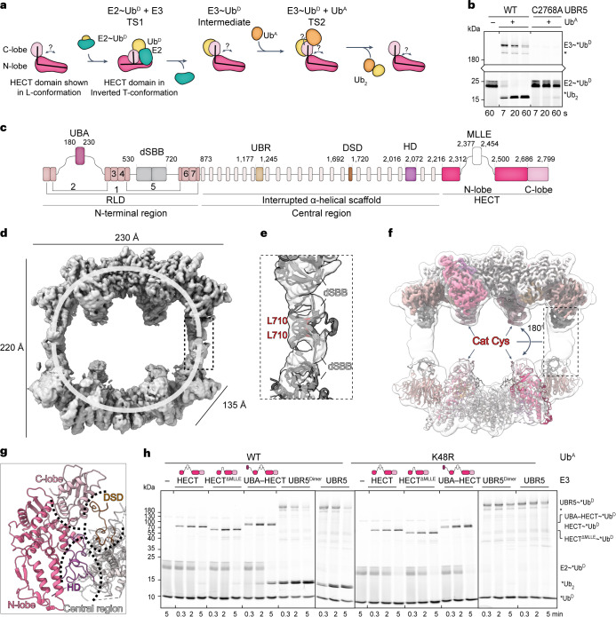
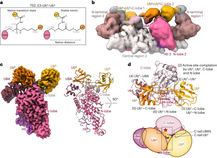
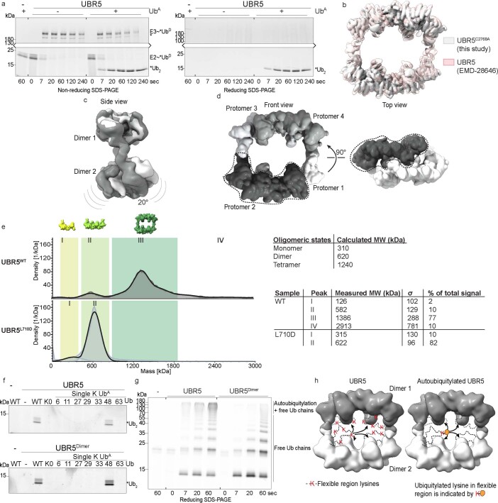
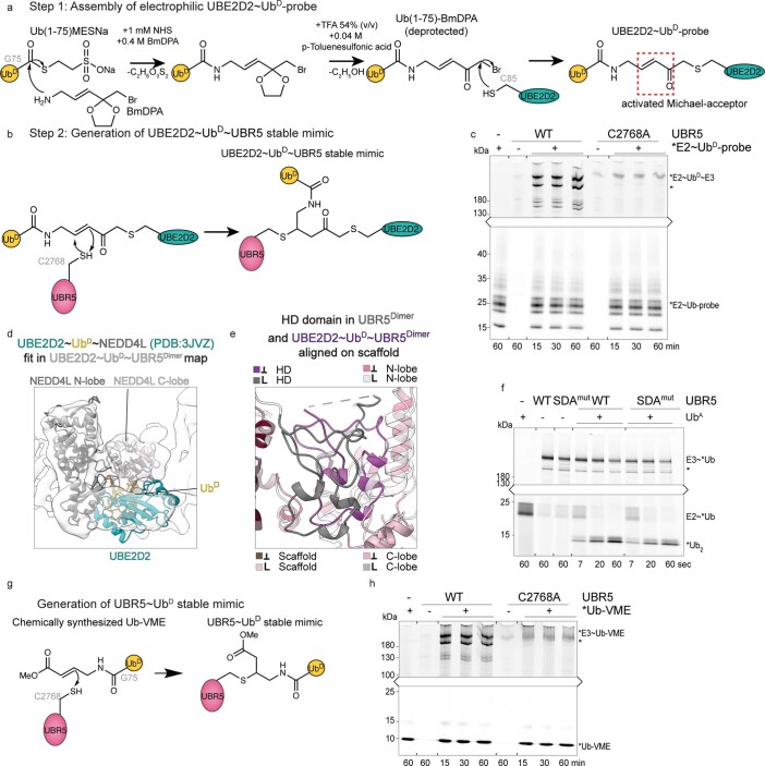
Ubiquitin (Ub) chain formation by homologous to E6AP C-terminus (HECT)-family E3 ligases regulates vast biology, yet the structural mechanisms remain unknown. We used chemistry and cryo-electron microscopy (cryo-EM) to visualize stable mimics of the intermediates along K48-linked Ub chain formation by the human E3, UBR5. The structural data reveal a ≈ 620 kDa UBR5 dimer as the functional unit, comprising a scaffold with flexibly tethered Ub-associated (UBA) domains, and elaborately arranged HECT domains. Chains are forged by a UBA domain capturing an acceptor Ub, with its K48 lured into the active site by numerous interactions between the acceptor Ub, manifold UBR5 elements and the donor Ub. The cryo-EM reconstructions allow defining conserved HECT domain conformations catalyzing Ub transfer from E2 to E3 and from E3. Our data show how a full-length E3, ubiquitins to be adjoined, E2 and intermediary products guide a feed-forward HECT domain conformational cycle establishing a highly efficient, broadly targeting, K48-linked Ub chain forging machine.


Conflict of interest statement

B.A.S. is a member of the scientific advisory boards of Interline Therapeutics and BioTheryX and co-inventor of intellectual property licensed to Cinsano. The remaining authors declare no competing interest.

Figures

Fig. 1
Fig. 1. Cryo-EM of UBR5 reveals oligomeric scaffold elaborately arranging HECT domains.
a, Cartoon depicting HECT E3-mediated Ub chain formation. b, Di-Ub synthesis assay testing activity of UBR5 WT and C2768A mutant. Fluorescently labeled K48R UbD (*UbD) is tracked through the cascade. The resulting di-Ub product is labeled *Ub2. Only upper and lower portions of the nonreducing gels showing *UbD-linked moieties are illustrated and connected for clarity. Throughout this work, single asterisk marks a UBR5-degradation-product that remains catalytically active. The assay was performed five times with similar results. c, UBR5 domains based on cryo-EM structure. d, Cryo-EM map of UBR5C2768A highlighting the two U-shaped units. Dotted box indicates dSBB domains shown in e as mediating tetramerization between the two dimeric units. e, AlphaFold2 model of dSBB domains with L710, in red. f, Transparent low-pass filtered map of tetrameric UBR5C2768A superimposed on UBR5Dimer density (top half) and experimentally derived coordinates (lower half). Rotation of 180° across the two halves is indicated. Dotted box indicates the position of dSBB domains, not well-resolved in the UBR5Dimer density. g, Close-up showing DSD and HD domain interactions with HECT domain C-lobe and N-lobe, respectively. h, Di-Ub synthesis assay testing various versions of UBR5:structurally redefined HECT domain with or without (HECTΔMLLE) MLLE insertion, with the UBA domain connected by a 15-residue linker, dimeric or WT full-length UBR5. Linkage-specificity was confirmed by comparing WT UbA or K48R UbA. The assay was performed twice with similar results. Source data
Fig. 2
Fig. 2. Cryo-EM map visualizing UbD transfer from E2 to UBR5.
a, Chemical structures of native TS1 and stable mimic for UbD transfer from E2 to UBR5. E2-to-E3 catalytic Cys distance is increased by three atoms in stable mimic compared to native TS1. b, Cryo-EM map of UBE2D2~UbD~UBR5Dimer. c, Model of E2~UbD (PDB: 3JVZ)-bound UBR5 HECT domain. Catalytic Cys of UBE2D2 (C85) and UBR5 (C2768) are labeled. d, Left, close-up of E2~UbD~UBR5Dimer model over E2-UBR5 interface. Right, di-Ub synthesis assay testing this interface using E2 F62A variant (UBE2D3 used). Three replicates showed similar results. A catalytically active UBR5-degradation-product is marked with an asterisk in d and e. e, Left, close-up of E2~UbD~UBR5Dimer model over C-lobe-UbD-interface. Right, di-Ub synthesis assay to test interface by steric clash (A2790W). Three replicates showed similar results. f, Superposition of apo-UBR5 or E2~UbD~UBR5Dimer aligned on scaffold N-lobes in gray and pink. UBR5 is depicted up to residue 2686 for clarity. HECT conformations are indicated with colored boxes and symbols (representation also used in subsequent figures). g, Docking E2~UbD on apo-UBR5 shows clashing in this conformation, indicated with intersecting red arcs. h, Rotation of the C-lobe between apo-UBR5 (gray) and TS1 (pink), aligned on N-lobes. i, Left, close-up of UBR5 SDA loop and UbD’s F4 patch in E2~UbD~UBR5Dimer model. Right, polyubiquitylation assay testing activity of SDA mutant (HLL1362-1364DDD). Three replicates showed similar results. Source data
Fig. 3
Fig. 3. Cryo-EM of stable mimic of UBR5Dimer~UbD intermediate.
a, Chemical structures of TS1 reaction product UBR5~UbD and chemically stable mimic. The stable mimic using Ub-VME to couple UbD to UBR5Dimer’s catalytic Cys maintains native number of atoms between the E3 Cys and UbD G75. b, Cryo-EM map of stable mimic representing UBR5Dimer~UbD. c, Model of UBR5Dimer~UbD from fitting coordinates for UBR5Dimer and UbD into the density. d, Overlay of HECT domain from UBR5Dimer~UbD (L-conformation) in pink/orange and from E2~UbD~UBR5Dimer (Inverted T-conformation) in gray/yellow indicates conformational change after UbD transfer from E2 to UBR5. For clarity, only the UBR5 HECT domain and UbD are shown. e, Left, frame 1 and right, frame 20 of 3D-VA performed on UBR5~UbD. UBR5Dimer N-terminal and central region, HECT domain N-lobe and C-lobe, and UbD were individually fitted into density.
Fig. 4
Fig. 4. Cryo-EM structure visualizing HECT E3-mediated linkage-specific Ub chain formation.
a, Chemical structures of native TS2 for Ub chain formation and chemically stable mimic. TS2 is mimicked by the installation of electrophilic moiety between the C-terminus of Ub (truncated at G75, representing UbD, yellow) and a Cys replacement for acceptor Ub’s K48 (UbA, orange). This traps UBR5Dimer’s catalytic Cys via a stable three-way cross-link while maintaining the native number of atoms between E3 catalytic Cys, G75 in UbD and the α-carbon for residue 48 in UbA. b, Initial low-resolution cryo-EM map of stable mimic of the UBR5Dimer~UbD~UbA complex (TS2). c, Local refined map (left) and atomic model (right) for catalytic complex mediating K48-linked Ub chain formation, wherein a UBA domain recruits UbA and residue 48 is placed at the HECT~UbD active site. For clarity, the remaining density corresponding to the scaffold is hidden. d, Structure above and cartoon below, indicating interfaces establishing the catalytic geometry for K48-linked Ub chain formation.
Fig. 5
Fig. 5. Elaborate interactions in HECT E3-mediated K48-linked Ub chain formation.
a, Cartoon of polyubiquitylation complex, highlighting regions tested in bh. b, Left, UBR5 C-lobe and UbD noncovalent interface. c, Left, multilayered active site assembly: UBR5’s C-terminal tail, UbD’s C-terminus linked to UBR5’s catalytic Cys, UbA and UBR5 N-lobe. Dotted lines represent electrostatic contacts. Di-Ub synthesis assays test effects of mutating UBR5’s C-terminus and UbA’s D58. Three repetitions gave similar results. A catalytically active UBR5-degradation-product is marked with an asterisk. d, Left, UBA domain interactions with UbA. Right, di-Ub synthesis assays, performed four times gave similar results. e, Left, UbA interactions with UBR5’s C-lobe. Right, di-Ub synthesis assay testing effects of mutating UbA’s A46 or UBR5’s Y2773 in interface. Three replicates showed similar results. f, Left, UbA interactions with UBR5 N-lobe. Right, di-Ub synthesis assay testing mutations of UbA R54, or UBR5’s E2287. Two replicates gave similar results. g, Left, close-up of LOL. Right, di-Ub synthesis assay testing effects of LOL mutated to Ala. Three replicates gave similar results. h, Left, close-up of UbA showing its residues that could be linked to other Ubs for branched chain formation. Right, tri-Ub synthesis assay with UBR5 and UBR5Dimer, testing di-Ubs with indicated linkages as acceptors. Coomassie-stained gels show di-Ub input. Assays performed three times gave similar results. Source data
Fig. 6
Fig. 6. Feed-forward mechanism of UBR5 forming K48-linked Ub chains.
a, UBR5 forms an oval tetramer composed of two dimeric, U-shaped multidomain assemblies, each containing a dimeric scaffold flexibly tethering UBA domains and connecting to HECT domains. Apo HECT domains adopt L-configuration, stabilized by HD and DSD domains, which is incompatible with E2~UbD-binding. b, E2~UbD binding to HECT domain drives reorientation to Inverted Tconfiguration. c, Release of E2 after UbD-transfer allows re-establishing scaffold connections with HECT domain in L-configuration. d, UbA’s K48 and UbD’s C-terminus linked to UBR5 are juxtaposed through multiple interactions, involving UBR5 regions and the Ubs being adjoined. e, Oligomerization could allow avid binding and modification of multiple Ub moieties within a chain.
Extended Data Fig. 1
Extended Data Fig. 1. Functional characterization of UBR5 and UBR5Dimer.
a, Autoubiquitylation assay of UBR5. E2~*UbD was added to UBR5 or UBR5 with UbA. SDS-PAGE shows isopeptide-linked UbD-bound products (reducing) or additionally thioester-bonded products (nonreducing). Two replicates were performed with similar results. b, Overlay of UBR5WT map (EMD-28646) with UBR5C2768A (this study). c, Breathing motion of UBR5C2768A during 3D classification. d, Monomers within tetramer are shaded individually, one is further highlighted using a dotted line. e, Molecular weights of UBR5WT and UBR5L710D assemblies determined by mass photometry. Measured and calculated MWs for distinct oligomeric states are shown. Measurements in triplicates gave comparable graphs. f, Di-Ub synthesis assay testing linkage-specificity of UBR5WT and UBR5Dimer. UbA with all lysines (WT), all lysines mutated to arginine (K0) or all but one Lys mutated to Arg, was used. Triplicates showed similar results. g, Free Ub chain formation in pulse-chase format for UBR5 and UBR5Dimer. Independent triplicates showed similar results. h, Low-pass filtered map of tetrameric UBR5 with dimers shaded. Left, unmodified UBR5 with flexible regions indicated by a dotted line, containing several lysines. Reachability of different HECT domains in cis or trans indicated by arrows. Right, autoubiquitylated UBR5. Accessibility of HECT domains in cis or trans for this Ub to serve as UbA is indicated. Source data
Extended Data Fig. 2
Extended Data Fig. 2. Structural characterization of UBR5Dimer.
a, Views of UBR5Dimer structure, termed ‘side’, ‘top’, ‘front’ and ‘bottom’, used in subsequent figures. b, Bottom-view of UBR5Dimer showing domains of protomers 1 and 2. c, Structure of 7-bladed β-propeller (RLD). First blade starts at UBR5’s N-terminus, and is completed by two more C-terminal β-strands. The second blade has three antiparallel strands and a ~270 residue insertion including the UBA domain (plum, not visible in UBR5Dimer map). The remaining blades are conventional, except for a >220-residue insertion containing dSBB domains, between the fifth and sixth blade. d, Analysis of scaffold module. α-helical bundles in protomers form intertwined dimerization-interface with one helix (1912-1928) particularly inserted into the other protomer. e, Left, structural analysis of a substrate recognition module: UBR domain is positioned within the scaffold, facing the HECT domain in trans. C1196/C1199/H1216/H1219, C1215/C1234/C1240/C1211, and C1211/C1179/C1208/C1232 coordinate three zinc ions. Right, cryo-EM map over UBR domain shows unassigned density in the canonical peptide-binding cleft indicated by overlay with crystal structure of UBR2s UBR domain bound to a substrate peptide (PDB: 3NY3). f, L-shaped HECT domain (white) compared with the previously annotated domain (N-lobe in pink, C-lobe in light pink). g, Structural analysis of HECT-stabilizing module. Left, zoom into DSD domain (brown) meandering between scaffold and HECT domain C-lobe. Right, zoom-in on HD domain (purple), interacting with the central region and the HECT domain N-lobe and contacting the interlobe linker connecting N- and C-lobe.
Extended Data Fig. 3
Extended Data Fig. 3. Chemical biology tools to visualize UbD transfer from E2 to UBR5 and subsequent UBR5~UbD intermediate.
a, Stable mimic representing UbD transfer from E2 to UBR5 (TS1). Reaction scheme, first step: installation of an electrophile between UbD C-terminus (UbD’s C-terminal G76 is deleted to approach near-native geometry) and active site Cys of E2 UBE2D2 (other cysteines mutated, see Methods). b, Reaction scheme, second step: the UBE2D2~UbD probe reacted with UBR5Dimer generating UBE2D2~UbD~UBR5Dimer with E2, UbD, and UBR5Dimer linked at a single atom. c, Dependence for UBE2D2~UbD~UBR5Dimer complex formation on UBR5s catalytic Cys was tested using fluorescent UBE2D2 in the probe. Two replicates showed similar results. d, Crystal structure of UBE2D2~UbD~HECTNEDD4L (PDB: 3JVZ), containing E2~UbD oxyester linkage with native distances fit into UBE2D2~UbD~UBR5Dimer map. e, Close-up of HD domain in apo UBR5Dimer and UBE2D2~UbD~UBR5Dimer (grey and purple), aligned on scaffold. HECT domain conformations are indicated. f, Di-Ub-synthesis assay for the SDA-mutant. Triplicates of this assay showed comparable results. g, Reaction scheme to generate stable mimic of UBR5~UbD. Fully synthetic Ub-VME was reacted with UBR5Dimer. h, UBR5 catalytic Cys dependence for forming UBR5~UbD complex was tested using a fluorescent version of Ub-VME. The specificity was tested twice. Source data
Extended Data Fig. 4
Extended Data Fig. 4. Chemical biology tools to visualize K48-linked Ub chain formation by UBR5.
a, Stable mimic representing TS2, UbD transfer from UBR5 to UbA, was generated in two steps. Reaction scheme for the first step: installation of electrophile between UbD C-terminus (here, UbD’s C-terminal G76 is deleted to obtain native distances) and a Cys replacement for K48 in UbA. b, Reaction scheme for second step to generate TS2 mimic: the UbD~UbA-probe was reacted with UBR5Dimer to generate a UBR5Dimer~UbD~UbA complex wherein the C-terminus of UbD, residue 48 of UbA, and UBR5’s catalytic Cys are linked at a single atom. c, Structural superposition of UBR5’s C-lobe bound to UbD and UbA in TS2 and HUWE1’s C-lobe bound to UbD (PDB: 6XZ1). The C-terminus of HUWE1 overlays with UBR5’s C-terminus in TS2. The ultimate residue was resolved for HUWE1 and provides a lead to UBR5’s ultimate residue. d, Di-Ub synthesis assay testing pH-dependency of LOL-caused deficiency. E2~*UbD was added to WT or LOL-mutant UBR5 at indicated pH. This experiment was performed twice with comparable results. Source data
Extended Data Fig. 5
Extended Data Fig. 5. Conserved step-by-step conformational trajectory for HECT E3-catalyzed Ub transfer cascades.
a, Structural superposition of UBE2D2~UbD-bound HECT domains of UBR5 and NEDD4L (PDB: 3JVZ). The structures suggest E2 transfers UbD to HECT domains in the Inverted T-configuration. Colored boxes indicate the conformation of the respective complex. b, Structural superposition of UbD linked to HECT domain of UBR5 (TS1 model) and NEDD4 (PDB: 4BBN). This represents the UbD-linked HECT domain in the Inverted T-configuration, immediately after UbD transfer from E2. c, Structural superposition of UbD-loaded HECT domains of UBR5 (from UBR5Dimer~UbD intermediate) and HUWE1~UbD (PDB: 6XZ1), both in L-configuration. d, Structural superposition of HECT domains representing ubiquitylation complexes: K48-linked Ub chain formation by UBR5, and UbD transfer to a peptide substrate for Rsp5 (PDB: 4LCD). The structures suggest HECT domains transfer UbD to downstream targets - either substrates or acceptor Ub during polyubiquitylation - in the L-configuration.

Similar articles

Cited by

References

    1. Komander D, Rape M. The ubiquitin code. Annu. Rev. Biochem. 2012;81:203–229. doi: 10.1146/annurev-biochem-060310-170328. - DOI - PubMed
    1. Husnjak K, Dikic I. Ubiquitin-binding proteins: decoders of ubiquitin-mediated cellular functions. Annu. Rev. Biochem. 2012;81:291–322. doi: 10.1146/annurev-biochem-051810-094654. - DOI - PubMed
    1. Chau V, et al. A multiubiquitin chain is confined to specific lysine in a targeted short-lived protein. Science. 1989;243:1576–1583. doi: 10.1126/science.2538923. - DOI - PubMed
    1. Spence J, Sadis S, Haas AL, Finley D. A ubiquitin mutant with specific defects in DNA repair and multiubiquitination. Mol. Cell. Biol. 1995;15:1265–1273. doi: 10.1128/MCB.15.3.1265. - DOI - PMC - PubMed
    1. Galan JM, Haguenauer-Tsapis R. Ubiquitin lys63 is involved in ubiquitination of a yeast plasma membrane protein. EMBO J. 1997;16:5847–5854. doi: 10.1093/emboj/16.19.5847. - DOI - PMC - PubMed

Publication types