. 2023 Aug 24;18(1):620.
doi: 10.1186/s13018-023-04092-x.

PPARγ activation suppresses chondrocyte ferroptosis through mitophagy in osteoarthritis

Affiliations

PPARγ activation suppresses chondrocyte ferroptosis through mitophagy in osteoarthritis

Xiang Xue et al. J Orthop Surg Res. .

Abstract

Background: Osteoarthritis (OA) is a prevalent disease plaguing the elderly. Recently, chondrocyte ferroptosis has been demonstrated to promote the progression of OA. Peroxisome proliferator-activated receptor-γ (PPARγ) is an important factor in maintaining cartilage health. However, the relationship between PPARγ and chondrocyte ferroptosis in OA and its mechanism is completely unclear.

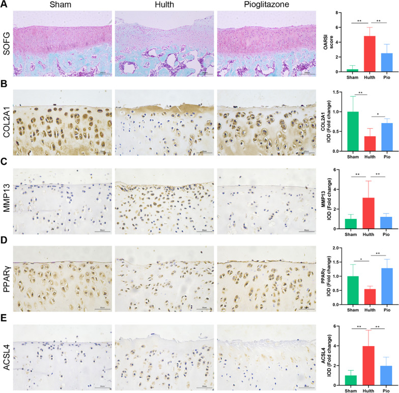
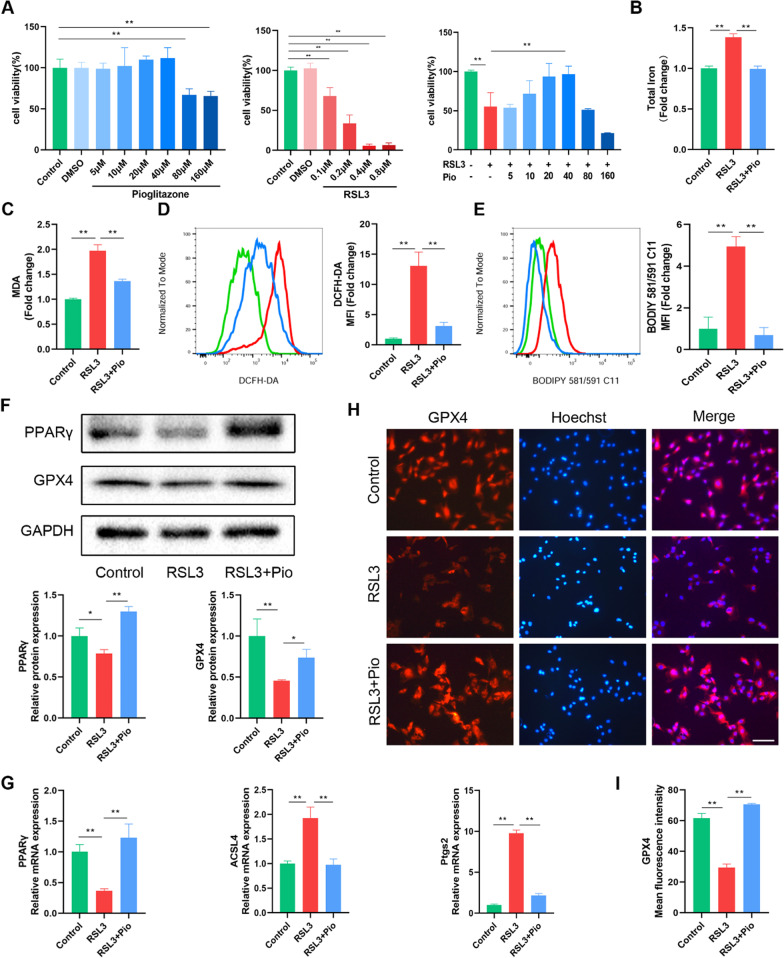
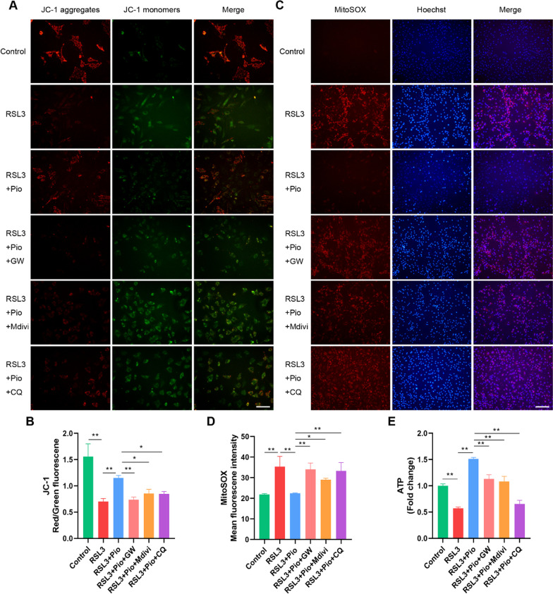
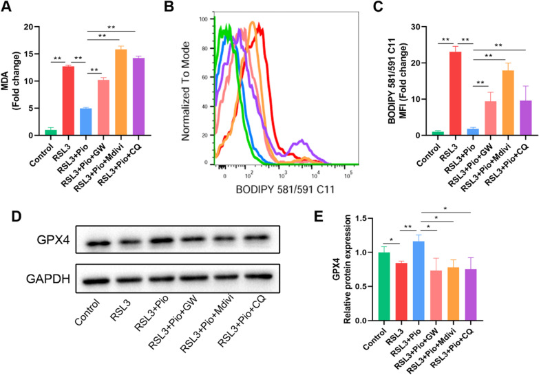
Methods: We established a surgically induced knee OA rat model to investigate PPARγ and chondrocyte ferroptosis in OA. Rat knee specimens were collected for Safranin O/Fast Green staining and immunohistochemical staining after administered orally placebo or pioglitazone (PPARγ agonist) for 4 weeks. We used RSL3 to establish a chondrocyte ferroptosis model cultured in vitro to study the role of PPARγ activation toward ferroptosis, mitochondrial function, and PTEN-induced putative kinase 1 (Pink1)/Parkin-dependent mitophagy. GW9662 (PPARγ antagonist), Mdivi-1 (mitophagy inhibitor), and chloroquine (mitophagy inhibitor) were employed to investigate the mechanism of PPARγ-Pink1/Parkin-dependent mitophagy in the inhibition of ferroptosis.

Results: We found that PPARγ activation by pioglitazone attenuated not only OA but also inhibited the expression of the ferroptosis marker acyl-CoA synthetase long-chain family member 4 (ACSL4) at the same time in rats. Furthermore, in vivo and in vitro data indicated that PPARγ activation restored Pink1/Parkin-dependent mitophagy, improved mitochondrial function, inhibited chondrocyte ferroptosis, and delayed the progression of OA.

Conclusions: The present study demonstrated that PPARγ activation attenuates OA by inhibiting chondrocyte ferroptosis, and this chondroprotective effect was achieved by promoting the Pink1/Parkin-dependent mitophagy pathway.

Keywords: Chondrocyte; Ferroptosis; Mitophagy; Osteoarthritis; PPARγ; Pink1.


Conflict of interest statement

All authors declare that they have no competing interests.

Figures

Fig. 1
Fig. 1
PPARγ activation attenuates osteoarthritis in rats, accompanied by inhibition of ferroptosis. A Cartilage sections were stained by Safranin O/fast green staining and evaluated with OARSI score (n = 6; Scale bars, 200 µm). BE Representative pictures of COL2A1, MMP13, PPARγ, and ACSL4 immunohistochemical staining in rat cartilage samples. Quantitative analysis results are on the right (n = 6; Scale bars, 50 µm). Data were expressed as mean ± S.D. *p < 0.05; **p < 0.01
Fig. 2
Fig. 2
PPARγ activation attenuates RSL3-induced ferroptosis in rat chondrocytes. A Rat chondrocytes were treated for 24 h with pioglitazone (5, 10, 20, 40, 80, 160 μM), RSL3 (0.1, 0.2, 0.4, 0. 8 μM) for 4 h or pretreated with pioglitazone (5, 10, 20, 40, 80, 160 μM) for 24 h and then cultured for 4 h combine with RSL3 (0.2 μM). The CCK-8 test was used to determine cell viability (n = 3). B, C Intracellular total iron and MDA were measured using commercial kits, respectively (n = 3). D, E ROS and LPO were labeled with DCFH-DA and BODIPY 581/591 C11 dyes, respectively. Fluorescence signal detected by flow cytometry. The fold change of mean fluorescence intensity (MFI) was presented (n = 3). F The protein levels of PPARγ and GPX4 were determined by western blot (n = 3). G The mRNA PPARγ, ACSL4, and Ptgs2 expression levels were detected using RT-qPCR (n = 3). H, I Immunofluorescence staining of GPX4 in rat chondrocytes (n = 3; Scale bars, 100 µm). Data were expressed as mean ± S.D. *p < 0.05; **p < 0.01
Fig. 3
Fig. 3
PPARγ activation attenuates chondrocyte mitochondrial damage caused by ferroptosis. A, B JC-1 images and the ratio of red/green fluorescence in chondrocytes (n = 3, Scale bars, 100 µm). C, D MitoSOX staining was used to detect mtROS levels. Mean fluorescence intensities were used for statistical analysis (n = 3, Scale bars, 200 µm). E ATP production was measured using commercial kits (n = 3). Data were expressed as mean ± S.D. *p < 0.05; **p < 0.01
Fig. 4
Fig. 4
PPARγ activation restores impaired mitophagy. AD Pink1, Parkin, and LC3B, with GAPDH as an internal control detected by western blot. Statistical analysis of western blotting is located on the right (n = 3). EH Representative pictures of Pink1 and Parkin immunohistochemical staining in rat cartilage samples. Quantitative analysis results are on the right (n = 6, Scale bars, 50 µm). Data were expressed as mean ± S.D. *p < 0.05; **p < 0.01
Fig. 5
Fig. 5
PPARγ activates Pink1/Parkin-dependent mitophagy in chondrocytes. AE PPARγ, Pink1, Parkin, and LC3B, with GAPDH as an internal control detected by western blot. Statistical analysis of western blotting is located on the right (n = 3). Data were expressed as means ± S.D. *p < 0.05; **p < 0.01
Fig. 6
Fig. 6
PPARγ mitigates chondrocyte mitochondrial damage through mitophagy. A, B JC-1 images and the ratio of red/green fluorescence in chondrocytes (n = 3; Scale bars, 100 µm). C, D MitoSOX staining was used to detect mtROS levels. Mean fluorescence intensities were used for statistical analysis (n = 3; Scale bars, 200 µm). E ATP production was measured using commercial kits (n = 3). Data were expressed as means ± S.D. *p < 0.05; **p < 0.01
Fig. 7
Fig. 7
Inhibition of PPARγ-mediated mitophagy aggravates chondrocyte ferroptosis. A MDA were measured using commercial kits (n = 3). BC LPO is labeled with BODIPY 581/591 C11 dye and assayed by flow cytometry. The fold change of MFI was presented (n = 3). D, E Western blot analysis of GPX4, with GAPDH as an internal control. Statistical analysis of western blotting is located on the right (n = 3). Data were expressed as means ± S.D. *p < 0.05; **p < 0.01
Fig. 8
Fig. 8
Schematic diagram of the potential mechanisms by which PPARγ activation suppresses chondrocyte ferroptosis through mitophagy in osteoarthritis. Activation of PPARγ by pioglitazone promotes the upregulation of Pink1 and Parkin, decreasing mtROS production, and restoring mitochondrial function, thereby suppressing the level of lipid peroxidation and ultimately inhibiting chondrocyte ferroptosis, alleviating OA symptoms

Similar articles

Cited by

References

    1. Glyn-Jones S, Palmer AJ, Agricola R, Price AJ, Vincent TL, Weinans H, et al. Osteoarthr Lancet. 2015;386(9991):376–387. - PubMed
    1. van den Berg WB. Osteoarthritis year 2010 in review: pathomechanisms. Osteoarthr Cartil. 2011;19(4):338–341. - PubMed
    1. Chen X, Song QL, Li ZH, Ji R, Wang JY, Cao ML, et al. Pterostilbene ameliorates oxidative damage and ferroptosis in human ovarian granulosa cells by regulating the Nrf2/HO-1 pathway. Arch Biochem Biophys. 2023:109561. - PubMed
    1. Liu H, Zhao Z, Yan M, Zhang Q, Jiang T, Xue J. Calycosin decreases cerebral ischemia/reperfusion injury by suppressing ACSL4-dependent ferroptosis. Arch Biochem Biophys. 2023;734:109488. - PubMed
    1. Yao X, Sun K, Yu S, Luo J, Guo J, Lin J, et al. Chondrocyte ferroptosis contribute to the progression of osteoarthritis. J Orthop Translat. 2021;27:33–43. - PMC - PubMed

Substances