. 2023 Aug 7;14(33):8823-8830.
doi: 10.1039/d3sc03593j. eCollection 2023 Aug 23.

Ligand-protected nanocluster-mediated photoswitchable fluorescent nanoprobes towards dual-color cellular imaging

Affiliations

Ligand-protected nanocluster-mediated photoswitchable fluorescent nanoprobes towards dual-color cellular imaging

Wencheng Zhong et al. Chem Sci. .

Abstract

Development of robust multi-color photoswitchable fluorescent probes is critical for many optical applications, but it remains a challenge to rationally design these probes. Here, we report a new design of Förster resonance energy transfer-based dual-color photoswitchable fluorescent nanoparticles (DPF NPs) by taking advantage of the distinct properties of ligand-protected gold nanoclusters (AuNCs). Detailed photophysical studies revealed that ultrasmall-sized AuNCs not only act as the FRET donors due to their intrinsic fluorescence properties, but also play a significant role in regulating the photochromic and aggregate properties of spiropyran through ligand-spiropyran interactions. These DPF NPs exhibit a high fluorescence on/off ratio (∼90%) for both green and red fluorescence emission, and good reversibility during cycled photo-stimulation. Cell imaging experiments showed that DPF NPs could specifically accumulate in lipid droplets, and enable photoswitchable dual-color imaging in living cells. Moreover, by labeling mitochondria with a green-emitting marker, we demonstrated that DPF NPs can distinguish different targets based on dynamic and static fluorescence signals at the sub-cellular level in two emission channels reliably. This study provides a new strategy for designing robust photoswitchable fluorescent probes by modulating the properties of photochromic dyes through ligand-protected nanoclusters, which can be generalized for the development of other photoswitch systems towards advanced optical applications.


Conflict of interest statement

The authors declare no conflict of interest.

Figures

Scheme 1
Scheme 1. (A) Schematic diagram of the design principle of DPF NPs, and (B) illustration of differentiating static and dynamic fluorescence signals from different sub-cellular compartments by employing DPF NPs as probes.
Fig. 1
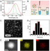
Fig. 1. (A) Fluorescence emission spectra of AuNCs in aqueous solution (excitation wavelength: 405 nm) and absorption spectra of SP/MC in ethanol. (B) Scheme of the preparation of DPF NPs. (C) Representative TEM image of DPF NPs in high-angle annular dark-field (HADDF) mode. The inset photograph is the enlarged version of several DPF NPs. (D) Size distribution histogram of DPF NPs based on TEM images. (E) EDS element mapping of Au and S in an individual DPF NP.
Fig. 2
Fig. 2. (A) Fluorescence emission spectra of DPF NPs before and after UV irradiation. Excitation wavelength: 405 nm (inset photograph: photocontrolled dual-color fluorescence switching of DPF NPs in aqueous solution). (B) Time-dependent fluorescence intensity change of DPF NPs at 530 nm and 645 nm under UV irradiation. (C) Fluorescence spectra of DPF NPs during several 365 nm/520 nm light irradiation cycles. Excitation wavelength: 405 nm. (D) Change of fluorescence intensity at 530 nm and 645 nm upon cycled irradiation 10 times.
Fig. 3
Fig. 3. Absorption spectra of (A) SP@DSPE-mPEG, (B) DPF NPs in aqueous solution, and (C) SP/MC in ethanol. (D) Scheme of the proposed arrangement of MC on the surface of Arg/ATT-AuNCs.
Fig. 4
Fig. 4. Photochromic kinetics of (A) spiropyran in ethanol, (B) spiropyran in DPF NPs and (C) SP-BSA in aqueous solution during the coloring and discoloring process. (D) Comparison of rate constants of SP → MC (k1) and MC → SP (k2) based on data in A–C.
Fig. 5
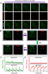
Fig. 5. (A) Time-dependent fluorescence images of DPF NP-incubated HeLa cells under UV irradiation in different emission channels (scale bar: 10 μm). (B) Fluorescence intensity changes of green and red channels versus the irradiation time based on fluorescence images. (C) Dual-fluorescence signal change in HeLa cells upon cycled irradiation with UV/vis light (scale bar: 10 μm). (D) Changes in the fluorescence intensity of green and red channel upon cycled irradiation with UV/vis light.
Fig. 6
Fig. 6. (A) Time-dependent fluorescence images of HeLa cells upon labelling with DPF NPs and MitoTracker™ Green under UV irradiation in different emission channels (scale bar: 10 μm). (B) Fluorescence images of HeLa cells upon labelling with DPF NPs and MitoTracker™ Green under cycled irradiation with UV/vis light in green and red channels. White circles in the images indicate two representative lipid droplets identified based on the dynamic signals. Changes in the fluorescence intensity of the (C) green channel and (D) red channel in mitochondria (upper) and lipid droplets (lower) of HeLa cells upon cycled irradiation with UV/vis light.

References

    1. Liu Y. Liang S. Yuan C. Best A. Kappl M. Koynov K. Butt H. J. Wu S. Adv. Funct. Mater. 2021;31:2103908. doi: 10.1002/adfm.202103908. - DOI
    1. Imato K. Momota K. Kaneda N. Imae I. Ooyama Y. Chem. Mater. 2022;34:8289–8296. doi: 10.1021/acs.chemmater.2c01809. - DOI
    1. Chai J. Zhao Y. Xu L. Li Q. Hu X. Y. Guo D. S. Liu Y. Angew. Chem., Int. Ed. 2022;61:e202116073. - PubMed
    1. Kim Y. Thangam R. Yoo J. Heo J. Park J. Y. Kang N. Lee S. Yoon J. Mun K. R. Kang M. Min S. Kim S. Y. Son S. Kim J. Hong H. Bae G. Kim K. Lee S. Yang L. Lee J. Y. Kim J. Park S. Kim D. H. Lee K. B. Jang W. Y. Kim B. H. Paulmurugan R. Cho S. W. Song H. C. Kang S. J. Sun W. Zhu Y. Lee J. Kim H. J. Jang H. S. Kim J. S. Khademhosseini A. Kim Y. Kim S. Kang H. Adv. Mater. 2022;34:2205498. doi: 10.1002/adma.202205498. - DOI - PubMed
    1. Kim D. Aktalay A. Jensen N. Uno K. Bossi M. L. Belov V. N. Hell S. W. J. Am. Chem. Soc. 2022;144:14235–14247. doi: 10.1021/jacs.2c05036. - DOI - PMC - PubMed