. 2023 Aug 9:17:1242245.
doi: 10.3389/fnana.2023.1242245. eCollection 2023.

The nuclei of the lateral lemniscus: unexpected players in the descending auditory pathway

Affiliations

The nuclei of the lateral lemniscus: unexpected players in the descending auditory pathway

Mario Gómez-Martínez et al. Front Neuroanat. .

Abstract

Introduction: In the mammalian auditory pathway, the nuclei of the lateral lemniscus (NLL) are thought to be exclusively involved in the bottom-up transmission of auditory information. However, our repeated observation of numerous NLL neurons labeled after injection of retrograde tracers into the superior olivary complex (SOC) led us to systematically investigate with retrograde tracers the descending projections from the NLL to the SOC of the rat.

Methods: We performed large injections of FluoroGold into the SOC to determine NLL contributions to descending projections, and focal injections of biotinylated dextran amine (BDA) to pinpoint the specific nuclei of the SOC innervated by each NLL.

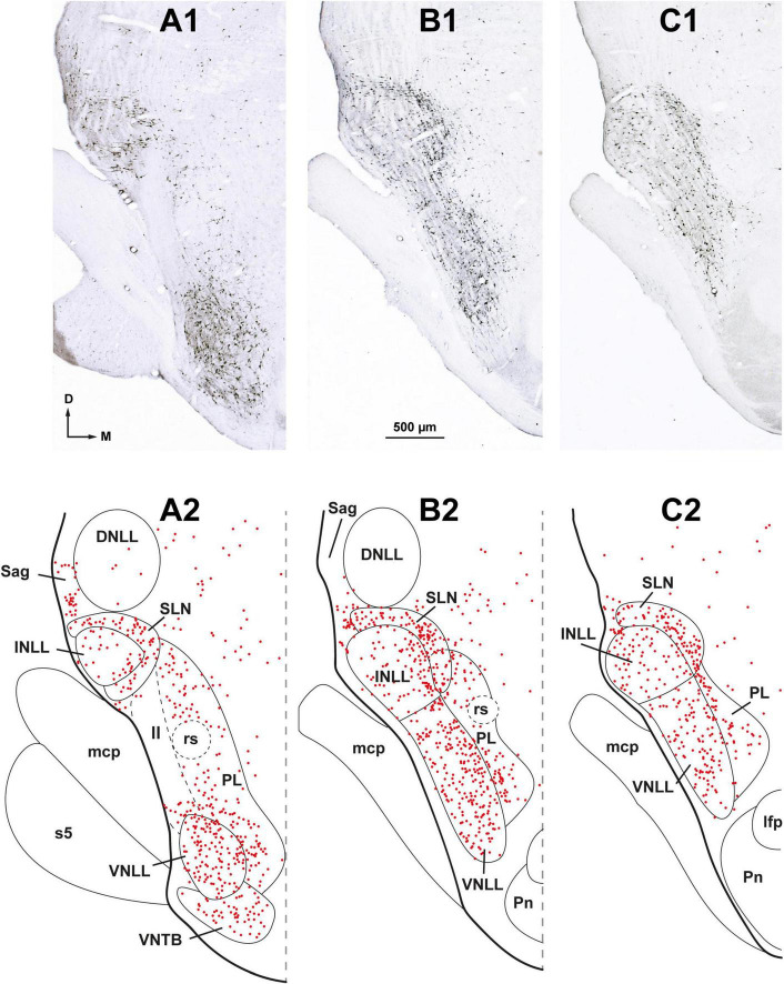
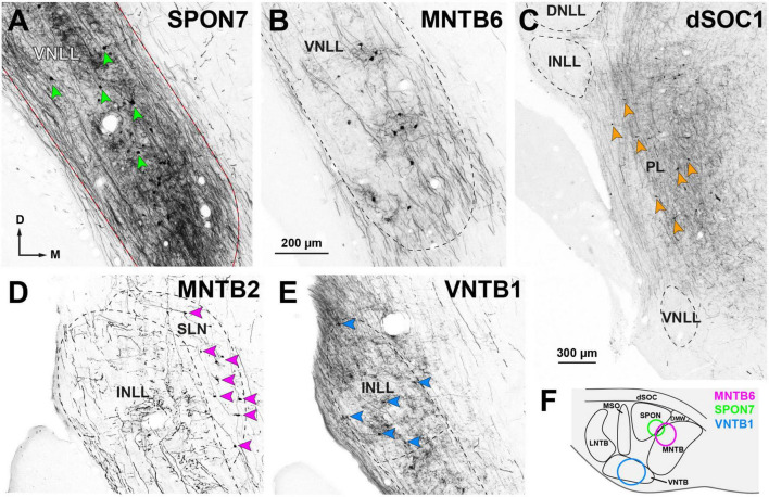
Results: The SOC is innervated by thousands of neurons distributed across four nuclei or regions associated with the lateral lemniscus: the ipsilateral ventral and intermediate nuclei of the lateral lemniscus (VNLL and INLL); the medial paralemniscal region (PL) of both sides; and the ipsilateral semilunar nucleus (SLN), a previously unrecognized nucleus that wraps around the INLL dorsally, medially, and caudally and consists of small, flat neurons. In some experiments, at least 30% of neurons in the VNLL and INLL were retrogradely labeled. All nuclei of the SOC, except the medial and lateral superior olives, are innervated by abundant lemniscal neurons, and each SOC nucleus receives a unique combination of lemniscal inputs. The primary target of the projections from the VNLL is the ventral nucleus of the trapezoid body (VNTB), followed by the superior paraolivary nucleus (SPON), and the medial nucleus of the trapezoid body (MNTB). The INLL selectively innervates the VNTB. The PL innervates dorsal periolivary regions bilaterally. The SLN preferentially innervates the MNTB and may provide the first identified non-calyceal excitatory input to MNTB neurons.

Discussion: Our novel findings have strong implications for understanding acoustic information processing in the initial stages of the auditory pathway. Based on the proportion of lemniscal neurons involved in all the projections described, the NLL should be considered major players in the descending auditory pathway.

Keywords: FluoroGold (FG); biotinylated dextran amine (BDA); medial nucleus of the trapezoid body (MNTB); paralemniscal; semilunar nucleus; superior olivary complex; superior paraolivary nucleus (SPON); ventral nucleus of the trapezoid body (VNTB).


Conflict of interest statement

The authors declare that the research was conducted in the absence of any commercial or financial relationships that could be construed as a potential conflict of interest.

Figures

FIGURE 1
FIGURE 1
Parcellation of the superior olivary complex (SOC) and nuclei of the lateral lemniscus (NLL). (A) Schematic representation of the parcellation of the rat SOC as it appears in coronal sections through the caudal half of the complex. In addition to the six classical nuclei (lateral and medial superior olives [LSO and MSO, respectively], superior paraolivary nucleus [SPON], and medial, ventral and lateral nuclei of the trapezoid body [MNTB, VNTB and LNTB, respectively]), the complex includes: the dorsomedial wedge (DMW), sandwiched between SPON and MNTB; the dorsal ribbon (dSOC), located dorsal to SPON, DMW, and MNTB; and the dorsal periolivary nucleus (DPO), located above LSO. (B) Schematic representation of the parcellation of the NLL as they appear in coronal sections through their mid-rostrocaudal level. In the rat, there are six NLL: the three classical nuclei (ventral [VNLL], intermediate [INLL] and dorsal [DNLL]); the nucleus sagulum (Sag), which lies lateral to the DNLL; the previously unrecognized semilunar nucleus (SLN), which covers the INLL medially, dorsally and caudally; and the medial paralemniscal region (PL), which lies medial to the VNLL and SLN. Other abbreviations: 7n, root of the facial nerve; lfp, longitudinal fascicle of the pons; mcp, middle cerebellar peduncle; Pn, pontine nuclei; Pr5VL, principal sensory trigeminal nucleus, ventrolateral part; py, pyramidal tract; rs, rubrospinal tract. The calibration bar in (B), uncorrected for shrinkage, also applies to (A).
FIGURE 2
FIGURE 2
Following a large injection of FluoroGold into the rat superior olivary complex (SOC), abundant neurons are retrogradely labeled in all nuclei known to innervate the SOC. Micrographs of coronal sections from case FG01. (A) Section through the center of a large injection site of FluoroGold (outlined in yellow) that affected all nuclei of the SOC. (B–E) Neurons labeled in the ipsilateral (B,C) and contralateral (D,E) cochlear nuclei. Notice the abundance of labeled neurons in the posteroventral (PVCN) and anteroventral (AVCN) cochlear nuclei of both sides, which contrasts with the paucity of neurons in the dorsal cochlear nucleus (DCN). (F) Widespread distribution of the abundant neurons labeled in the ipsilateral inferior colliculus (IC). (G) Small multipolar neurons labeled in the contralateral ventral nucleus of the trapezoid body (VNTB). (H,I) Neurons labeled in the locus coeruleus (LC) of both sides. (J) Neurons labeled in the ipsilateral ventral tectal longitudinal column (TLCv). (K,L) Neurons labeled in the subparafascicular nucleus of the thalamus (SPF) of both sides. (M,N) Pyramidal neurons labeled in layer 5 of the auditory cortex of both sides. For other abbreviations, see Figure 1. Calibration bars uncorrected for shrinkage. Calibration bar in (D) also applies to (B,C,E). Calibration bar in (I) also applies to (H). Calibration bar in (K) also applies to (G,J,L). Calibration bar in (N) also applies to (M).
FIGURE 3
FIGURE 3
Neurons retrogradely labeled in the nuclei of the lateral lemniscus (NLL) following a large injection of FluoroGold into the ipsilateral superior olivary complex. Case FG01, whose injection site is shown in Figure 2A. (A1–C1) Low magnification micrographs of three coronal sections through the NLL. The sections are arranged from caudal to rostral and are regularly spaced by intervals of 320 μm. (A2–C2) Plots of the distribution of labeled neurons in the corresponding sections shown above. Each red dot represents one labeled neuron. Abundant labeled neurons are widely distributed throughout the ventral nucleus of the lateral lemniscus (VNLL), the intermediate nucleus of the lateral lemniscus (INLL), the semilunar nucleus (SLN), and the medial paralemniscal region (PL). Notice that the SLN wraps around the INLL. ll, lateral lemniscus; s5, sensory root of the trigeminal nerve. For other abbreviations, see Figure 1. Calibration bar in (B1), uncorrected for shrinkage, applies to all panels.
FIGURE 4
FIGURE 4
Neurons retrogradely labeled in the nuclei of the lateral lemniscus (NLL) following a large injection of FluoroGold into the contralateral superior olivary complex. Case FG01, whose injection site is shown in Figure 2A. (A1–C1) Low magnification micrographs of three coronal sections through the NLL. The sections are ordered from caudal to rostral and are regularly separated by intervals of 320 μm. (A2–C2) Plots of the distribution of the labeled neurons in the corresponding sections shown above. Each red dot represents one labeled neuron. Most labeled neurons are found in the medial paralemniscal region (PL); the ventral and the intermediate nuclei of the lateral lemniscus (VNLL, INLL) are almost completely devoid of labeled neurons. The asterisks indicate a fiducial mark used to align the sections dorsoventrally. ll, lateral lemniscus; s5, sensory root of the trigeminal nerve. For other abbreviations, see Figure 1. Calibration bar in (B1), uncorrected for shrinkage, applies to all panels.
FIGURE 5
FIGURE 5
Details of neurons labeled in the ipsilateral (A–C, E–G) and contralateral (D) nuclei of the lateral lemniscus (NLL) following a large injection of FluoroGold into the superior olivary complex. Case FG01, whose injection site is shown in Figure 2A. Micrographs of coronal sections. (A) In the ipsilateral intermediate nucleus of the lateral lemniscus (INLL), labeled neurons are widely distributed. Most of the labeled neurons are multipolar and arranged horizontally. (B) In the ventral half of the ipsilateral ventral nucleus of the lateral lemniscus (VNLL), labeled neurons are more densely packed and lack a preferred orientation. Bushy neurons intermingle with multipolar neurons. Blue arrows indicate typical bushy neurons and red arrows indicate typical multipolar neurons. (C) In the ipsilateral semilunar nucleus (SLN), most labeled neurons appear flattened and horizontally oriented. Notice that the SLN includes the region dorsal to the INLL, formerly known as the horizontal cell group. The neurons labeled in the SLN show more extensive dendritic filling than those labeled in the INLL. (D) In the contralateral medial paralemniscal region (PL), labeled neurons are concentrated near the medial border of the VNLL. Most neurons appear multipolar and lack a preferred dendritic orientation. The boxed area is shown at a higher magnification in Figure 6G. (E) In the ipsilateral sagulum (Sag), labeled neurons are predominantly small, and their dendrites show a vertical alignment, in contrast to the horizontal orientation of neurons in the SLN. (F) Typical bushy neuron labeled in the ipsilateral VNLL. (G) Typical multipolar neurons in the ipsilateral INLL. Calibration bars uncorrected for shrinkage. Calibration bar in (C) also applies to (A,B,D). Calibration bar in (G) also applies to (F).
FIGURE 6
FIGURE 6
High magnification micrographs of neurons labeled in the ipsilateral (A–F) and contralateral (G) nuclei of the lateral lemniscus following the injection of FluoroGold into the superior olivary complex. Coronal sections. (A) Multipolar neuron of the ventral nucleus of the lateral lemniscus (VNLL). (B) Small cluster of bushy neurons from the ventral portion of the VNLL. (C) Flat, horizontally oriented neurons from the dorsal part of the semilunar nucleus (SLN). (D) Polymorphic neurons from the ventral portion of the ipsilateral medial paralemniscal region (PL). (E) A rare bushy-like neuron of the intermediate nucleus of the lateral lemniscus (INLL). (F) Multipolar neuron of the INLL. (G) Neurons labeled in the contralateral PL. The location of this microscopic field is indicated by the boxed area in Figure 5D. The brown staining in panels (A–E) was obtained with a plain peroxidase reaction using diaminobenzidine as the chromogen. In the sections shown in (F,G), the peroxidase reaction was enhanced with nickel salts, hence the black color. The calibration bar in (G), uncorrected for shrinkage, applies to all images.
FIGURE 7
FIGURE 7
Neurons retrogradely labeled in the nuclei of the lateral lemniscus (NLL) following smaller injections of FluoroGold into the ipsilateral superior olivary complex (SOC). Micrographs of coronal sections. (A1,A2) Case FG02. Following a relatively dorsal injection of FluoroGold into the medial half of the SOC (inset), abundant neurons are labeled in the ventral nucleus of the lateral lemniscus (VNLL), the semilunar nucleus (SLN), and the medial paralemniscal region (PL). Section A1 is 160 μm more caudal than section A2. (B) Case FG03. Following a very medial injection that mainly affected the medial nucleus of the trapezoid body (MNTB) (inset, purple outline), most labeled neurons were found in the SLN. (C) Case FG04. Very caudal section through the intermediate nucleus of the lateral lemniscus (INLL). Notice that the labeled neurons of the SLN surround the INLL. The injection site is shown in the inset, green outline. (D) Case FG05. Following a very medial and dorsal injection that affected mostly the dorsal ribbon of the SOC (dSOC) (inset), most labeled neurons were found in the PL, and the VNLL was almost devoid of labeling. (E) Case FG06. Following an injection centered in the SOC that spared the dSOC (inset), abundant neurons were labeled in the VNLL, and the PL contained scarce labeled neurons. Notice that the labeling pattern in this case is complementary to that of case FG05, shown in (D). (F) Case FG07. Following an injection into the medial half of the SOC that affected mainly the superior paraolivary nucleus (SPON), the ventral nucleus of the trapezoid body (VNTB), and the MNTB (inset), abundant neurons were labeled in the INLL and SLN. For other abbreviations, see Figure 1. Calibration bars uncorrected for shrinkage. Calibration bar in (A1) also applies to (A2,B,C), and calibration bar in (F) also applies to (D,F).
FIGURE 8
FIGURE 8
Injection sites of BDA into individual nuclei of the superior olivary complex (SOC). Micrographs of cresyl violet-counterstained, coronal sections through the center of the injection site in: (A) The rostral portion of the ventral nucleus of the trapezoid body (VNTB). (B) The medial half of the superior paraolivary nucleus (SPON). The dense plexus of labeled fibers in the medial limb of the lateral superior olive (LSO) is formed by collaterals of the axons of MNTB neurons that innervate the injection site (see also Saldaña et al., 2009, their Figures 1A, B; Viñuela et al., 2011, their Figure 1A). (C) The caudal portion of the medial nucleus of the trapezoid body (MNTB). (D) The dorsal periolivary nucleus (DPO). (E) The lateral region of the dorsal ribbon of the SOC (dSOC). (F) The caudal region of the lateral nucleus of the trapezoid body (LNTB). (G) The central limb of the LSO. The dense vertical plexus of labeled fibers in the SPON is formed by collaterals of the axons of MNTB neurons that innervate the injection site (see also Warr et al., 1997, their Figure 3A). (H) The caudal tail of the medial superior olive (MSO). The white stars indicate the center of the injection sites. These and other injection sites are depicted schematically in Figure 11. Other abbreviations: DMW, dorsomedial wedge of the SOC. Calibration bar in (A), uncorrected for shrinkage, applies to all micrographs.
FIGURE 9
FIGURE 9
Neurons retrogradely labeled in the nuclei of the lateral lemniscus (NLL) following the injection of BDA into individual nuclei of the ipsilateral superior olivary complex (SOC). Micrographs of coronal sections. (A) Case SPON7. Injection into the superior paraolivary nucleus (SPON) resulted in abundant neurons labeled in the ventral nucleus of the lateral lemniscus (VNLL), embedded in a dense plexus of labeled axons. In this and other panels, red arrowheads point to some of the labeled neurons. (B) Case MNTB6. The injection into the medial nucleus of the trapezoid body (MNTB) resulted in fewer labeled neurons and axons within the VNLL. (C) Case dSOC1. Neurons labeled in the medial paralemniscal region (PL) after injection into the dorsal ribbon of the SOC (dSOC). (D) Case MNTB2. Following injection into the MNTB, numerous neurons were labeled in the semilunar nucleus (SLN). The intermediate nucleus of the lateral lemniscus (INLL) contained labeled axons, but no labeled cell bodies. (E) Case VNTB1. Injection into the ventral nucleus of the trapezoid body (VNTB) resulted in numerous labeled INLL neurons. (F) Schematic representation of the injection sites of the cases depicted in (A,B,E), located in the rostral half of the SOC. The injection sites of the cases depicted in (C,D) affected the caudal half of the SOC and are shown in Figure 11. Calibration bar in (B), uncorrected for shrinkage, applies to all panels.
FIGURE 10
FIGURE 10
Average number of retrogradely labeled neurons in each nucleus of the lateral lemniscus (NLL) following the injection of BDA into individual nuclei of the superior olivary complex (SOC). For each experimental group, the numbers represent the average values of the three cases with the most labeled neurons; these cases are indicated with asterisks in Table 3. In the histograms, the color of each bar identifies the NLL in which the neurons were labeled, according to the inset. (A,B) Illustrate the numbers of neurons labeled ipsilaterally and contralaterally, respectively. For other abbreviations, see Figure 1.
FIGURE 11
FIGURE 11
Distribution of the neurons labeled retrogradely in the nuclei of the lateral lemniscus (NLL) following the injection of BDA into individual nuclei of the superior olivary complex (SOC). Cases are segregated in rows according to the SOC nucleus that received the injection site: (A) ventral nucleus of the trapezoid body (VNTB); (B) superior paraolivary nucleus (SPON); (C) medial nucleus of the trapezoid body (MNTB); (D) dorsal periolivary nucleus (DPO); (E) dorsal ribbon of the SOC (dSOC); (F) lateral nucleus of the trapezoid body (LNTB); (G) medial and/or lateral superior olive (MSO and/or LSO). In each row, the left panel illustrates schematically the location of several injection sites, identified by colors. The three middle panels of each row show the distribution of the neurons labeled in the ipsilateral NLL in the case whose injection site is depicted in red. The panel on the right shows the distribution of the neurons labeled contralaterally in the same case. Each red dot represents one labeled neuron. Other abbreviations: DMW, dorsomedial wedge of the SOC; KF, Kölliker-Fuse nucleus. Calibration bar in (G), uncorrected for shrinkage, applies to all schemes of the SOC.

References

    1. Abercrombie M. (1946). Estimation of nuclear population from microtome sections. Anat. Rec. 94 239–247. 10.1002/ar.1090940210 - DOI - PubMed
    1. Adams J. C., Warr W. B. (1976). Origins of axons in the cat’s acoustic striae determined by injection of horseradish peroxidase into severed tracts. J. Comp. Neurol. 170 107–121. 10.1002/cne.901700108 - DOI - PubMed
    1. Albrecht O., Dondzillo A., Mayer F., Thompson J. A., Klug A. (2014). Inhibitory projections from the ventral nucleus of the trapezoid body to the medial nucleus of the trapezoid body in the mouse. Front. Neural Circuits 8:83. 10.3389/fncir.2014.00083 - DOI - PMC - PubMed
    1. Antunes F. M., Malmierca M. S. (2021). Corticothalamic pathways in auditory processing: Recent advances and insights from other sensory systems. Front. Neural Circuits 15:721186. 10.3389/fncir.2021.721186 - DOI - PMC - PubMed
    1. Aparicio M. A., Saldaña E. (2014). The dorsal tectal longitudinal column (TLCd): A second longitudinal column in the paramedian region of the midbrain tectum. Brain Struct. Funct. 219 607–630. 10.1007/s00429-013-0522-x - DOI - PMC - PubMed

LinkOut - more resources