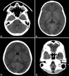
Tradeoffs between Radiation Exposure to the Lens of the Eyes and Diagnostic Image Quality in Pediatric Brain Computed Tomography
- PMID: 37622039
- PMCID: PMC10445673
- DOI: 10.4103/jmss.jmss_19_22
Tradeoffs between Radiation Exposure to the Lens of the Eyes and Diagnostic Image Quality in Pediatric Brain Computed Tomography
Abstract
Background: Computed tomography (CT) of the brain is associated with radiation exposure to the lens of the eyes. Therefore, it is necessary to optimize scan settings to keep radiation exposure as low as reasonably achievable without compromising diagnostic image information. The aim of this study was to compare the effectiveness of the five practical techniques for lowering eye radiation exposure and their effects on diagnostic image quality in pediatric brain CT.
Methods: The following scan protocols were performed: reference scan, 0.06-mm Pbeq bismuth shield, 30% globally lowering tube current (GLTC), reducing tube voltage (RTV) from 120 to 90 kVp, gantry tilting, and combination of gantry tilting with bismuth shielding. Radiation measurements were performed using thermoluminescence dosimeters. Objective and subjective image quality was evaluated.
Results: All strategies significantly reduced eye dose, and increased the posterior fossa artifact index and the temporal lobe artifact index, relative to the reference scan. GLTC and RTV increased image noise, leading to a decrease signal-to-noise ratio and contrast-to-noise ratio. Except for bismuth shielding, subjective image quality was relatively the same as the reference scan.
Conclusions: Gantry tilting may be the most effective method for reducing eye radiation exposure in pediatric brain CT. When the scanner does not support gantry tilting, GLTC might be an alternative.
Keywords: Brain computed tomography; eye lens; image quality; radiation exposure.
Copyright: © 2023 Journal of Medical Signals & Sensors.
Conflict of interest statement
There are no conflicts of interest.
Figures


Similar articles
-
Lens dose in routine head CT: comparison of different optimization methods with anthropomorphic phantoms.AJR Am J Roentgenol. 2015 Jan;204(1):117-23. doi: 10.2214/AJR.14.12763. AJR Am J Roentgenol. 2015. PMID: 25539246
-
Organ-based tube current modulation and bismuth eye shielding in pediatric head computed tomography.Pediatr Radiol. 2022 Dec;52(13):2584-2594. doi: 10.1007/s00247-022-05410-x. Epub 2022 Jul 15. Pediatr Radiol. 2022. PMID: 35836016
-
Signal-to-noise ratio and dose to the lens of the eye for computed tomography examination of the brain using an automatic tube current modulation system.Emerg Radiol. 2017 Jun;24(3):233-239. doi: 10.1007/s10140-016-1470-6. Epub 2016 Dec 2. Emerg Radiol. 2017. PMID: 27909929
-
Radiation dose and risk to the lens of the eye during CT examinations of the brain.J Med Imaging Radiat Oncol. 2019 Dec;63(6):786-794. doi: 10.1111/1754-9485.12950. Epub 2019 Sep 13. J Med Imaging Radiat Oncol. 2019. PMID: 31520467 Review.
-
Use of bismuth shield for protection of superficial radiosensitive organs in patients undergoing computed tomography: a literature review and meta-analysis.Radiol Phys Technol. 2019 Mar;12(1):6-25. doi: 10.1007/s12194-019-00500-2. Epub 2019 Feb 21. Radiol Phys Technol. 2019. PMID: 30790174 Review.
Cited by
-
A systematic review of radiation dose to the eye lens from pediatric brain computed tomography scans.Pediatr Radiol. 2025 Aug 15. doi: 10.1007/s00247-025-06371-7. Online ahead of print. Pediatr Radiol. 2025. PMID: 40815377 Review.
-
Bismuth Shielding in Head Computed Tomography-Still Necessary?J Clin Med. 2023 Dec 20;13(1):25. doi: 10.3390/jcm13010025. J Clin Med. 2023. PMID: 38202032 Free PMC article.
References
-
- World Health Organization. Communicating Radiation Risks in Paediatric Imaging. Information to Support Healthcare Discussions About Benefit and Risk. 2016. [Last accessed on 2020 Sep 04]. Available from: http://apps.who.int/iris/bitstream/10665/205033/1/9789241510349_eng.pdf?....
-
- Harbron RW, Ainsbury EA, Barnard SG, Lee C, McHugh K, Berrington de González A, et al. Radiation dose to the lens from CT of the head in young people. Clin Radiol. 2019;74:816.e9–17. - PubMed
-
- Raissaki M, Perisinakis K, Damilakis J, Gourtsoyiannis N. Eye-lens bismuth shielding in paediatric head CT: Artefact evaluation and reduction. Pediatr Radiol. 2010;40:1748–54. - PubMed
-
- Karami V, Zabihzadeh M. Prevalence of radiosensitive organ shielding in patients undergoing computed tomography examinations: An observational service audit in Ahvaz, Iran. Asian Biomed. 2015;9:771–5.
LinkOut - more resources
Full Text Sources
