

Integrated Biosignal Analysis to Provide Biomarkers for Recognizing Time Perception Difficulties
- PMID: 37622046
- PMCID: PMC10445675
- DOI: 10.4103/jmss.jmss_24_22
Integrated Biosignal Analysis to Provide Biomarkers for Recognizing Time Perception Difficulties
Abstract
Background: Time perception refers to the capability to recognize the passage of time. The cerebellum is located at the back of the brain, underlying the occipital and temporal lobes. Dyschronometria is a cerebellar dysfunction, in which a person cannot precisely estimate the amount of time that has passed. Cardiac indicators such as heart rate (HR) variability have been associated with mental function in healthy individuals. Moreover, time perception has been previously studied concerning cardiac signs. Human time perception is influenced by various factors such as attention and drowsiness. An electroencephalogram (EEG) is a suitable modality for evaluating cortical reactions due to its affordability and usefulness. Because EEG has a high sequential outcome, it offers valuable data to explore variability in psychological situations. An electrocardiogram (ECG) records electrical signals from the heart to examine various heart conditions. The electromyography (EMG) technique detects electrical impulses produced by muscles.
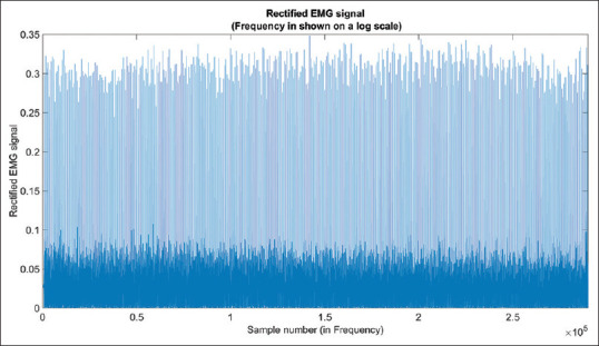
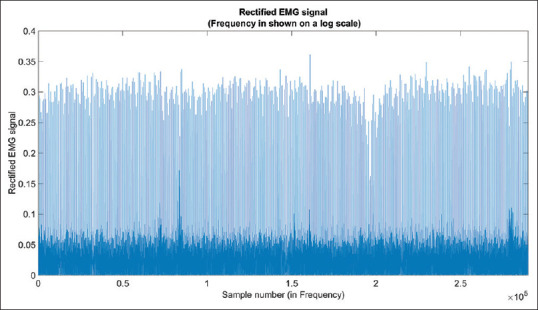
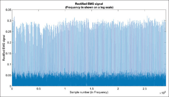
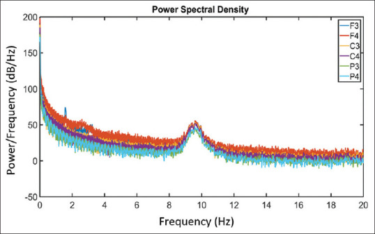
Methods: EEG, ECG, and EMG are integrated during time perception. This study evaluated the human body's time perception through the neurological, cardiovascular, and muscular systems using a simple neurofeedback exercise after time perception tasks. The three biosignals which are EEG, ECG, and EMG were investigated to use them as biomarkers for recognizing time perception difficulty as the main goal of the study. Five healthy college students with no health issues participated, and their EEG, ECG, and EMG were recorded while relaxing and performing a time wall estimation task and neurofeedback training. Previous research has shown the relationship between EEG frequency bands and the frontal center during time perception. Investigating the connection between ECG, EEG, and EMG under time perception conditions is significant.
Results: The results show that ECG (HR), EEG (Delta wave), and EMG (root mean square) are critical features in time perception difficulties.
Conclusion: The ability and outcomes of multiple biomarkers might allow for improved diagnosis and monitoring of the progress of any treatment applications such as biofeedback training. Furthermore, those biomarkers could be used as useful for evaluating and treating dyschronometria.
Keywords: Electrocardiogram; electroencephalogram; electromyography; time perception.
Copyright: © 2023 Journal of Medical Signals & Sensors.
Conflict of interest statement
There are no conflicts of interest.
Figures

Similar articles
-
The Investigation of the Relationship Between Individual Pain Perception, Brain Electrical Activity, and Facial Expression Based on Combined EEG and Facial EMG Analysis.J Pain Res. 2025 Jan 3;18:21-32. doi: 10.2147/JPR.S477658. eCollection 2025. J Pain Res. 2025. PMID: 39776765 Free PMC article.
-
Recording human electrocorticographic (ECoG) signals for neuroscientific research and real-time functional cortical mapping.J Vis Exp. 2012 Jun 26;(64):3993. doi: 10.3791/3993. J Vis Exp. 2012. PMID: 22782131 Free PMC article.
-
EMG biofeedback training in adult attention-deficit/hyperactivity disorder: An active (control) training?Behav Brain Res. 2017 Jun 30;329:58-66. doi: 10.1016/j.bbr.2017.04.021. Epub 2017 Apr 22. Behav Brain Res. 2017. PMID: 28442359
-
Stress detection using ECG and EMG signals: A comprehensive study.Comput Methods Programs Biomed. 2020 Sep;193:105482. doi: 10.1016/j.cmpb.2020.105482. Epub 2020 May 5. Comput Methods Programs Biomed. 2020. PMID: 32408236 Review.
-
Biofeedback and Neurofeedback for Anxiety Disorders: A Quantitative and Qualitative Systematic Review.Adv Exp Med Biol. 2020;1191:265-289. doi: 10.1007/978-981-32-9705-0_16. Adv Exp Med Biol. 2020. PMID: 32002934
References
-
- Ackerman S. Major structures and functions of the brain. Discov Brain. 1992:13–33.
LinkOut - more resources
Full Text Sources