Review
. 2023 Aug 3;13(8):786.
doi: 10.3390/bios13080786.

Recent Advances in Quantum Dot-Based Lateral Flow Immunoassays for the Rapid, Point-of-Care Diagnosis of COVID-19

Affiliations
Review

Recent Advances in Quantum Dot-Based Lateral Flow Immunoassays for the Rapid, Point-of-Care Diagnosis of COVID-19

Seyyed Mojtaba Mousavi et al. Biosensors (Basel). .

Abstract

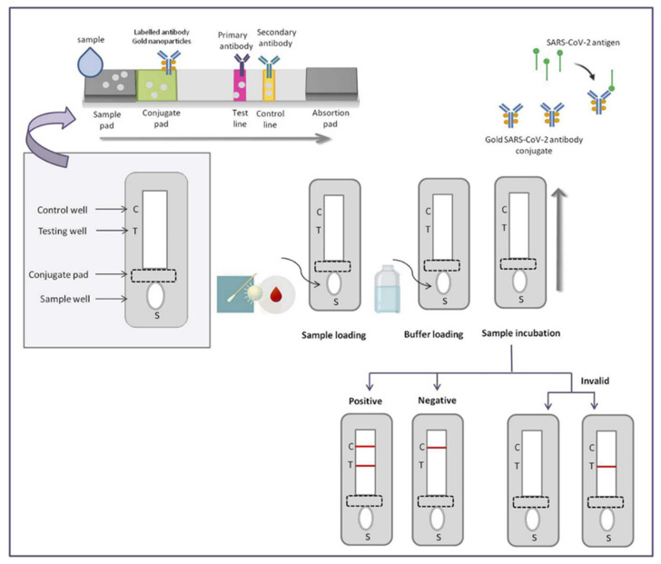
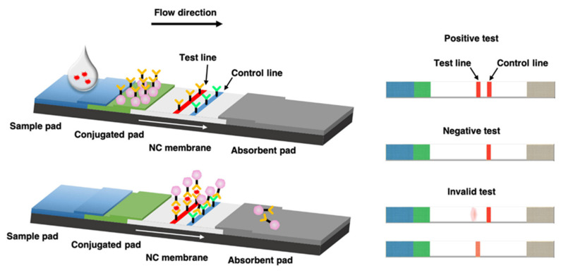
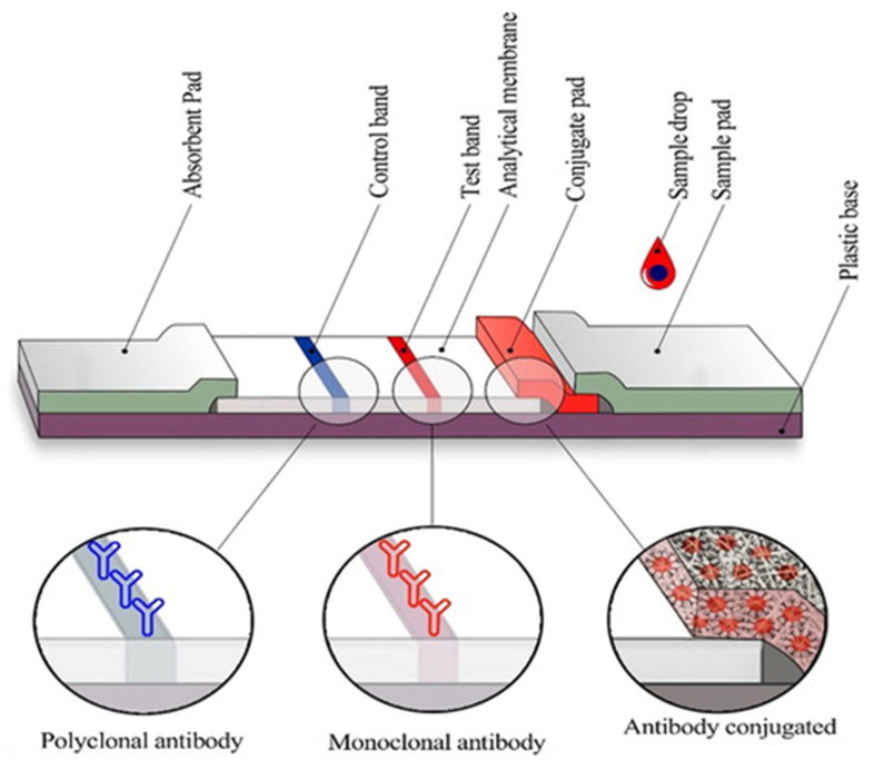
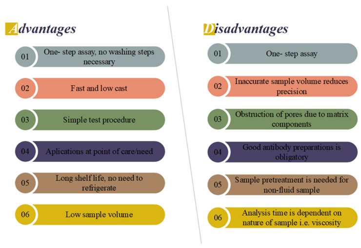
The COVID-19 pandemic has spurred demand for efficient and rapid diagnostic tools that can be deployed at point of care to quickly identify infected individuals. Existing detection methods are time consuming and they lack sensitivity. Point-of-care testing (POCT) has emerged as a promising alternative due to its user-friendliness, rapidity, and high specificity and sensitivity. Such tests can be conveniently conducted at the patient's bedside. Immunodiagnostic methods that offer the rapid identification of positive cases are urgently required. Quantum dots (QDs), known for their multimodal properties, have shown potential in terms of combating or inhibiting the COVID-19 virus. When coupled with specific antibodies, QDs enable the highly sensitive detection of viral antigens in patient samples. Conventional lateral flow immunoassays (LFAs) have been widely used for diagnostic testing due to their simplicity, low cost, and portability. However, they often lack the sensitivity required to accurately detect low viral loads. Quantum dot (QD)-based lateral flow immunoassays have emerged as a promising alternative, offering significant advancements in sensitivity and specificity. Moreover, the lateral flow immunoassay (LFIA) method, which fulfils POCT standards, has gained popularity in diagnosing COVID-19. This review focuses on recent advancements in QD-based LFIA for rapid POCT COVID-19 diagnosis. Strategies to enhance sensitivity using QDs are explored, and the underlying principles of LFIA are elucidated. The benefits of using the QD-based LFIA as a POCT method are highlighted, and its published performance in COVID-19 diagnostics is examined. Overall, the integration of quantum dots with LFIA holds immense promise in terms of revolutionizing COVID-19 detection, treatment, and prevention, offering a convenient and effective approach to combat the pandemic.

Keywords: COVID-19; detection; lateral flow immunoassay; performance; point-of-care testing; quantum dots.


Conflict of interest statement

The authors declare no conflict of interest.

Figures

Figure 2
Figure 2
Basic principles of the novel SARS-CoV-2 coronavirus. Reproduced with permission from Ref. [54].
Figure 9
Figure 9
(A) Revelation of a quantum dot (QD)-based lateral flow immunoassay with graphene oxide (GO) for pathogen detection; absorbent pad (AP), antibody–quantum dot immunocomplex (Ab–QD), and sample pad (SP). Reproduced with permission from Ref. [157]. © 2019 American Dairy Science Association®. (B) (i) The absorption and photoluminescent spectra undergo progressive changes as the CdxZn1—xS composition is consecutively altered. (ii) The quantum yields exhibit a systematic evolution with each additional shell monolayer. (iii) The photoluminescent quantum yields display distinct variations following the successive precipitation of the ZnSe/3CdSe (represented by black squares) and ZnSe/3CdSe/CdxZn1—xS/ZnS (represented by red dots) core/shell QDs. Reproduced with permission from Ref. [158]. Copyright © 2023 Elsevier B.V. or its licensors or contributors. ScienceDirect® is a registered trademark of Elsevier B.V (License Number: 5566100011830). (C) Procedure for the simultaneous detection of AFB1 and ZEN using QB-ICA. Reproduced with permission from Ref. [156]. Copyright © 2018 Elsevier B.V. All rights reserved (License Number: 5566081465711).
Figure 1
Figure 1
Schematic representation of a POC detection method for COVID-19 using LFIA. Reproduced with permission from Ref. [9]. © 2021 Elsevier B.V. All rights reserved (License Number: 5566111027872).
Figure 3
Figure 3
(A) Indirect detection methods to detect anti-SARS-CoV-2 antibodies. Reproduced with permission from Ref. [61]. (B) Lateral flow assay (LFA) detects SARS-CoV-2 antigens. Reproduced with permission from Ref [62]. © 2022 The Author(s). Published by Elsevier B.V. on behalf of King Saud University.
Figure 4
Figure 4
Schematic of the lateral flow immunoassay (LFIA) for the detection of COVID-19. Reproduced with permission from Ref. [71]. © 2022 The Author(s). Published by Elsevier Masson SAS.
Figure 5
Figure 5
Schematic illustration of LFIA test based on antigen detection. Reproduced with permission from Ref. [91].
Figure 6
Figure 6
Typical configuration of a lateral flow immunoassay test strip. Reproduced with permission from Ref. [107]. Copyright © 2023 Informa UK Limited.
Figure 7
Figure 7
Advantages and disadvantages of the LFIA.
Figure 8
Figure 8
An LFIA (Lateral Flow Immunoassay) system based on the cellular receptor, ACE2. (A) The schematic showcases the recognition process between ACE2 and SARS-CoV-2. ACE2, which is a type 1 membrane protein expressed in the lung, heart, kidneys, and intestine, serves as the cellular receptor for the virus. (B) Showcase of the components of the ACE2-based LFIA. The LFIA system consists of a sample pad, conjugate pad, nitrocellulose membrane, and absorbent pad. The nitrocellulose membrane features a test line that contains ACE2, enabling the detection of the SARS-CoV-2 spike antigen. In addition, the control line utilizes an anti-IgG antibody for validation purposes within the LFIA system. Reproduced with permission from Ref. [118]. Copyright 2020 Elsevier (License Number: 5566101395195).

References

    1. WHO . Statement on the Second Meeting of the International Health Regulations Emergency Committee regarding the Outbreak of Novel Coronavirus (2019-nCoV) WHO; Geneva, Switzerland: 2005.
    1. Zhu N., Zhang D., Wang W., Li X., Yang B., Song J., Zhao X., Huang B., Shi W., Lu R. A novel coronavirus from patients with pneumonia in China, 2019. N. Engl. J. Med. 2020;382:727–733. doi: 10.1056/NEJMoa2001017. - DOI - PMC - PubMed
    1. Mousavi S.M., Hashemi S.A., Rahmanian V., Kalashgrani M.Y., Gholami A., Omidifar N., Chiang W.-H. Highly sensitive flexible SERS-based sensing platform for detection of COVID-19. Biosensors. 2022;12:466. doi: 10.3390/bios12070466. - DOI - PMC - PubMed
    1. Corman V.M., Landt O., Kaiser M., Molenkamp R., Meijer A., Chu D.K., Bleicker T., Brünink S., Schneider J., Schmidt M.L. Detection of 2019 novel coronavirus (2019-nCoV) by real-time RT-PCR. Eurosurveillance. 2020;25:2000045. doi: 10.2807/1560-7917.ES.2020.25.3.2000045. - DOI - PMC - PubMed
    1. Klein D. Quantification using real-time PCR technology: Applications and limitations. Trends Mol. Med. 2002;8:257–260. doi: 10.1016/S1471-4914(02)02355-9. - DOI - PubMed