Review
. 2023 Aug 15;13(8):823.
doi: 10.3390/bios13080823.

Electrochemical Wearable Biosensors and Bioelectronic Devices Based on Hydrogels: Mechanical Properties and Electrochemical Behavior

Affiliations
Review

Electrochemical Wearable Biosensors and Bioelectronic Devices Based on Hydrogels: Mechanical Properties and Electrochemical Behavior

Mohsen Saeidi et al. Biosensors (Basel). .

Abstract

Hydrogel-based wearable electrochemical biosensors (HWEBs) are emerging biomedical devices that have recently received immense interest. The exceptional properties of HWEBs include excellent biocompatibility with hydrophilic nature, high porosity, tailorable permeability, the capability of reliable and accurate detection of disease biomarkers, suitable device-human interface, facile adjustability, and stimuli responsive to the nanofiller materials. Although the biomimetic three-dimensional hydrogels can immobilize bioreceptors, such as enzymes and aptamers, without any loss in their activities. However, most HWEBs suffer from low mechanical strength and electrical conductivity. Many studies have been performed on emerging electroactive nanofillers, including biomacromolecules, carbon-based materials, and inorganic and organic nanomaterials, to tackle these issues. Non-conductive hydrogels and even conductive hydrogels may be modified by nanofillers, as well as redox species. All these modifications have led to the design and development of efficient nanocomposites as electrochemical biosensors. In this review, both conductive-based and non-conductive-based hydrogels derived from natural and synthetic polymers are systematically reviewed. The main synthesis methods and characterization techniques are addressed. The mechanical properties and electrochemical behavior of HWEBs are discussed in detail. Finally, the prospects and potential applications of HWEBs in biosensing, healthcare monitoring, and clinical diagnostics are highlighted.

Keywords: biocompatible polymer; electroactive hydrogel; electrochemistry; flexible biosensors; mechanical behavior.


Conflict of interest statement

The authors declare no conflict of interest.

Figures

Figure 3
Figure 3
(a) PEDOT:PSS hydrogel integrated with Prussian blue nanoparticles for electrochemical glucose biosensing working diagram. Reprinted with permission from Elsevier from ref. [51], and (b) The functionalization of the working electrode made of gold with GOx through two steps of electropolymerization and Nafion treatment. Finally, the electrical signals produced during the enzymatic reaction are monitored for measuring the glucose level. Reprinted with permission from Elsevier from ref. [122].
Figure 5
Figure 5
(aI) Schematic illustration of PVA–CNF organohydrogel, (aII) Tensile stress–strain curves for PVA–CNF organohydrogels with varying amounts of CNF. Reprinted with permission from Wiley from ref. [193], (bI) Schematic illustration of the DN agar/AAc–Fe3+ hydrogel, (bII) The comparison of tensile stress–strain curves between the SN agar, SN AAc, and DN agar/AAc–Fe3+ hydrogels. Reprinted with permission from ACS from ref. [196], (cI) The formation of Ca2+/SA/PAM DN hydrogels using homogeneous Ca2+ cross-linking strategies, (cII) Tensile stress–strain curves of Ca2+/SA/PAM DN hydrogels at different incubation times in HCl. Reprinted with permission from RSC from ref. [197], (dI) Formation mechanism of DAMFC/ChN/gel hybrid composite hydrogel, and (dII) Swelling behavior of lyophilized DAMFC/ChN/gel hybrid composite hydrogels. Reprinted with permission from Elsevier from ref. [141].
Figure 1
Figure 1
(a) Chemical structures of conductive polymers. Reprinted with permission from ACS from ref. [40], (the schematic representation of various physical cross-linking mechanisms observed in hydrogels including (bI) ionic interaction, (bII) hydrophobic interaction, (bIII) cross-linking junction formed through cooling, and (bIV) complex coacervation. Reprinted with permission from ACS from ref. [41].
Figure 2
Figure 2
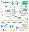
Schematic representation of (a) multifunctional polyvinyl alcohol/MXene/polyaniline hydrogel. Reprinted with permission from RSC from ref. [68], (b) AuNPs@MoS2-QDs composite with an agarose hydrogel-based stable visual platform in the presence of H2O2. The right diagram shows the UV–vis absorbance spectra of the hydrogel at various glucose levels (0, 2, 4, 5, 8, 10, 11, 12 mM) in serum. Reprinted with permission from Elsevier from ref. [69], (c) a flexible three-electrode system based on Ni-Co MOF-coated Au/PDMS hydrogel as a wearable electrochemical biosensor for glucose detection. Reprinted with permission from RSC from ref. [70], and (d) porous PVA/BTCA/β-CD/GOx/AuNPs composite integrated with PVA polymer nanofiber hydrogel synthesized by electrospinning method as a wearable glucose-responsive biosensor. Reprinted with permission from Nature from ref. [71].
Figure 4
Figure 4
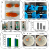
(a) The self-healing ability of the DN hydrogel. Reprinted with permission from ACS from ref. [159], (b) Comparing 25%PVA/EG hydrogel with PVA hydrogel at 25 °C and −20 °C. Reprinted with permission from Elsevier from ref. [160], (c) MXene/PMN hydrogels can adhere to a variety of surfaces, including (i) glass, (ii) PET, (iii) metal, and (iv) porcine skin. Reprinted with permission from Elsevier from ref. [161], (d) Porcine skin adhesion to G-P-C@agarose gels with different glycerol contents. Reprinted with permission from ACS from ref. [162], (e) Cell viability of NIH3T3 cells on Day 1 and Day 5, cultured with PDDA/CNF hydrogels and the control group. Reprinted with permission from Elsevier from ref. [163], and (f) Observation of the inhibition zone on the culture dishes for control and PNAg-hydrogel samples relative to E. coli and S. aureus. Reprinted with permission from Elsevier from ref. [164].
Figure 6
Figure 6
(aI) Schematic diagram of the preparation, (aII) The swelling kinetics curve of the N-CNTs/P(AAc–co–AAm) composite hydrogel at pH = 7. Reprinted with permission from RSC from ref. [76], (bI) Loading–unloading curve of PVA/P(AAc–co–AAm)/PDA@CNTs composite hydrogel after ten cycles of 120% strain stretching, (bII) The amount of energy that dissipated from ten cycles stretching of the composite hydrogel at 120% tensile strain. Reprinted with permission from Nature from ref. [192], (cI) The composite hydrogel’s proposed molecular structure, (cII) The comparison of the storage modulus (G′) and loss modulus (G″) of hydrogels with different mass ratios at 25 °C. Reprinted with permission from Elsevier from ref. [220], and (d) Schematic illustration of the preparation of ink for extrusion printing prepared from PEDOT:PSS functionalized with MXene. Reprinted with permission from Wiley from ref. [66].
Figure 7
Figure 7
(aI) Two layers of gelatin hydrogels integrated on the gate electrode surface, comprising a platinum layer and a carbon layer, (aII) The amperometric response to changes in UA concentrations in a PBS solution. Reprinted with permission from Wiley from ref. [248], (bI) The amperometric response of the PEDOT:PSS/DF/PB/GOx sensor during a glucose selectivity test, (bII) The calibration curve for the PEDOT:PSS/DF/PB/GOx sensor used for glucose detection. Reprinted with permission from Elsevier from ref. [51], (cI) The hydrogel-based ring case, including the carbon working electrode (WE 1), Ag/AgCl reference electrode (RE), carbon/PB working electrode (WE 2), and carbon counter electrode (CE), (cII) The SWV response for different concentrations of MPOx from 0 to 1.25 mM in 0.1 M PBS, with 0.25 mM increments (black, brown, purple, green, blue and red lines show 0, 0.25, 0.5,0.75, 1 and 1.25 mM of MPOx, respectively). The inset shows the corresponding calibration plot. Reprinted with permission from ACS from ref. [249], (dI) A schematic representation of the peptide/AuNPs hydrogel electrode used for the catalysis of DA, (dII) The DPV curves of the peptide/AuNPs hydrogel electrode in the presence (red) and absence (black) of 1.0 mM DA in 10 mM PBS, (dIII) The DPV curves of 1.0 mM DA recorded at the bare GCE (black), peptide/GCE hydrogel (blue), and AuNPs/peptide/GCE hydrogel (red). Reprinted with permission from Elsevier from ref. [250].
Figure 8
Figure 8
(aI) Schematic illustration of an HWEB using hydrogel-elastomer materials, (aII) OCP curve for HWEB at various Na+ concentrations shown insets is the calibration plot for the sensor. Reprinted with permission from Elsevier from ref. [265], (bI) the surface reaction of the GOx-chitosan on platinum electrode, and (bII) GOx-chitosan on platinum macroelectrode OCP in a blank solution of DPBS (green), and in a solution of DPBS containing glucose with various concentrations (blue). Reprinted with permission from Elsevier from ref. [266].
Figure 9
Figure 9
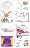
(aI) Schematic illustration of contact lens consisting of a PI and PDMS hybrid substrate, (aII) The relative resistance change response of the contact lens sensors with different glucose concentrations, inset is calibration curves of the glucose sensor. Reprinted with permission from Science from ref. [261], (bI) A schematic illustration of a packaged smart contact lens consisting of NFC chip, capacitor, resistor, antenna and cortisol sensor, and (bII) Change in relative resistance with increasing concentrations of cortisol in the buffer (black) and aqueous solution of artificial tears (red). Reprinted with permission from Science from ref. [262].
Figure 10
Figure 10
(aI) Schematic for Ti3C2Tx MXene-loaded/LBG-based cortisol biomarker detection, (aII) Nyquist plot for cortisol biomarker detection Ti3C2Tx MXene-loaded/LBG-based using EIS method. Reprinted with permission from Elsevier from ref. [264], (bI) lab-on-a-patch platform exploded view, and (bII) The Nyquist plots for the stretchable impedimetric biosensor (black, red, blue, purple, green and dark blue curves show cortisol concentrations of 0.001, 0.01, 0.1, 1, 10 and 1000 ng mL−1, respectively). Reprinted with permission from Elsevier from ref. [263].
Figure 11
Figure 11
(aI) Schematic illustration of the hydrogel-based patch for glucose monitoring, (aII) Working mechanism diagram using PVA/BTCA/β-CD/GOx/AuNPs nanofibrous hydrogel. Reprinted with permission from Nature from ref. [71], (bI) GOx-functionalized hydrogel tattoo, (bII) Working mechanism diagram. Reprinted with permission from ACS from ref. [273], (cI) using hydrogel in epidermal electrochemical microfluidic biosensor for monitoring of various analytes in sweat, and (cII) Working mechanism diagram. Reprinted with permission from Springer from Ref. [274].
Figure 12
Figure 12
(aI) swellable microneedle patch made of hydrogel, (aII) Working mechanism diagram. Reprinted with permission from Wiley from ref. [284], (bI) a smart contact lens for tear glucose level measurement, (bII) Working mechanism diagram. Reprinted with permission from Science from ref. [276], (cI) hydrogel-paper patch that simultaneously detects electrophysiology and biochemical changes during exercise, and (cII) Working mechanism diagram, by using origami techniques (where steps (1), (2), and (3) represent the folding process), a flat piece was transformed into a 3D configuration of wearable biosensor including a region for collecting sweat, an area where glucose sensing takes place, and a zone for evaporation. Reprinted with permission from Elsevier from ref. [277].

Similar articles

Cited by

References

    1. Kim J., Campbell A.S., Esteban-Fernández Ávila B., Wang J. Wearable Biosensors for Healthcare Monitoring. Nat. Biotechnol. 2019;37:389–406. doi: 10.1038/s41587-019-0045-y. - DOI - PMC - PubMed
    1. Zhu P., Peng H., Rwei A.Y. Flexible, Wearable Biosensors for Digital Health. Med. Nov. Technol. Devices. 2022;14:100118. doi: 10.1016/j.medntd.2022.100118. - DOI
    1. Haleem A., Javaid M., Singh R.P., Suman R., Rab S. Biosensors Applications in Medical Field: A Brief Review. Sens. Int. 2021;2:100100. doi: 10.1016/j.sintl.2021.100100. - DOI
    1. Singh S.U., Chatterjee S., Lone S.A., Ho H.-H., Kaswan K., Peringeth K., Khan A., Chiang Y.-W., Lee S., Lin Z.-H. Advanced Wearable Biosensors for the Detection of Body Fluids and Exhaled Breath by Graphene. Microchim. Acta. 2022;189:236. doi: 10.1007/s00604-022-05317-2. - DOI - PMC - PubMed
    1. Chen S., Qi J., Fan S., Qiao Z., Yeo J.C., Lim C.T. Flexible Wearable Sensors for Cardiovascular Health Monitoring. Adv. Healthc. Mater. 2021;10:2100116. doi: 10.1002/adhm.202100116. - DOI - PubMed

LinkOut - more resources