Review
. 2023 Aug 6;9(8):632.
doi: 10.3390/gels9080632.

Polymeric Nanoparticles and Nanogels: How Do They Interact with Proteins?

Affiliations
Review

Polymeric Nanoparticles and Nanogels: How Do They Interact with Proteins?

Amirhossein Sadeghi et al. Gels. .

Abstract

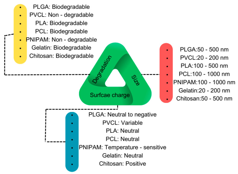
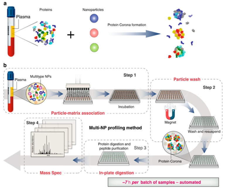
Polymeric nanomaterials, nanogels, and solid nanoparticles can be fabricated using single or double emulsion methods. These materials hold great promise for various biomedical applications due to their biocompatibility, biodegradability, and their ability to control interactions with body fluids and cells. Despite the increasing use of nanoparticles in biomedicine and the plethora of publications on the topic, the biological behavior and efficacy of polymeric nanoparticles (PNPs) have not been as extensively studied as those of other nanoparticles. The gap between the potential of PNPs and their applications can mainly be attributed to the incomplete understanding of their biological identity. Under physiological conditions, such as specific temperatures and adequate protein concentrations, PNPs become coated with a "protein corona" (PC), rendering them potent tools for proteomics studies. In this review, we initially investigate the synthesis routes and chemical composition of conventional PNPs to better comprehend how they interact with proteins. Subsequently, we comprehensively explore the effects of material and biological parameters on the interactions between nanoparticles and proteins, encompassing reactions such as hydrophobic bonding and electrostatic interactions. Moreover, we delve into recent advances in PNP-based models that can be applied to nanoproteomics, discussing the new opportunities they offer for the clinical translation of nanoparticles and early prediction of diseases. By addressing these essential aspects, we aim to shed light on the potential of polymeric nanoparticles for biomedical applications and foster further research in this critical area.

Keywords: disease diagnosis; nanogels; nanoproteomics; polymeric nanoparticles; protein corona.


Conflict of interest statement

The authors declare that they have no conflict of interest.

Figures

Figure 1
Figure 1
Pattern of a protein corona around a nanoparticle. Reused with permission from MDPI [26].
Figure 5
Figure 5
Drug loading on a PEG-PLA nanoparticle and its application in biomedicine [53]. Reused with permission from Elsevier.
Figure 2
Figure 2
Schematic presentation of conventional polymeric nanoparticle routes. (a) The diagram showcases the nanoprecipitation process. The inset, which is an enlarged image, elucidates the formation of nanoparticles (depicted as yellow spheres). This formation is attributed to the disparity in surface tension between the aqueous phase (characterized by high surface tension) and the organic phase (characterized by low surface tension). (b) emulsification-solvent evaporation technique. (c) emulsification solvent diffusion method. (d) salting-out technique. (e) emulsion polymerization method. Reused with permission from MDPI [31].
Figure 3
Figure 3
Schematic overview of the most used biocompatible polymers at the nanoscale for biomedical applications.
Figure 4
Figure 4
Chemical structure of polymers that are mostly utilized in nanoparticle synthesis for biomedical applications.
Figure 6
Figure 6
Biological parameters influence nanoparticle–biomolecule interactions.
Figure 7
Figure 7
The surface of a polymeric nanoparticle manages the protein–NP interaction.
Figure 8
Figure 8
Nanogels encounter a diverse array of proteins and biomolecules, their interplay being heavily influenced by the surface and bulk characteristics of these nanoscale gel-like structures. The intricate interplay between nanogels and the surrounding media is dictated by their finely tuned surface and bulk properties, imparting a crucial role in the regulation of their interactions.
Figure 9
Figure 9
(a) Schematic presentation of the most common methods in chromatography-based protein analysis. (b) Quantitative bar chart demonstrating the efficacy and selectivity of chromatography-based protein analysis methods.
Figure 10
Figure 10
Schematic illustration of automated nanoproteomics [83]. Reused with permission from Nature. (a) Incubation of nanoparticles with plasma and protein corona formation. (b) automated multi-NP profiling method and Nanoproteomics.

References

    1. Cedervall T., Lynch I., Lindman S., Berggård T., Thulin E., Nilsson H., Dawson K.A., Linse S. Understanding the nanoparticle–protein corona using methods to quantify exchange rates and affinities of proteins for nanoparticles. Proc. Natl. Acad. Sci. USA. 2007;104:2050–2055. doi: 10.1073/pnas.0608582104. - DOI - PMC - PubMed
    1. Cai R., Chen C. The Crown and the Scepter: Roles of the Protein Corona in Nanomedicine. Adv. Mater. 2019;31:1805740. doi: 10.1002/adma.201805740. - DOI - PubMed
    1. Xiao W., Gao H. The impact of protein corona on the behavior and targeting capability of nanoparticle-based delivery system. Int. J. Pharm. 2018;552:328–339. doi: 10.1016/j.ijpharm.2018.10.011. - DOI - PubMed
    1. Chen X., Chen J., Huang N. The structure, formation, and effect of plasma protein layer on the blood contact materials: A review. Biosurf. Biotribol. 2022;8:1–14. doi: 10.1049/bsb2.12029. - DOI
    1. Ishak N.A.I.M., Kamarudin S.K., Timmiati S.N., Karim N.A., Basri S. Biogenic platinum from agricultural wastes extract for improved methanol oxidation reaction in direct methanol fuel cell. J. Adv. Res. 2021;28:63–75. doi: 10.1016/j.jare.2020.06.025. - DOI - PMC - PubMed

LinkOut - more resources