. 2023 Aug 14;45(8):6717-6727.
doi: 10.3390/cimb45080425.

Length-Dependent Translation Efficiency of ER-Destined Proteins

Affiliations

Length-Dependent Translation Efficiency of ER-Destined Proteins

Hana Sahinbegovic et al. Curr Issues Mol Biol. .

Abstract

Gene expression is a fundamental process that enables cells to produce specific proteins in a timely and spatially dependent manner. In eukaryotic cells, the complex organization of the cell body requires precise control of protein synthesis and localization. Certain mRNAs encode proteins with an N-terminal signal sequences that direct the translation apparatus toward a specific organelle. Here, we focus on the mechanisms governing the translation of mRNAs, which encode proteins with an endoplasmic reticulum (ER) signal in human cells. The binding of a signal-recognition particle (SRP) to the translation machinery halts protein synthesis until the mRNA-ribosome complex reaches the ER membrane. The commonly accepted model suggests that mRNA that encodes a protein that contains an ER signal peptide continuously repeats the cycle of SRP binding followed by association and dissociation with the ER. In contrast to the current view, we show that the long mRNAs remain on the ER while being translated. On the other hand, due to low ribosome occupancy, the short mRNAs continue the cycle, always facing a translation pause. Ultimately, this leads to a significant drop in the translation efficiency of small, ER-targeted proteins. The proposed mechanism advances our understanding of selective protein synthesis in eukaryotic cells and provides new avenues to enhance protein production in biotechnological settings.

Keywords: endoplasmic reticulum; mRNA; proteosynthesis; ribosome stalling; signal peptide.


Conflict of interest statement

The authors declare no conflict of interest.

Figures

Figure 1
Figure 1
Single Ig domain abrogates translation. (a) Schematic representation of the translation efficiency reporter system with various stalling cassettes. (b) Normalized GFP:RFP fluorescence ratio of the translation efficiency reporter containing the control (K)0 and stalling (K)20 cassettes (reference, purple) and repeats of 1, 2 and 4 IgLλ domains (IgL, green). Significance was compared using the two-tailed Student’s t-test; * = p, 0.05. Error bars represent the mean ± SD. (c) X-ray structures of IgLλ (green, PDB code: 1CD0) and Filamin A (blue, PDB code: 3ISW). (d) Normalized GFP:RFP fluorescence ratio of the translation efficiency reporter containing the control and stalling cassettes (K)0 and (K)20 (reference, purple) and repeats of 1, 2 and 4 FLNA Ig-like domains (FLNA, blue). Significance was compared using the two-tailed Student’s t-test; * p = 0.05, ns = non-significant. Error bars represent the mean ± SD.
Figure 2
Figure 2
Evaluation of ribosome stalling and ER SP on translation efficiency of short mRNA. (a) Normalized GFP:RFP fluorescence ratio of the translation efficiency reporter containing a single repeat of the IgLλ domain (IgL) together with co-expression of empty vector (light green) or ZNF598 (dark green). Significance was compared using the two-tailed Student’s t-test, ns = non-significant. Error bars represent the mean ± SD. (b) Ubiquitin pull-down (Ub-PD) and whole cell lysate (WCL) from a cell expressing the translation efficiency reporter containing the control (K)0 and stalling (K)20 cassettes (reference) and a single repeat of the IgLλ domain (IgL1x). (c) Normalized GFP:RFP fluorescence ratio of the translation efficiency reporter containing the control (K)0 and stalling (K)20 cassettes (purple), a single repeat of IgL (green) and FLNA Ig-like domains with deletion (ΔSP IgL) and insertion of SP (SP FLNA). Significance was compared using the two-tailed Student’s t-test; * p = 0.05, ns = non-significant. Error bars represent the mean ± SD.
Figure 3
Figure 3
Analysis of transcript and protein abundance. (a) Average expression of human genes (transcripts per million reads) from 32 tissues in 122 individuals. Genes are divided into groups based on length of open reading frame. (b) Average abundance of human proteins (parts per million) and whole organism (integrated). Proteins are divided into groups based on length of relative coding sequence.
Figure 4
Figure 4
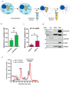
Membrane localization of short vs. long mRNAs. (a) Schematic representation of the fractionation assay. (b) The relative ratio of free (cytosolic) and membrane-bound (ER-associated) mRNAs encoding 1 and 2 IgLλ domains. Significance was compared using the two-tailed Student’s t-test; * = p, 0.05. Error bars represent the mean ± SD. (c) The relative ratio of free (cytosolic) and membrane-bound (ER-associated) endogenous mRNAs encoding short (<400 nt, n = 8 genes) and long (>400 nt, n = 9 genes) proteins containing ER-destined SPs. Significance was compared using the two-tailed Student’s t-test; * = p, 0.05. Error bars represent the mean ± SD. (d) WB analysis of fractionation samples. Cyt—cytosolic fraction, Mem—membrane fraction, WCL—whole cell lysate. GAPDH—cytosolic marker; calnexin, ERP5, PRDX4—ER markers; PCNA—nuclear marker. (e) Polysome analysis using Ribo Mega-SEC. Cell lysates from HEK293 cells transfected with plasmids encoding 1 and 2 IgLλ domains were separated using Agilent Bio SEC-5 2000 Å column. Retention time is indicated on the x-axis and UV absorbance at 260 nm is shown on the y-axis.
Figure 5
Figure 5
Schematic model of length-dependent translation efficiency of ER-destined proteins. The proposed model of SP-dependent localization of short vs. long ER-targeted mRNA (blue: mRNA, green: ribosome, orange: SRP, red: nascent polypeptide, purple: translocon, yellow: ER).

References

    1. Munro A., Jackson R., Korner A. Studies on the Nature of Polysomes. Biochem. J. 1964;92:289–299. doi: 10.1042/bj0920289. - DOI - PMC - PubMed
    1. Thompson M.K., Gilbert W.V. MRNA Length-Sensing in Eukaryotic Translation: Reconsidering the “Closed Loop” and Its Implications for Translational Control. Curr. Genet. 2017;63:613–620. doi: 10.1007/s00294-016-0674-3. - DOI - PMC - PubMed
    1. Panda A., Martindale J., Gorospe M. Polysome Fractionation to Analyze MRNA Distribution Profiles. Bio-Protocol. 2017;7:e2126. doi: 10.21769/BioProtoc.2126. - DOI - PMC - PubMed
    1. Arpat A.B., Liechti A., De Matos M., Dreos R., Janich P., Gatfield D. Transcriptome-Wide Sites of Collided Ribosomes Reveal Principles of Translational Pausing. Genome Res. 2020;30:985–999. doi: 10.1101/gr.257741.119. - DOI - PMC - PubMed
    1. Shoemaker C.J., Green R. Translation Drives MRNA Quality Control. Nat. Struct. Mol. Biol. 2012;19:594–601. doi: 10.1038/nsmb.2301. - DOI - PMC - PubMed

LinkOut - more resources