. 2023 Jul 28;11(8):182.
doi: 10.3390/dj11080182.

Remineralization of Early Enamel Lesions with Apatite-Forming Salt

Affiliations

Remineralization of Early Enamel Lesions with Apatite-Forming Salt

Clifton M Carey. Dent J (Basel). .

Abstract

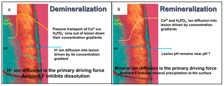
Objectives: This study sought to evaluate the remineralization of ex vivo human teeth using commercially available artificial saliva, SalivaMAX®, a supersaturated calcium phosphate rinse (SSCPR).

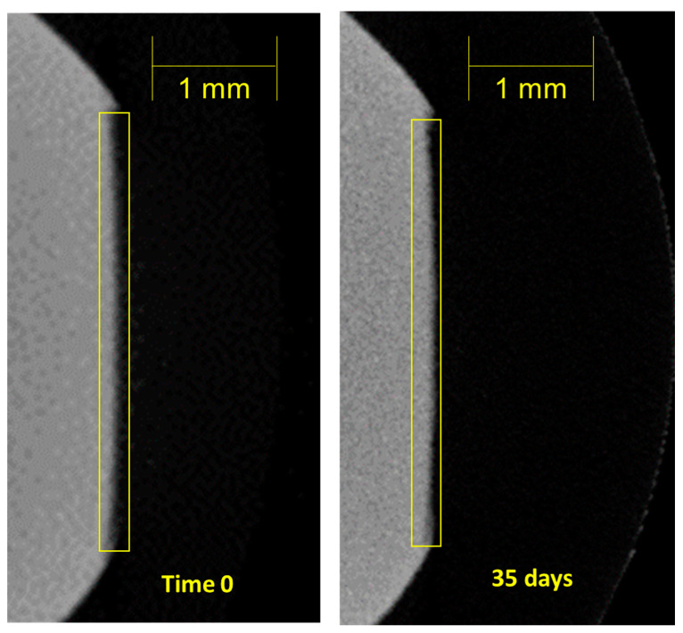
Methods: early enamel lesions were artificially induced on ex vivo human teeth by chemical means. The teeth were exposed to the SSCPR for two minutes (experimental) or dH2O (control) four times per day for a total of 35 days. At time points of 0, 2.5, 21, and 35 days, micro-CT was utilized to determine the mineral density profile across the lesion and evaluate lesion depth. The relative percent remineralization was calculated from the initial lesion depth (Time 0) at each evaluation time. Student's t-test was used to compare the extent of remineralization between the SSCPR and control groups for statistical significance at each time. To evaluate the changes in percent remineralization over time, a two-way ANOVA was used.

Results: At Time 0 and 2.5 days, there was no difference in the percent remineralization between the SSCPR and control groups (p > 0.05). After 21 days, the teeth exposed to the SSCPR remineralized 56.7 ± 3.7%, while the control only remineralized 10.7 ± 11.0% (p < 0.0001). At day 35, the remineralization was 73.7 ± 5.4% and 18.2 ± 10.8% (p < 0.0001) for the SSCPR and control groups, respectively.

Conclusions: A marked increase in remineralization occurred with the use of the SSCPR. Notably, the remineralization of the SSCPR occurred deep within the tooth and progressed toward the surface over time.

Keywords: SalivaMAX; calcium phosphate; early enamel lesion; remineralization; rinse.


Conflict of interest statement

The author declares no conflict of interest. While the funders of the study suggested a remineralization evaluation, they did not have a role in the study design; in the collection, analyses, or interpretation of data; in the writing of the manuscript; or in the decision to publish the results.

Figures

Figure 1
Figure 1
A polarized light micrograph of a white spot lesion with the mechanisms for demineralization (A) where an acid attack causes dissolution of enamel mineral at the lesion boundary followed by passive diffusion of calcium and phosphate ions out of the tooth, and (B) the mechanism for remineralization where calcium and phosphate ions diffuse from a higher concentration outside of the tooth into the WSL where precipitation occurs on existing apatite mineral at the lesion boundary progressing to the enamel surface.
Figure 2
Figure 2
Box and whisker plot showing all the data points for the measurements of lesion depth (μm) during the experiments. The “X” in each box is the average, and the data points are shown along with the standard deviation for each group. The experimental samples were significantly remineralized (p < 0.0001), while the control samples did not significantly remineralize (p > 0.05).
Figure 3
Figure 3
This is a figure that shows the relative percent lesion remineralization over time where the Boundary advances toward the original pre-demineralized surface. After 21 days of exposure to the SSCPR rinse, the % remineralization of the experimental teeth was significantly greater than the controls. The small amount of remineralization of the control samples came from the calcium phosphate-containing saliva-like solution.
Figure 4
Figure 4
This is a figure that shows the relative densities of a single example from the experimental group (A) and the control group (B) over time. The initial edge of the enamel (Boundary) is defined as 80% mineral density of the enamel prior to demineralization, which is set at 0 μm in the graphs. Figure 4A and Figure 5 show that the mineral density increased from the deepest part of the lesion toward the surface over time rather than developing a mineral-dense layer at the surface.
Figure 5
Figure 5
This is a figure that shows the pattern of remineralization of a sample. At Time 0, the tooth surface outlined by the box is mostly missing compared to the same tooth surface at 35 days of remineralization. The SSCPR rinse has deposited significant amounts of calcium and phosphate in the lesion to restore the lesion.

References

    1. Roopa K.B., Pathak S., Poornima P., Neena I.E. White spot lesions: A literature review. J. Pediatr. Dent. 2015;3:1–7. doi: 10.4103/2321-6646.151839. - DOI
    1. Ando M., Fontana M., Eckert G., Arthur R., Zhang H., Zero D. Objective and quantitative assessment of caries lesion activity. J. Dent. 2018;78:76–82. doi: 10.1016/j.jdent.2018.08.009. - DOI - PMC - PubMed
    1. Sheiham A., James W.P.T. Diet and dental caries: The pivotal role of free sugars reemphasized. J. Dent. Res. 2015;94:1341–1347. doi: 10.1177/0022034515590377. - DOI - PubMed
    1. Sundararaj D., Venkatachalapathy S., Tandon A., Pereira A. Critical evaluation of incidence and prevalence of white spot lesions during fixed orthodontic appliance treatment: A meta-analysis. J. Int. Soc. Prev. Community Dent. 2015;5:433–439. doi: 10.4103/2231-0762.167719. - DOI - PMC - PubMed
    1. Hopcraft M.S., Tan C. Xerostomia: An update for clinicians. Aust. Dent. J. 2010;55:238–244. doi: 10.1111/j.1834-7819.2010.01229.x. - DOI - PubMed

LinkOut - more resources