. 2023 Oct 31;14(5):e0045123.
doi: 10.1128/mbio.00451-23. Epub 2023 Aug 25.

SARS-CoV-2 ORF8 accessory protein is a virulence factor

Affiliations

SARS-CoV-2 ORF8 accessory protein is a virulence factor

M Bello-Perez et al. mBio. .

Abstract

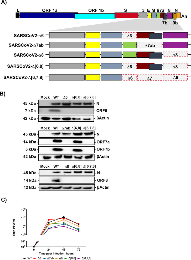
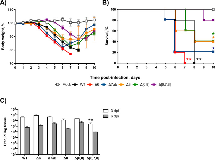
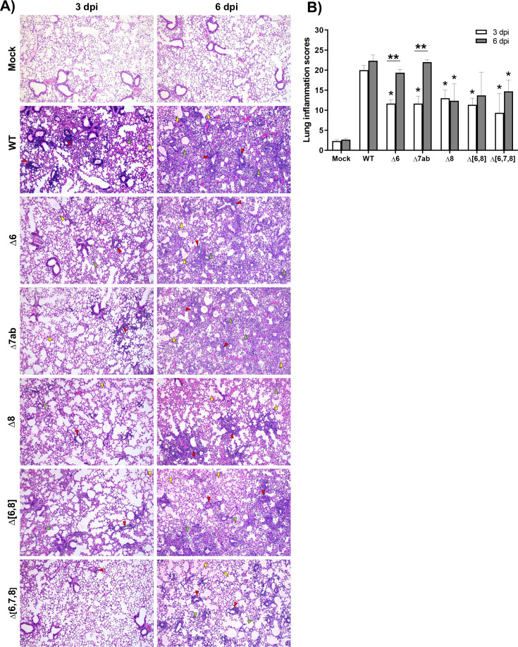
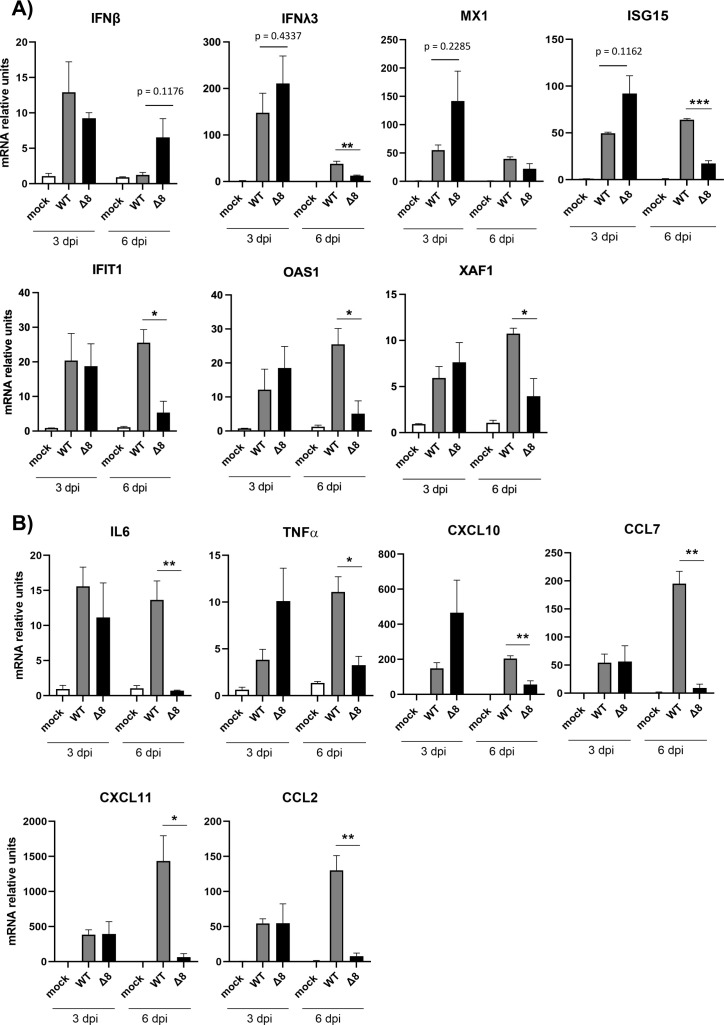
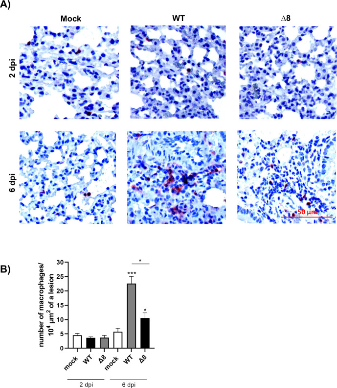
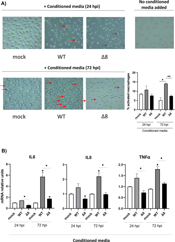
The relevance of severe acute respiratory syndrome coronavirus 2 (SARS-CoV-2) ORF8 in the pathogenesis of COVID-19 is unclear. Virus natural isolates with deletions in ORF8 were associated with wild milder disease, suggesting that ORF8 might contribute to SARS-CoV-2 virulence. This manuscript shows that ORF8 is involved in inflammation and in the activation of macrophages in two experimental systems: humanized K18-hACE2 transgenic mice and organoid-derived human airway cells. These results identify ORF8 protein as a potential target for COVID-19 therapies.

Keywords: SARS-CoV-2; accessory proteins; coronavirus; viral pathogenesis; virulence.


Conflict of interest statement

The authors declare no conflict of interest.

Figures

Fig 1
Fig 1
Generation and characterization of SARS-CoV-2 deletion mutants of accessory genes. (A) Diagram of deletions of accessory genes engineered in each mutant virus. Letters above or below boxes indicate viral genes. L, leader sequence; An, poly-A tail. Red dashed line indicates deleted regions. The shadowed area shows the 3′-end viral genes. (B) Immunoblot analysis of expression of accessory proteins 6, 7a, 7b, and 8 with specific antibodies in lysates from Vero E6 cells infected at an MOI 1 for 24 hours with the indicated mutants. βActin was used as a loading control. (C) Growth kinetics of SARS-CoV-2 deletion mutants. Vero E6/TMPRSS2 cells were infected at an MOI of 0.001, and the infections followed for 72 hours. Results are represented as a mean ± SEM.
Fig 2
Fig 2
Evaluation of virulence of SARS-CoV-2 deletion mutants of accessory genes in K18-hACE2 mice. Twenty-six-week-old female K18-hACE2 mice were intranasally mock-infected or infected with 105 PFU/animal of each virus. Weight loss (A) and survival (B) were monitored daily for 10 days. Weight loss is represented as the mean ± SDs of the mean (n = 5 mice per group). Lung samples were obtained at 3 and 6 dpi. (C) Viral titers were determined by plaque assays in Vero E6 cells at 4 and 6 dpi. Mean ± SDs of the mean are represented (n = 3). **P < 0.01; *P < 0.05.
Fig 3
Fig 3
Histopathological evaluation of lungs of K18-hACE2 mice infected with SARS-CoV-2 deletion mutants of accessory genes. (A) Representative lung histopathological sections (H&E staining) from K18-hACE2 mice mock infected, infected with wild-type virus (WT), or infected with recombinant viruses (∆6, ∆7ab, ∆8, ∆[6,8], and ∆[6,7,8]) and euthanized at 3 and 6 dpi (magnification: 10×). Yellow arrowheads, diffuse thickening of the alveolar septae, moderate alveolar edema; green arrowheads, moderate mononuclear cell infiltrates within alveolar spaces; and red arrowheads, multifocal perivascular and peribronchiolar mononuclear infiltrates. (B) Lung inflammation scores examined in lung samples (left lobes) taken from K18-hACE2 mice (n = 3/group) intranasally mock infected, infected with wild-type virus (WT), or infected with recombinant viruses (∆6, ∆7ab, ∆8, ∆[6,8], and ∆[6,7,8]) and euthanized at 3 and 6 dpi. Mean and SEM of cumulative histopathological lesion scores. Unpaired t-test: *P < 0.05; **P < 0.01.
Fig 4
Fig 4
Innate immune response in the lungs of K18-hACE2 mice infected with SARS-CoV-2-WT and SARS-CoV-2-∆8. Total RNA was collected from lung samples of mice infected with WT or deletion mutant at 3 and 6 dpi. mRNAs of genes encoding interferon-related genes (IFN-β, IFN-λ3, MX1, ISG-15, IFIT1, OAS1, and XAF1) (A) or pro-inflammatory response genes (IL-6, TNF-α, CXCL10, CCL2, CCL7, and CXCL11) (B) were quantified by RT-qPCR using specific TaqMan assays. Relative mRNA levels were referred to expression in mock-infected mice. Results show means from n = 3 animals per group. Error bars represent SEMs. *P < 0.05; **P < 0.01; ***P < 0.001.
Fig 5
Fig 5
Viral titers of SARS-CoV-2-WT and SARS-CoV-2-∆8 in the lungs and nasal turbinates of infected mice. Viral titers were obtained from lungs (A) and nasal turbinates (B) samples at 1, 2, and 4 dpi. Viral titers are represented as the mean ± SEM. *P < 0.05.
Fig 6
Fig 6
Innate immune response in the lungs of mice infected with the virulent SARS-CoV-2-WT and the attenuated mutant SARS-CoV-2-∆8 at early times post-infection. Total RNA was extracted from lung samples of mice infected at 1, 2, and 4 dpi. mRNA-encoding genes related to the interferon system (IFN-β, IFN-λ3, MX1, ISG-15, IFIT1, OAS1, and XAF1) (A) or the pro-inflammatory response (IL-6, TNF-α, CXCL10, CCL2, CCL7, and CXCL11) (B) were quantified by RT-qPCR using specific TaqMan assays. Relative mRNA levels were based on comparison with mock-infected mice. Results show means from n = 3 animals per group. Error bars represent SEMs. *P < 0.05; **P < 0.01; ***P < 0.001; ****P < 0.001.
Fig 7
Fig 7
Characterization of SARS-CoV-2-WT and SARS-CoV-2-∆8 in two-dimensional (2D) human airway organoids. Representative data of replication kinetics in terms of viral titers (A) and RNA (B) studied in organoids from three different donors. (C) Proportion of replication of each virus in 2D airway organoids infected with a 1:1 ratio.
Fig 8
Fig 8
Innate immune response in organoid-derived human airway cells infected with SARS-CoV-2-∆8 and SARS-CoV-2. Total RNA was extracted from cells infected with WT or deletion mutant at 24 and 72 hpi. mRNA-encoding genes related to the interferon system (IFN-β, IFN-λ, ISG-15, IFIT1, and OAS1) (A) or the pro-inflammatory response (IL-6, CXCL10, CXCL11, and CCL7) (B) were quantified by RT-qPCR using specific TaqMan assays. Relative mRNA levels were based on comparison with mock-infected mice. Results show means from n = 2 donors. Error bars represent SEMs. *P < 0.05; **P < 0.01; ***P < 0.001.
Fig 9
Fig 9
Immunohistochemical stainings for macrophages in mice lungs. (A) Representative images of pulmonary macrophages observed in mice euthanized at 2 and 6 dpi. (B). Quantification of macrophages. Bars represent means of three mice. Error bars represent SEMs. *P < 0.05; **P< 0.01; ***P < 0.001.
Fig 10
Fig 10
Evaluation of human macrophages’ activation. (A) Human macrophages were treated with conditioned media from mock-infected, infected with SARS-CoV-2-WT or SARS-CoV-2-∆8 organoids for 24 hours, and the cellular morphology was analyzed with a microscope. Activated macrophages were marked with arrows. (B) Quantitative reverse transcription PCR (RT-PCR) analysis of the relative expression of IL-6, IL-8, and TNF-α mRNA. Error bars represent SEMs. *P < 0.05; **P < 0.01; ***P < 0.001.

Similar articles

Cited by

References

    1. Zhou P, Yang XL, Wang XG, Hu B, Zhang L, Zhang W, Si HR, Zhu Y, Li B, Huang CL, Chen HD, Chen J, Luo Y, Guo H, Jiang RD, Liu MQ, Chen Y, Shen XR, Wang X, Zheng XS, Zhao K, Chen QJ, Deng F, Liu LL, Yan B, Zhan FX, Wang YY, Xiao GF, Shi ZL. 2020. A pneumonia outbreak associated with a new coronavirus of probable bat origin. Nature 588:270–273. doi:10.1038/s41586-020-2951-z - DOI - PMC - PubMed
    1. Gorbalenya AE, Baker SC, Baric RS, de Groot RJ, Drosten C, Gulyaeva AA, Haagmans BL, Lauber C, Leontovich AM, Neuman BW, Penzar D, Perlman S, Poon LLM, Samborskiy DV, Sidorov IA, Sola I, Ziebuhr J, Coronaviridae Study Group of the International Committee on Taxonomy of Viruses . 2020. The species severe acute respiratory syndrome-related coronavirus: classifying 2019-nCoV and naming it SARS-CoV-2. Nat Microbiol 5:536–544. doi:10.1038/s41564-020-0695-z - DOI - PMC - PubMed
    1. Enjuanes L, Zuñiga S, Castaño-Rodriguez C, Gutierrez-Alvarez J, Canton J, Sola I. 2016. Molecular basis of coronavirus virulence and vaccine development. Adv Virus Res 96:245–286. doi:10.1016/bs.aivir.2016.08.003 - DOI - PMC - PubMed
    1. Menachery VD, Mitchell HD, Cockrell AS, Gralinski LE, Yount BL, Graham RL, McAnarney ET, Douglas MG, Scobey T, Beall A, Dinnon K, Kocher JF, Hale AE, Stratton KG, Waters KM, Baric RS, Racaniello VR. 2017. MERS-CoV accessory ORFs play key role for infection and pathogenesis. mBio 8:e00665-17. doi:10.1128/mBio.00665-17 - DOI - PMC - PubMed
    1. Dediego ML, Pewe L, Alvarez E, Rejas MT, Perlman S, Enjuanes L. 2008. Pathogenicity of severe acute respiratory coronavirus deletion mutants in hACE-2 transgenic mice. Virology 376:379–389. doi:10.1016/j.virol.2008.03.005 - DOI - PMC - PubMed

Substances