. 2023 Jul 28;9(8):797.
doi: 10.3390/jof9080797.

The Extracellular Lipopeptides and Volatile Organic Compounds of Bacillus subtilis DHA41 Display Broad-Spectrum Antifungal Activity against Soil-Borne Phytopathogenic Fungi

Affiliations

The Extracellular Lipopeptides and Volatile Organic Compounds of Bacillus subtilis DHA41 Display Broad-Spectrum Antifungal Activity against Soil-Borne Phytopathogenic Fungi

Dhabyan Mutar Kareem Al-Mutar et al. J Fungi (Basel). .

Abstract

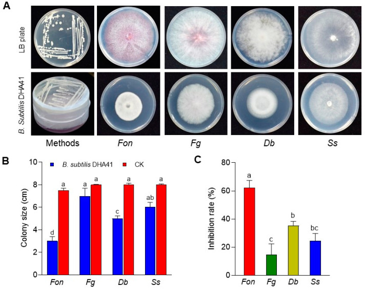
Fusarium oxysporum f. sp. niveum (Fon) is a devastating soil-borne fungus causing Fusarium wilt in watermelon. The present study investigated the biochemical mechanism underlying the antifungal activity exhibited by the antagonistic bacterial strain DHA41, particularly against Fon. Molecular characterization based on the 16S rRNA gene confirmed that DHA41 is a strain of Bacillus subtilis, capable of synthesizing antifungal lipopeptides, such as iturins and fengycins, which was further confirmed by detecting corresponding lipopeptide biosynthesis genes, namely ItuB, ItuD, and FenD. The cell-free culture filtrate and extracellular lipopeptide extract of B. subtilis DHA41 demonstrated significant inhibitory effects on the mycelial growth of Fon, Didymella bryoniae, Sclerotinia sclerotiorum, Fusarium graminearum, and Rhizoctonia solani. The lipopeptide extract showed emulsification activity and inhibited Fon mycelial growth by 86.4% at 100 µg/mL. Transmission electron microscope observations confirmed that the lipopeptide extract disrupted Fon cellular integrity. Furthermore, B. subtilis DHA41 emitted volatile organic compounds (VOCs) that exhibited antifungal activity against Fon, D. bryoniae, S. sclerotiorum, and F. graminearum. These findings provide evidence that B. subtilis DHA41 possesses broad-spectrum antifungal activity against different fungi pathogens, including Fon, through the production of extracellular lipopeptides and VOCs.

Keywords: Fusarium oxysporum f. sp. niveum; antifungal activity; lipopeptide; soil-borne pathogens; volatile organic compounds.


Conflict of interest statement

The authors declare no conflict of interest.

Figures

Figure 1
Figure 1
Molecular characterization of the antagonistic bacterial strain DHA41. The phylogenetic tree of strain DHA41’s 16S rRNA gene (bold text), with those from other Bacillus species, was constructed using the neighbor joining method with a 1000-bootstrap approach. The Escherichia coli 16S rRNA gene sequence (MN318323.1) was used as an outgroup in the tree. The scale bar indicates that the sequences represented in the tree differ by an average of 0.050 nucleotide substitutions per site.
Figure 2
Figure 2
Identification of extracellular lipopeptides produced by B. subtilis DHA41 by matrix-assisted laser desorption/ionization–time-of-flight mass spectrometry.
Figure 3
Figure 3
Detection of the lipopeptide biosynthesis genes in B. subtilis DHA41 (A) and analysis of the emulsification index of B. subtilis DHA41 on different organic oil substrates (B). The amplicon sizes are indicated above the bands in (A).
Figure 4
Figure 4
Antifungal activity of cell-free supernatant from B. subtilis DHA41 culture against F. oxysporum f. sp. nevium (Fon), S. sclerotiourm (Ss), and D. byroniae (Db). (A) Fungal colonies grown on potato dextrose agar supplemented with 60 µL of cell-free supernatant or sterile Luria–Bertani medium. (B) The inhibition rate of the cell-free supernatant against Fon, Ss, and Db. The experiment in (A) was independently performed three times with similar results. The data presented in (B) are the means ± standard deviation from three independent experiments. Different letters above the columns indicate a significant difference at 95% confidence level according to the one-way analysis of variance test.
Figure 5
Figure 5
Antifungal activity of extracellular lipopeptide extract from B. subtilis DHA41 against F. oxysporum f. sp. nevium (Fon), S. sclerotiourm (Ss), D. byroniae (Db), R. solani (Rs), and F. graminearum (Fg). (A) Fungal colonies grown on potato dextrose agar supplemented with the extracellular lipopeptide extract (LP) or dimethyl sulfoxide (CK). (B) The inhibition zones of the LP against Fon, Ss, Db, Rs, and Fg. (C) The inhibition activity of different concentrations of the LP on Fon growth. The experiment in (A) was independently performed three times with similar results. The data presented in (B,C) are the means ± standard deviation from three independent experiments. Different letters above the columns indicate a significant difference at 95% confidence level according to the one-way analysis of variance test.
Figure 6
Figure 6
B. subtilis DHA41-produced lipopeptides affect cell viability in F. oxysporum f. sp. niveum. Lipopeptide extract- (30 µg/mL) or dimethyl sulfoxide-treated (CK) Fon mycelia (A) and conidia (B) were stained with fluorescein diacetate (FDA) and propidium iodide (PI) for 12 h, followed by detection of fluorescent signals using a Zeiss LSM 880 confocal laser microscope, with excitation at 488 nm for FDA and 561 nm for PI. Scale bar, 10 µm. The experiments were independently performed three times with similar results, and data from one representative experiment are shown.
Figure 7
Figure 7
Ultrastructural changes in mycelia and conidia of F. oxysporum f. sp. niveum after treatment with B. subtilis DHA41-produced lipopeptide extract (30 µg/mL). Mycelia (A) and conidia (B) of Fon were treated with lipopeptide extract or dimethyl sulfoxide (CK) for 12 h and then examined under a transmission electron microscope (TEM). Scale bars, representing 2 μm in (A) and 1 μm in (B), respectively, are shown at the bottom of the TEM images. The experiments were independently performed three times with similar results, and data from one representative experiment are shown.
Figure 8
Figure 8
Antifungal activity of B. subtilis DHA41-emitted volatile organic compounds against F. oxysporum f. sp. nevium (Fon), F. graminearum (Fg), D. byroniae (Db), and S. sclerotiourm (Ss). (A) Fungal colonies grown with or without B. subtilis DHA41 in the two-sealed-base-plate experiments. (B) Colony sizes of the tested fungi grown with B. subtilis DHA41. (C) Inhibition rates of colony growth of the tested fungi grown with B. subtilis DHA41. The experiment in (A) was independently performed three times with similar results. The data presented in (B,C) are the means ± standard deviation from three independent experiments. Different letters above the columns indicate a significant difference at 95% confidence level according to the one-way analysis of variance test.

References

    1. Coque J.J.R., Álvarez-Pérez J.M., Cobos R., González-García S., Ibáñez A.M., Diez Galán A., Calvo-Peña C. Advances in the control of phytopathogenic fungi that infect crops through their root system. Adv. Appl. Microbiol. 2020;111:123–170. - PubMed
    1. Gordon T.R. Fusarium oxysporum and the Fusarium wilt syndrome. Annu. Rev. Phytopathol. 2017;55:23–39. doi: 10.1146/annurev-phyto-080615-095919. - DOI - PubMed
    1. Lü G., Guo S., Zhang H., Geng L., Song F., Fei Z., Xu Y. Transcriptional profiling of watermelon during its incompatible interaction with Fusarium oxysporum f. sp. niveum. Eur. J. Plant Pathol. 2011;131:585–601. doi: 10.1007/s10658-011-9833-z. - DOI
    1. Mazzola M., Freilich S. Prospects for biological soilborne disease control: Application of indigenous versus synthetic microbiomes. Phytopathology. 2017;107:256–263. doi: 10.1094/PHYTO-09-16-0330-RVW. - DOI - PubMed
    1. Bonaterra A., Badosa E., Daranas N., Francés J., Roselló G., Montesinos E. Bacteria as biological control agents of plant diseases. Microorganisms. 2022;10:1759. doi: 10.3390/microorganisms10091759. - DOI - PMC - PubMed

LinkOut - more resources