

Surf Redfish-Based ZnO-NPs and Their Biological Activity with Reference to Their Non-Target Toxicity
- PMID: 37623718
- PMCID: PMC10455839
- DOI: 10.3390/md21080437
Surf Redfish-Based ZnO-NPs and Their Biological Activity with Reference to Their Non-Target Toxicity
Abstract
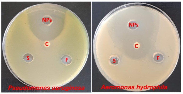
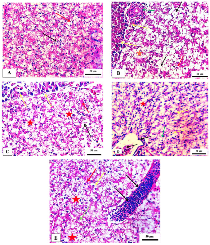
The marine environment is a rich source of bioactive compounds. Therefore, the sea cucumber was isolated from the Red Sea at the Al-Ain Al-Sokhna coast and it was identified as surf redfish (Actinopyga mauritiana). The aqueous extract of the surf redfish was utilized as an ecofriendly, novel and sustainable approach to fabricate zinc oxide nanoparticles (ZnO-NPs). The biosynthesized ZnO-NPs were physico-chemically characterized and evaluated for their possible antibacterial and insecticidal activities. Additionally, their safety in the non-target organism model (Nile tilapia fish) was also investigated. ZnO-NPs were spherical with an average size of 24.69 ± 11.61 nm and had a peak at 350 nm as shown by TEM and UV-Vis, respectively. XRD analysis indicated a crystalline phase of ZnO-NPs with an average size of 21.7 nm. The FTIR pattern showed biological residues from the surf redfish extract, highlighting their potential role in the biosynthesis process. DLS indicated a negative zeta potential (-19.2 mV) of the ZnO-NPs which is a good preliminary indicator for their stability. ZnO-NPs showed larvicidal activity against mosquito Culex pipiens (LC50 = 15.412 ppm and LC90 = 52.745 ppm) and a potent adulticidal effect to the housefly Musca domestica (LD50 = 21.132 ppm and LD90 = 84.930 ppm). Tested concentrations of ZnO-NPs showed strong activity against the 3rd larval instar. Topical assays revealed dose-dependent adulticidal activity against M. domestica after 24 h of treatment with ZnO-NPs. ZnO-NPs presented a wide antibacterial activity against two fish-pathogen bacteria, Pseudomonas aeruginosa and Aeromonas hydrophila. Histopathological and hematological investigations of the non-target organism, Nile tilapia fish exposed to 75-600 ppm ZnO-NPs provide dose-dependent impacts. Overall, data highlighted the potential applications of surf redfish-mediated ZnO-NPs as an effective and safe way to control mosquitoes, houseflies and fish pathogenic bacteria.
Keywords: ZnO-NPs; adulticidal; antibacterial; larvicidal; non-target organism; sea cucumber; surf redfish.
Conflict of interest statement
The authors declare no conflict of interest.
Figures


References
-
- Czyżowska A., Barbasz A. A review: Zinc oxide nanoparticles–friends or enemies? Int. J. Environ. Health Res. 2022;32:885–901. - PubMed
-
- Bai Z., Yan X., Chen X., Liu H., Shen Y., Zhang Y. ZnO nanowire array ultraviolet photodetectors with self-powered properties. Curr. Appl. Phys. 2013;13:165–169. doi: 10.1016/j.cap.2012.07.005. - DOI
-
- Khan Z.U.H., Sadiq H.M., Shah N.S., Khan A.U., Muhammad N., Hassan S.U., Tahir K., Khan F.U., Imran M., Ahmad N., et al. Greener synthesis of zinc oxide nanoparticles using Trianthema portulacastrum extract and evaluation of its photocatalytic and biological applications. J. Photochem. Photobiol. B Biol. 2019;192:147–157. doi: 10.1016/j.jphotobiol.2019.01.013. - DOI - PubMed
-
- Faizan M., Hayat S., Pichtel J. Sustainable Agriculture Reviews 41. Springer; Cham, Switzerland: 2020. Effects of zinc oxide nanoparticles on crop plants: A perspective analysis; pp. 83–99.
-
- Hasaballah A.I., El Naggar H.A., Abdelbary S., Bashar M.A.E., Selim T.A. Eco friendly Synthesis of Zinc Oxide Nanoparticles by Marine Sponge, Spongia offcinalis: Antimicrobial and Insecticidal Activities against the Mosquito Vectors, Culex pipiens and Anopheles pharoensis. BioNanoScience. 2021;12:89–104. doi: 10.1007/s12668-021-00926-2. - DOI
MeSH terms
Substances
LinkOut - more resources
Full Text Sources
Medical
Research Materials
Miscellaneous
