. 2023 Jul 27;12(8):984.
doi: 10.3390/pathogens12080984.

HPV and Other Risk Factors Involved in Pharyngeal Neoplasm-Clinical and Morphopathological Correlations in the Southwestern Region of Romania

Affiliations

HPV and Other Risk Factors Involved in Pharyngeal Neoplasm-Clinical and Morphopathological Correlations in the Southwestern Region of Romania

Carmen Aurelia Mogoantă et al. Pathogens. .

Abstract

Oropharyngeal squamous cell carcinoma (OPSCC) development is strongly associated with risk factors like smoking, chronic alcohol consumption, and the living environment, but also chronic human papilloma virus (HPV) infection, which can trigger cascade cellular changes leading to a neoplastic transformation. The prevalence of these factors differs among different world regions, and the prevention, diagnosis, and prognosis of OPSCC are highly dependent on them. We performed a retrospective study on 406 patients diagnosed with OPSCC in our region that were classified according to the tumor type, localization and diagnosis stage, demographic characteristics, risk factors, and histological and immunohistochemical features. We found that most of the patients were men from urban areas with a smoking habit, while most of the women in our study were diagnosed with tonsillar OPSCC and had a history of chronic alcoholism. During the immunohistochemical study, we analyzed the tumor immunoreactivity against anti-p16 and anti-HPV antibodies as markers of HPV involvement in tumor progression, as well as the correlation with the percentage of intratumoral nuclei immunomarked with the anti-Ki 67 antibody in serial samples. We observed that the percentage of Ki67-positive nuclei increased proportionally with the presence of intratumoral HPV; thus, active HPV infection leads to an increase in the rate of tumor progression. Our results support the implementation of strategies for OPSCC prevention and early diagnosis and can be a starting point for future studies aiming at adapting surgical and oncological treatment according to the HPV stage for better therapeutic results.

Keywords: cell proliferation; human papillomavirus; tumor suppressor gene.


Conflict of interest statement

The authors declare no conflict of interest.

Figures

Figure 1
Figure 1
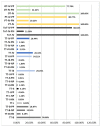
The progress of patients according to the group and stage (F: female; M: male; TT: palatine tonsil or tonsillar fossa tumor; ToT: tongue base tumor; PT: palatine veil or uvula; OT: tumors with other oropharyngeal locations; B: benign; Ma: malign; I, II, III, IV–staging; I: improved; C: cured; D: poor health state or death).
Figure 2
Figure 2
Percentage distribution of tumors in the 4 study groups according to the reactivity to anti-p16 and anti-HPV antibodies. TT: palatine tonsil or tonsillar fossa tumor; ToT: tongue base tumor; PT: palatine veil or uvula; OT: tumors with other oropharyngeal locations; I, II, III, IV–stages; NN: negative reaction for both the anti-p16 antibody and the anti-HPV antibody; NP: negative reaction for the anti-p16 antibody and positive for the anti-HPV antibody; PN: positive reaction for the anti-p16 antibody and negative for the anti-HPV antibody; PP: positive reaction for both the anti-p16 antibody and the anti-HPV antibody.
Figure 3
Figure 3
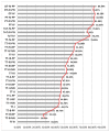
Percentage distribution of Ki67-positive nuclei according to the type and stage of tumors in the 4 groups. A gradual increase in the percentage of Ki67-positive nuclei directly proportional to the increase in tumor stage is observed (red line). TT: palatine tonsil or tonsillar fossa tumor; ToT: tongue base tumor; PT: palatine veil or uvula; OT: tumors with other oropharyngeal locations; B: benign; I, II, III, IV–staging; NN: negative reaction for both the anti-p16 antibody and the anti-HPV antibody; NP: negative reaction for the anti-p16 antibody and positive for the anti-HPV antibody; PN: positive reaction for the anti-p16 antibody and negative for the anti-HPV antibody; PP: positive reaction for both the anti-p16 antibody and the anti-HPV antibody.
Figure 4
Figure 4
Distribution of the total number of nuclei/field 40× and of the total number of nuclei immunmarked with the anti-Ki67 antibody/field 40×, depending on the type and stage of the tumors in the 4 groups. We observe a gradual increase in the number of Ki67-positive nuclei, directly proportional to the tumor stage (green line), while the total number of nuclei/field 40× remained quasi-constant (red line). TT: palatine tonsil or tonsillar fossa tumor; ToT: tongue base tumor; PT: palatine veil or uvula; OT: tumors with other oropharyngeal locations; B: benign; I, II, III, IV–stages; NN: negative reaction for both the anti-p16 antibody and the anti-HPV antibody; NP: negative reaction for the anti-p16 antibody and positive for the anti-HPV antibody; PN: positive reaction for the anti-p16 antibody and negative for the anti-HPV antibody; PP: positive reaction for both the anti-p16 antibody and the anti-HPV antibody.
Figure 5
Figure 5
(AJ)—Malignant tumor from the TT group. (A): Macroscopic image showing an infiltrative-vegetative tumor at the level of the left palatine tonsil (arrow and yellow border); (B): microscopic aspect of the tonsilar tumor in classical HE staining, 10×; (C): positive immunoreaction to the anti-p16 antibody 10×; (D): positive immunoreaction to the anti-HPV antibody 10×; (E): immunoreaction against the anti-Ki67 antibody of the tonsillar tumor area; the nuclei in the division phase can be identified, stained with brown, 40×; (FJ): malignant tumor from the OT group. (F): macroscopic image showing the presence of the tumor destroying the oropharynx (arrow and yellow border); (G): microscopic aspect of the oropharyngeal tumor in classical HE staining, 10×; (H): positive immunoreaction to the anti-p16 antibody of the oropharyngeal tumor area, 10×; (I): positive immunoreaction to the anti-HPV antibody of the oropharyngeal tumor area; (J): positive immunoreaction to the anti-Ki67 antibody of the oropharyngeal tumor area; we can identify the nuclei in the division stained with brown, 40×; TT: tongue base/tonsillar fossa tumor; OT: tumors with other oropharyngeal locations; HE: hematoxylin and eosin staining; HPV: human papillomavirus.

References

    1. IARC Working Group on the Evaluation of Carcinogenic Risks to Humans . Smokeless Tobacco and Some Tobacco-specific N-Nitrosamines. International Agency for Research on Cancer; Lyon, France: 2007. [(accessed on 1 April 2022)]. IARC Monographs on the Evaluation of Carcinogenic Risks to Humans, No. 89. Available online: https://www.ncbi.nlm.nih.gov/books/NBK326497/
    1. Plummer M., de Martel C., Vignat J., Ferlay J., Bray F., Franceschi S. Global burden of cancers attributable to infections in 2012: A synthetic analysis. Lancet Glob. Health. 2016;4:e609–e616. doi: 10.1016/S2214-109X(16)30143-7. - DOI - PubMed
    1. Ndiaye C., Mena M., Alemany L., Arbyn M., Castellsagué X., Laporte L., Bosch F.X., de Sanjosé S., Trottier H. HPV DNA, E6/E7 mRNA, and p16INK4a detection in head and neck cancers: A systematic review and meta-analysis. Lancet Oncol. 2014;15:1319–1331. doi: 10.1016/S1470-2045(14)70471-1. Erratum in Lancet Oncol. 2015, 16, e262. - DOI - PubMed
    1. Kreimer A.R., Clifford G.M., Boyle P., Franceschi S. Human papillomavirus types in head and neck squamous cell carcinomas worldwide: A systematic review. Cancer Epidemiol. Biomark. Prev. 2005;14:467–475. doi: 10.1158/1055-9965.EPI-04-0551. - DOI - PubMed
    1. Taberna M., Mena M., Pavón M.A., Alemany L., Gillison M.L., Mesía R. Human papillomavirus-related oropharyngeal cancer. Ann. Oncol. 2017;28:2386–2398. doi: 10.1093/annonc/mdx304. - DOI - PubMed

LinkOut - more resources