

Epilepsy Due to Solitary Calcified Cysticercus Granuloma
- PMID: 37623997
- PMCID: PMC10459524
- DOI: 10.3390/pathogens12081037
Epilepsy Due to Solitary Calcified Cysticercus Granuloma
Abstract
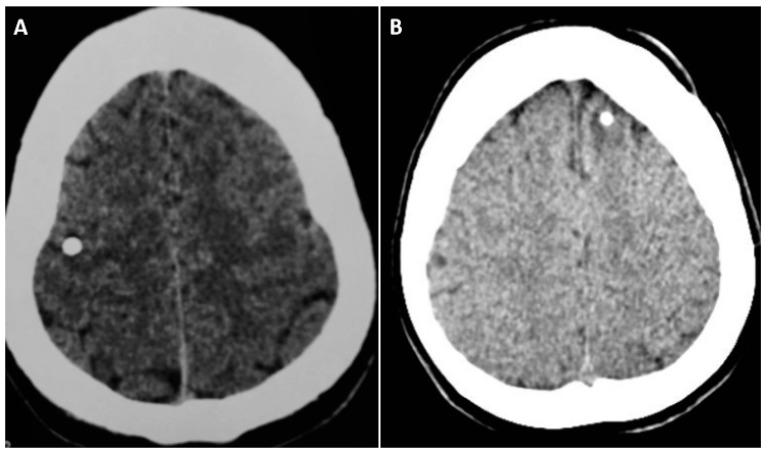
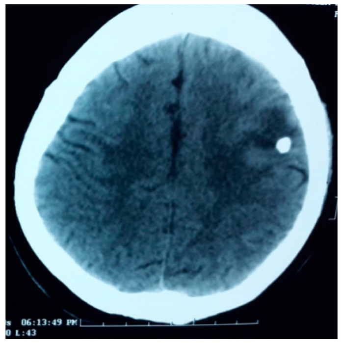
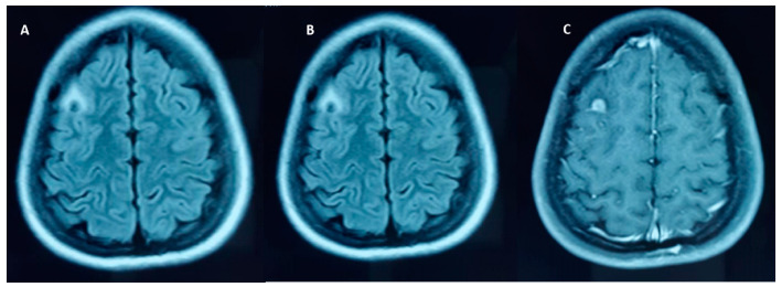
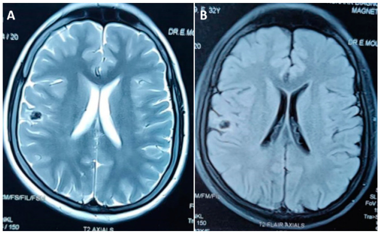
The calcified stage of the neurocysticercosis (NCC) is the common cause of acquired epilepsy in low and middle income countries in people aged > 20 years. Approximately 30% of adult onset seizures and epilepsy are attributable to NCC. In India and some of the Latin American countries, epilepsy due to solitary calcified NCC is the common adult onset epilepsy. The current evidence suggests that the calcified cysticercus granuloma is probably the epileptogenic focus. The mechanisms involved in the epileptogenic process are not well understood; Focal-onset seizures with or without impaired awareness are the common seizure type. Focal-onset seizure can evolve to bilateral tonic-clonic seizure. Seizure outcome with anti-seizure medication, most often with monotherapy, is very good. The seizure disorders associated with various stages of NCC can be preventable.
Keywords: anti-seizure medication; convulsive status epilepticus; focal-onset seizure; neurocysticercosis; solitary calcified cysticercus granulaoma; solitary calcified neurocysticercosis.
Conflict of interest statement
The authors declare no conflict of interest.
Figures
Similar articles
-
Seizure recurrence in patients with solitary cystic granuloma or single parenchymal cerebral calcification: a comparative evaluation.Seizure. 2013 Dec;22(10):840-5. doi: 10.1016/j.seizure.2013.07.001. Epub 2013 Jul 21. Seizure. 2013. PMID: 23880307
-
From seizures to epilepsy and its substrates: neurocysticercosis.Epilepsia. 2013 May;54(5):783-92. doi: 10.1111/epi.12159. Epilepsia. 2013. PMID: 23621876 Review.
-
Predictors of Lesion Calcification in Patients with Solitary Cysticercus Granuloma and New-Onset Seizures.Am J Trop Med Hyg. 2016 Sep 7;95(3):623-8. doi: 10.4269/ajtmh.16-0070. Epub 2016 Jul 18. Am J Trop Med Hyg. 2016. PMID: 27430545 Free PMC article.
-
Convulsive status epilepticus due to different evolutionary stages of neurocysticercosis - solitary cyticercus granuloma, low cyst load, and single calcific lesion in an endemic country: Clinical profile.Seizure. 2019 Oct;71:229-232. doi: 10.1016/j.seizure.2019.07.012. Epub 2019 Jul 24. Seizure. 2019. PMID: 31419720
-
Clinical presentation of neurocysticercosis-related epilepsy.Epilepsy Behav. 2017 Nov;76:151-157. doi: 10.1016/j.yebeh.2017.08.008. Epub 2017 Sep 5. Epilepsy Behav. 2017. PMID: 28882721 Review.
Cited by
-
The process of residual calcification following antiparasitic treatment in the pig model of neurocysticercosis is dynamic.PLoS Negl Trop Dis. 2025 May 5;19(5):e0013022. doi: 10.1371/journal.pntd.0013022. eCollection 2025 May. PLoS Negl Trop Dis. 2025. PMID: 40323912 Free PMC article.
-
Calcified Neurocysticercosis: Demographic, Clinical, and Radiological Characteristics of a Large Hospital-Based Patient Cohort.Pathogens. 2023 Dec 27;13(1):26. doi: 10.3390/pathogens13010026. Pathogens. 2023. PMID: 38251334 Free PMC article.
References
-
- Escobar A., Weidenheim K. The pathology of neurocysticercosis. In: Singh G., Prabhakar S., editors. Taenia solium Cysticercosis: From Basics to Clinical Science. CABI Publishing; Wallingford, UK: 2002. pp. 289–305.
LinkOut - more resources
Full Text Sources
