. 2023 Aug 16;12(8):1049.
doi: 10.3390/pathogens12081049.

Taurine, a Component of the Tear Film, Exacerbates the Pathogenic Mechanisms of Acanthamoeba castellanii in the Ex Vivo Amoebic Keratitis Model

Affiliations

Taurine, a Component of the Tear Film, Exacerbates the Pathogenic Mechanisms of Acanthamoeba castellanii in the Ex Vivo Amoebic Keratitis Model

Lizbeth Salazar-Villatoro et al. Pathogens. .

Abstract

Acanthamoeba spp. is the etiological agent of amoebic keratitis. In this study, the effect of taurine in physiological concentrations in tears (195 μM) on trophozoites of Acanthamoeba castellanii through the ex vivo amoebic keratitis model was evaluated. Trophozoites were coincubated with the Syrian golden hamster cornea (Mesocricetus auratus) for 3 and 6 h. Group 1: Control (-). Corneas coincubated with amoebic culture medium and taurine. Group 2: Control (+). Corneas coincubated with trophozoites without taurine. Group 3: Corneas coincubated with taurine 15 min before adding trophozoites. Group 4: Trophozoites coincubated 15 min with taurine before placing them on the cornea. Group 5: Corneas coincubated for 15 min with trophozoites; subsequently, taurine was added. Results are similar for both times, as evaluated by scanning electron microscopy. As expected, in the corneas of Group 1, no alterations were observed in the corneal epithelium. In the corneas of Group 2, few adhered trophozoites were observed on the corneal surface initiating migrations through cell junctions as previously described; however, in corneas of Groups 3, 4 and 5, abundant trophozoites were observed, penetrating through different corneal cell areas, emitting food cups and destabilizing corneal surface in areas far from cell junctions. Significant differences were confirmed in trophozoites adherence coincubated with taurine (p < 0.05). Taurine does not prevent the adhesion and invasion of the amoebae, nor does it favor its detachment once these have adhered to the cornea, suggesting that taurine in the physiological concentrations found in tears stimulates pathogenic mechanisms of A. castellanii.

Keywords: A. castellanii; pathogenic mechanisms; taurine in tear.


Conflict of interest statement

The authors declare no conflict of interest.

Figures

Figure 1
Figure 1
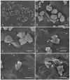
Scanning electron microscopy. Qualitative evaluation of the interaction of A. castellanii in the ex vivo model of amoebic keratitis. (A,B) Group 1, control corneas interacting for 3 and 6 h at 36.5 °C with amoebic culture medium to which 195 µM of taurine was added; an intact corneal epithelium is observed. (B) At higher magnification, well-defined cell junctions are visualized (arrows). (CF). Group 2, corneas coincubated with 2.5 × 10⁵ trophozoites of A. castellanii without taurine. (C) A considerable number of trophozoites adhered to the corneal surface are observed (arrowheads). (D) Trophozoites mainly attached to the cell junctions of the cornea; amoebae are observed in the process of cell division (arrow). (E,F) It is possible to see trophozoites emitting food cups (asterisks) and penetrating the cell junctions; the folding and lifting of the corneal cells are noted (arrow). Bar = 10 μm.
Figure 2
Figure 2
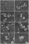
Hamster corneas coincubation with taurine, and subsequent interaction with trophozoites of A. castellanii (Group 3). (A) A considerable number of trophozoites are observed adhering and penetrating the corneal epithelium, causing evident damage to the corneal surface (arrowhead). (B) An abrasion zone (arrows) is observed adjacent to a group of trophozoites adhered to the corneal epithelium. (C,D) A large number of amoebae penetrating below the corneal cells, which probably causes the detachment of the superficial cells of the corneal epithelium (arrows), suggesting the occurrence of the process of phagocytosis coupled with the presence of food cups in some trophozoites (asterisks). (E,F) It is possible to observe areas devoid of cells and the presence of amoebae below the first cell layer (arrows). The detachment of cells from the corneal epithelium is evident at 3 and 6 h of interaction as is a visible alteration in the corneal surface (arrowheads). Bar = 10 μm.
Figure 3
Figure 3
A. castellanii trophozoites coincubated with taurine, and their subsequent interaction with hamster corneas (Group 4). (A) An abundant number of trophozoites adhered to the corneal surface are observed; areas with damage are visible (arrowheads). (B) A group of amoebae is found penetrating between the cell junctions of the corneal surface (arrows) by mechanical and/or enzymatic action; it is possible to appreciate the formation of a food cup of an amoeba surrounding a portion of a cell epithelial (arrowhead). (CE) the phagocytosis process is evident; some trophozoites were observed emitting food cups; one of them was prominent, the amoeba engulfing a cell on the surface of the cornea just at the cell junction, and another amoeba emitted very thin food cups, ingesting a portion of the corneal cell away from the cell junction (arrows); there were also visible abrasion areas far from cell junctions (asterisks). (F) It was possible to observe a trophozoite invading part of the amoebic body to penetrate deeper layers of the cornea (arrow). Similarly, an amoeba in the process of phagocytosis of a corneal epithelial cell can be seen (arrowhead). Bar = 10 μm.
Figure 4
Figure 4
Interaction of trophozoites of A. castellanii with corneas and subsequent taurine addition (Group 5). (A) A large number of adherent A. castellanii trophozoites, as well as areas of damage, are visible on the corneal surface (arrowheads). (B,C) Amoebae are observed adhering to the corneal surface, emitting their characteristic acanthopodia. The trophozoites of A. castellanii alone or in groups initiate the disorganization of the superficial cells of the cornea, which are observed to be folded (arrows) as a consequence of the mechanical action exerted by the amoebae; a food cup is observed (asterisk). (DG) Several trophozoites are in the process of phagocytosis of corneal epithelial cells in areas far from the cell junctions which have not yet been detached (arrow). It is possible to observe the development of various food cups (asterisks) and a great avidity of the trophozoites for cells of the corneal epithelium. (H) It was possible to observe the phagocytosis of an epithelial cell by two trophozoites (arrows) at the same time. Bar = 10 μm.
Chart 1
Chart 1
Quantitative evaluation of the interaction of A. castellanii with taurine in the ex vivo model of amoebic keratitis. Student’s t-test was performed using GraphPad Prism 6 software and indicated statistically significant differences in the number (#) of trophozoites that adhered to control Group 2 compared to experimental Groups 3, 4 and 5 coincubated with taurine * p < 0.05.

Similar articles

Cited by

References

    1. Lorenzo-Morales J., Khan N.A., Walochnik J. An update on Acanthamoeba keratitis: Diagnosis, pathogenesis and treatment. Parasite. 2015;22:10. doi: 10.1051/parasite/2015010. - DOI - PMC - PubMed
    1. Kot K., Łanocha-Arendarczyk N.A., Kosik-Bogacka D.I. Amoebas from the genus Acanthamoeba and their pathogenic properties. Ann. Parasitol. 2018;64:299–308. doi: 10.17420/ap6404.164. - DOI - PubMed
    1. Seal D. Treatment of Acanthamoeba keratitis. Expert Rev. Anti-Infect. Ther. 2003;1:205–208. doi: 10.1586/14787210.1.2.205. - DOI - PubMed
    1. Clarke D.W., Niederkorn J.Y. The pathophysiology of Acanthamoeba keratitis. Trends Parasitol. 2006;22:175–180. doi: 10.1016/j.pt.2006.02.004. - DOI - PubMed
    1. Visvesvara G.S., Moura H., Schuster F.L. Pathogenic and opportunistic free-living amoebae: Acanthamoeba spp., Balamuthia mandrillaris, Naegleria fowleri, and Sappinia diploidea. FEMS Immunol. Med. Microbiol. 2007;50:1–26. doi: 10.1111/j.1574-695X.2007.00232.x. - DOI - PubMed