

Bovine Papillomavirus Type 1 Infection in an Equine Congenital Papilloma
- PMID: 37624019
- PMCID: PMC10458069
- DOI: 10.3390/pathogens12081059
Bovine Papillomavirus Type 1 Infection in an Equine Congenital Papilloma
Abstract
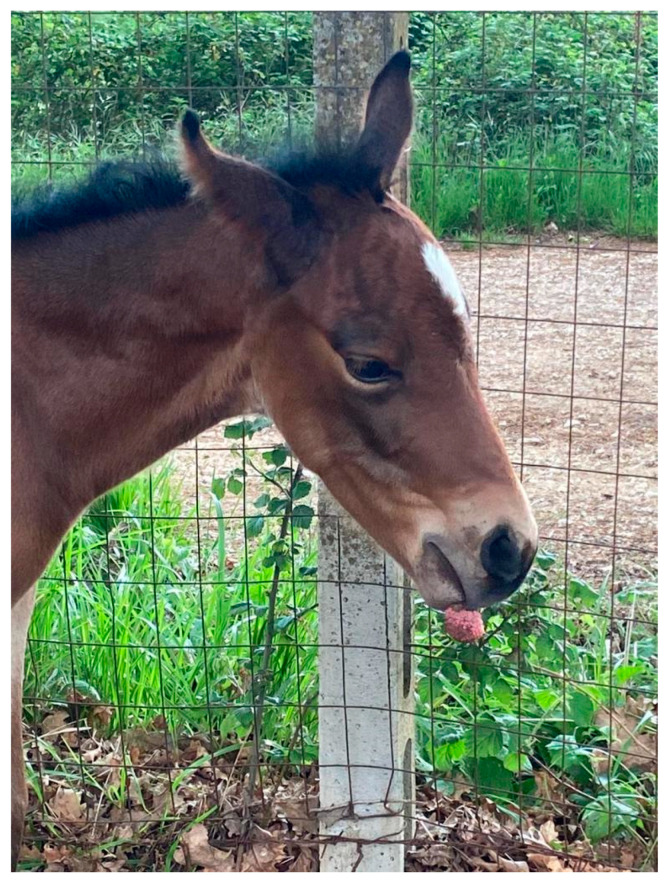
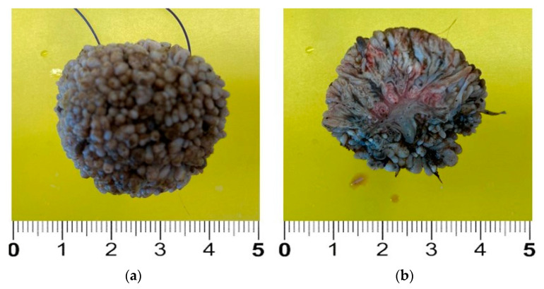
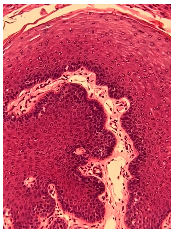
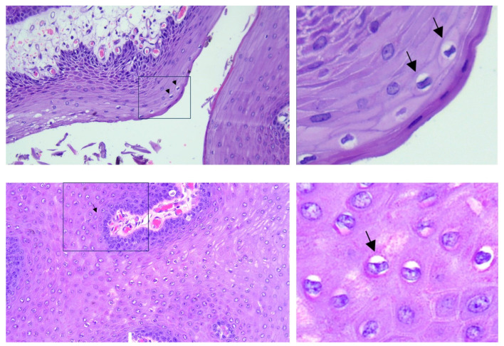
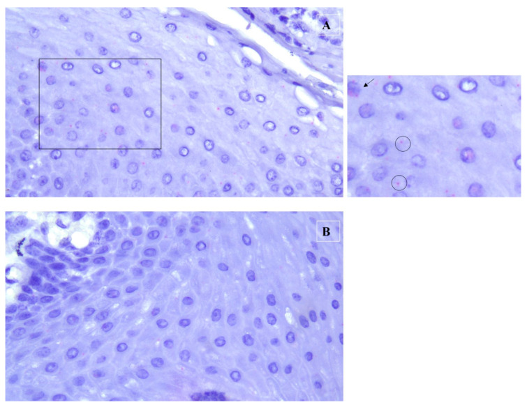
Papillomas are benign epithelial lesions protruding on the epithelial surfaces as finger-like or warty projections. These lesions are often caused by papillomavirus (PV) infection. Congenital papillomas have been reported in foals. However, to date, no evidence of PV infection has been provided. In the present paper, we describe the main clinical-pathological features of a congenital papilloma observed in a foal. In addition, biomolecular tests demonstrated BPV1 infection in the case under study. Such data stimulate further investigations, even on archived samples, aiming to clarifying the etiology of equine congenital papilloma and the clinical relevance, if any, of BPV1 vertical transmission in horses.
Keywords: bovine papillomaviruses; congenital papilloma; horse.
Conflict of interest statement
The authors declare no conflict of interest.
Figures
Similar articles
-
Genomic characterisation of bovine papillomavirus types 1 and 2 identified in equine sarcoids in Japan.Equine Vet J. 2021 Nov;53(6):1199-1209. doi: 10.1111/evj.13398. Epub 2021 Jan 8. Equine Vet J. 2021. PMID: 33300145
-
Evaluation of equine papillomas, aural plaques, and sarcoids for the presence of Equine papillomavirus DNA and Papillomavirus antigen.Can J Vet Res. 2007 Jan;71(1):28-33. Can J Vet Res. 2007. PMID: 17193879 Free PMC article.
-
No evidence of bovine papillomavirus type 1 or 2 infection in healthy equids.Equine Vet J. 2019 Sep;51(5):612-616. doi: 10.1111/evj.13061. Epub 2019 Jan 28. Equine Vet J. 2019. PMID: 30560998
-
Papillomavirus-like Particles in Equine Medicine.Viruses. 2023 Jan 25;15(2):345. doi: 10.3390/v15020345. Viruses. 2023. PMID: 36851559 Free PMC article. Review.
-
Anogenital-Associated Papillomaviruses in Animals: Focusing on Bos taurus Papillomaviruses.Pathogens. 2020 Nov 27;9(12):993. doi: 10.3390/pathogens9120993. Pathogens. 2020. PMID: 33260814 Free PMC article. Review.
Cited by
-
Fetal rhabdomyoma in a Thoroughbred filly.Vet Med Sci. 2024 Jul;10(4):e1534. doi: 10.1002/vms3.1534. Vet Med Sci. 2024. PMID: 38975617 Free PMC article.
-
Bovine papillomavirus vertical transmission: BPV diversity and expression in maternal and fetal tissues.Braz J Microbiol. 2024 Sep;55(3):2879-2884. doi: 10.1007/s42770-024-01394-y. Epub 2024 May 27. Braz J Microbiol. 2024. PMID: 38801639 Free PMC article.
References
-
- Kumar V., Abbas A.K., Fausto N. Neoplasia. In: Kumar V., Abbas A.K., Fausto N., editors. Pathologic Basis of Disease. 7th ed. Elsevier Saunders; Philadelphia, PA, USA: 2005. pp. 269–342.
-
- Mauldin E.A., Peters-Kennedy J. Integumentary System. In: Maxie M.G., editor. Pathology of Domestic Animals. 6th ed. Elsevier; Alpharetta, GA, USA: 2015. pp. 509–736.
-
- Scott D.W. Neoplastic diseases. In: Scott D.W., editor. Large Animal Dermatology. W.B. Saunders Company; Philadelphia, PA, USA: 1998. pp. 419–468.
Publication types
Grants and funding
LinkOut - more resources
Full Text Sources
