

CSF-1R+ Macrophages Control the Gut Microbiome-Enhanced Liver Invariant NKT Function through IL-18
- PMID: 37624046
- PMCID: PMC10529904
- DOI: 10.4049/jimmunol.2200854
CSF-1R+ Macrophages Control the Gut Microbiome-Enhanced Liver Invariant NKT Function through IL-18
Abstract
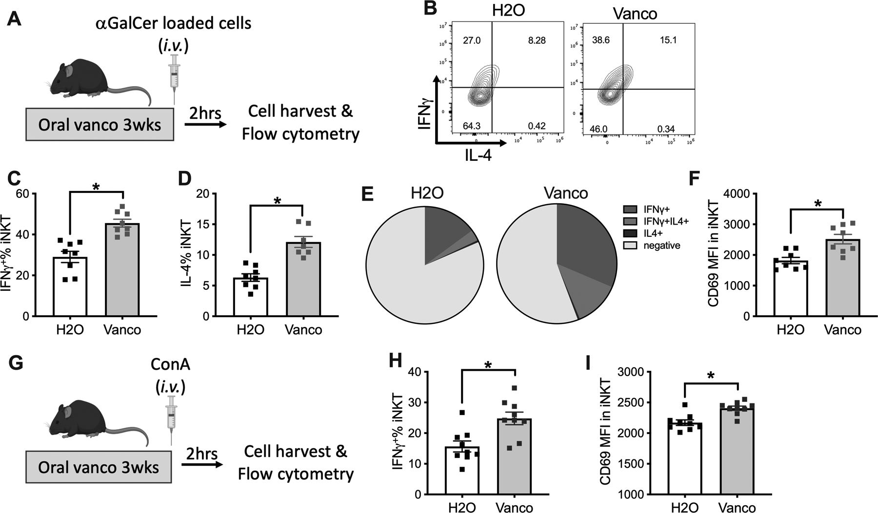
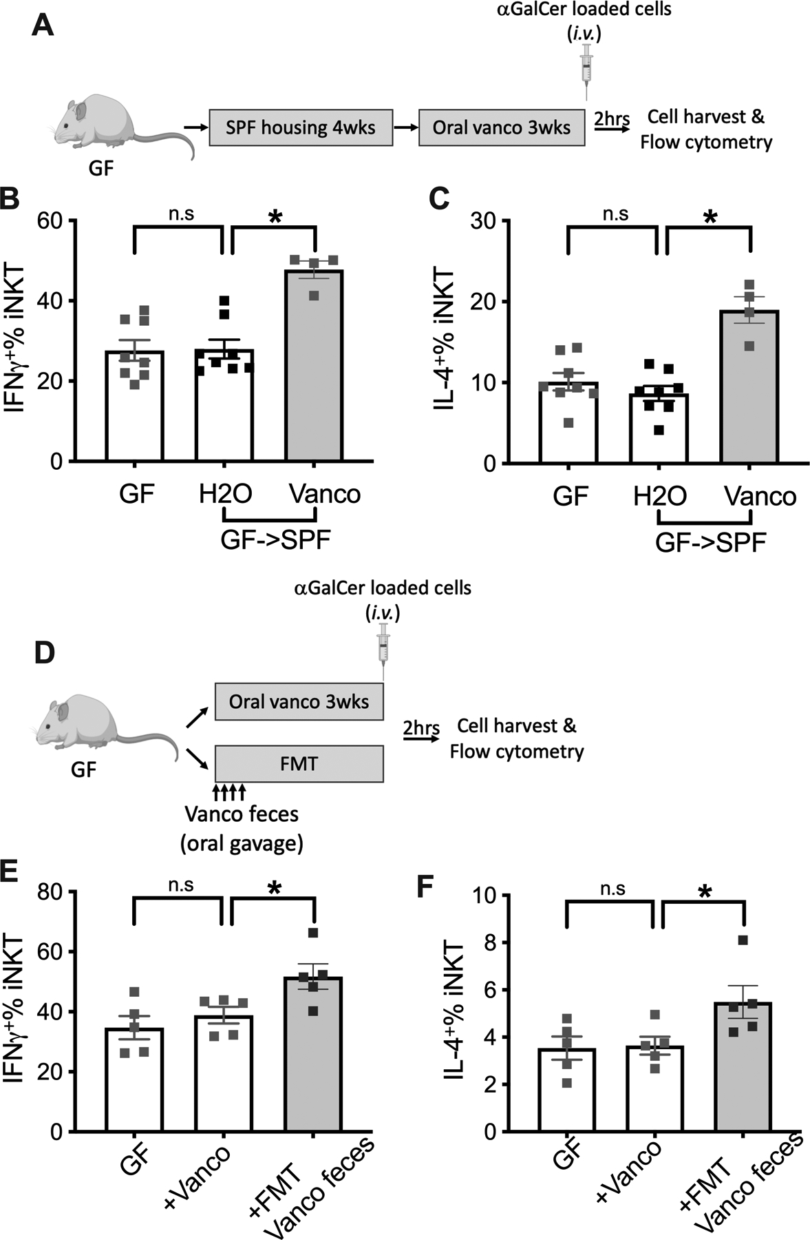
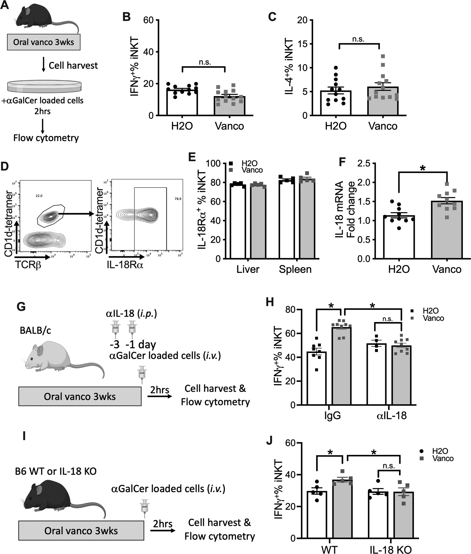
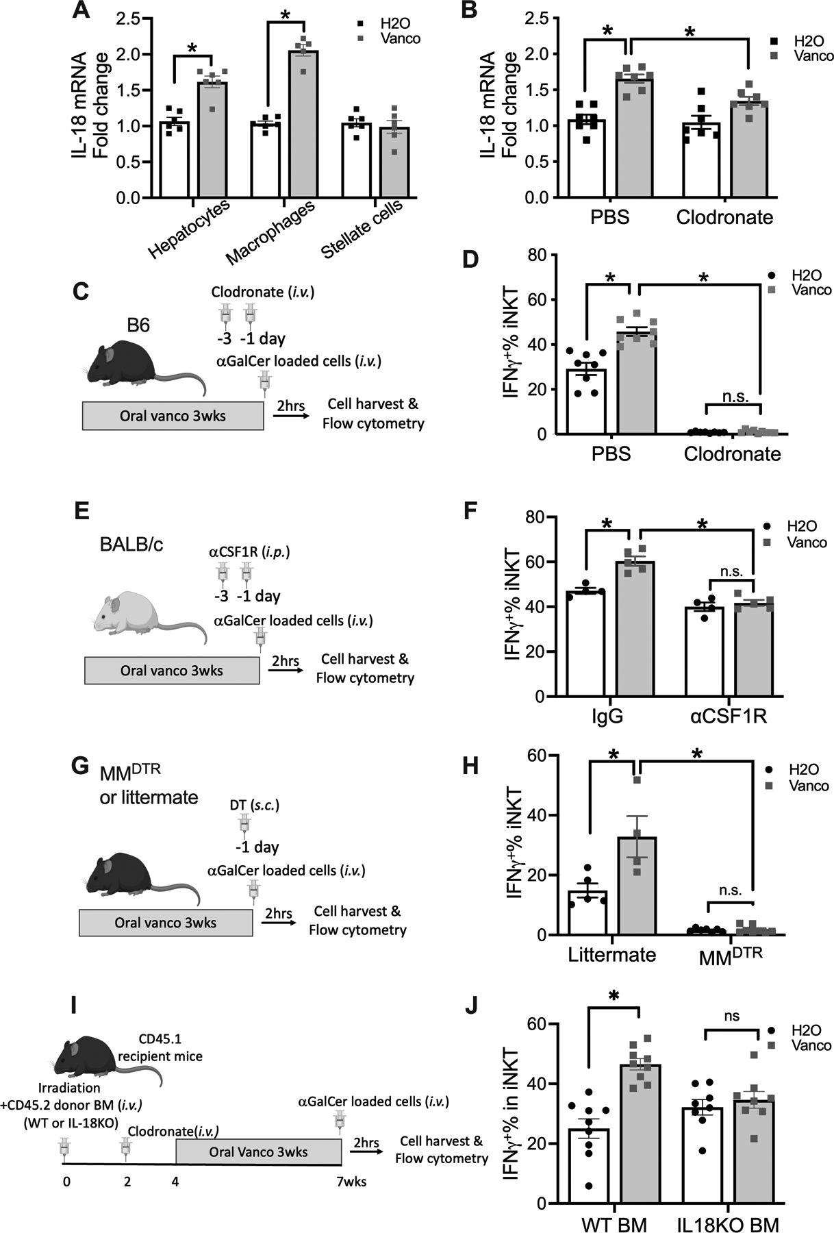
The gut microbiome is an important modulator of the host immune system. In this study, we found that altering the gut microbiome by oral vancomycin increases liver invariant NKT (iNKT) cell function. Enhanced iNKT cytokine production and activation marker expression were observed in vancomycin-treated mice following both Ag-specific and Ag-independent in vivo iNKT stimulations, with a more prominent effect in the liver than in the spleen. Fecal transplantation studies demonstrated that the iNKT functional regulation is mediated by altering the gut microbiome but uncoupled from the modulation of iNKT cell population size. Interestingly, when stimulated in vitro, iNKT cells from vancomycin-treated mice did not show increased activation, suggesting an indirect regulation. iNKT cells expressed high levels of IL-18 receptor, and vancomycin increased the expression of IL-18 in the liver. Blocking IL-18 by neutralizing Ab or using genetically deficient mice attenuated the enhanced iNKT activation. Liver macrophages were identified as a major source of IL-18. General macrophage depletion by clodronate abolished this iNKT activation. Using anti-CSF-1R depletion or LyzCrexCSF-1RLsL-DTR mice identified CSF-1R+ macrophages as a critical modulator of iNKT function. Vancomycin treatment had no effect on iNKT cell function in vivo in IL-18 knockout macrophage reconstituted mice. Together, our results demonstrate that the gut microbiome controls liver iNKT function via regulating CSF-1R+ macrophages to produce IL-18.
Conflict of interest statement
Conflict-of-interest statement
The authors declare no conflict of interest.
Figures
Similar articles
-
Activation of iNKT Cells Facilitates Liver Repair After Hepatic Ischemia Reperfusion Injury Through Acceleration of Macrophage Polarization.Front Immunol. 2021 Oct 6;12:754106. doi: 10.3389/fimmu.2021.754106. eCollection 2021. Front Immunol. 2021. PMID: 34691073 Free PMC article.
-
TCR-dependent and -independent activation underlie liver-specific regulation of NKT cells.J Immunol. 2011 Jan 15;186(2):838-47. doi: 10.4049/jimmunol.1001735. Epub 2010 Dec 10. J Immunol. 2011. PMID: 21148802 Free PMC article.
-
Activation of murine invariant NKT cells promotes susceptibility to candidiasis by IL-10 induced modulation of phagocyte antifungal activity.Eur J Immunol. 2016 Jul;46(7):1691-703. doi: 10.1002/eji.201545987. Epub 2016 May 27. Eur J Immunol. 2016. PMID: 27151377
-
Fecal IgA Levels and Gut Microbiota Composition Are Regulated by Invariant Natural Killer T Cells.Inflamm Bowel Dis. 2020 Apr 11;26(5):697-708. doi: 10.1093/ibd/izz300. Inflamm Bowel Dis. 2020. PMID: 31819985
-
Complex Network of NKT Cell Subsets Controls Immune Homeostasis in Liver and Gut.Front Immunol. 2018 Sep 11;9:2082. doi: 10.3389/fimmu.2018.02082. eCollection 2018. Front Immunol. 2018. PMID: 30254647 Free PMC article. Review.
Cited by
-
IFN-γ-dependent regulation of intestinal epithelial homeostasis by NKT cells.Cell Rep. 2024 Dec 24;43(12):114948. doi: 10.1016/j.celrep.2024.114948. Epub 2024 Nov 23. Cell Rep. 2024. PMID: 39580798 Free PMC article.
-
Porphyromonas gingivalis fuels colorectal cancer through CHI3L1-mediated iNKT cell-driven immune evasion.Gut Microbes. 2024 Jan-Dec;16(1):2388801. doi: 10.1080/19490976.2024.2388801. Epub 2024 Aug 12. Gut Microbes. 2024. PMID: 39132842 Free PMC article.
-
SPP1 + macrophages cause exhaustion of tumor-specific T cells in liver metastases.Nat Commun. 2025 May 7;16(1):4242. doi: 10.1038/s41467-025-59529-0. Nat Commun. 2025. PMID: 40335453 Free PMC article.
References
-
- Albillos A, de Gottardi A, and Rescigno M. 2020. The gut-liver axis in liver disease: Pathophysiological basis for therapy. J Hepatol 72: 558–577. - PubMed
-
- Cohen NR, Garg S, and Brenner MB. 2009. Antigen Presentation by CD1 Lipids, T Cells, and NKT Cells in Microbial Immunity. Adv Immunol 102: 1–94. - PubMed
-
- Kronenberg M 2005. Toward an understanding of NKT cell biology: progress and paradoxes. Annu Rev Immunol 23: 877–900. - PubMed
-
- Bendelac A, Savage PB, and Teyton L. 2007. The biology of NKT cells. Annu Rev Immunol 25: 297–336. - PubMed
Publication types
MeSH terms
Substances
Grants and funding
LinkOut - more resources
Full Text Sources
Molecular Biology Databases
Miscellaneous
