Review
. 2023 Nov 1;325(5):H1012-H1038.
doi: 10.1152/ajpheart.00288.2023. Epub 2023 Aug 25.

Impact of aging on vascular ion channels: perspectives and knowledge gaps across major organ systems

Affiliations
Review

Impact of aging on vascular ion channels: perspectives and knowledge gaps across major organ systems

Erik J Behringer. Am J Physiol Heart Circ Physiol. .

Abstract

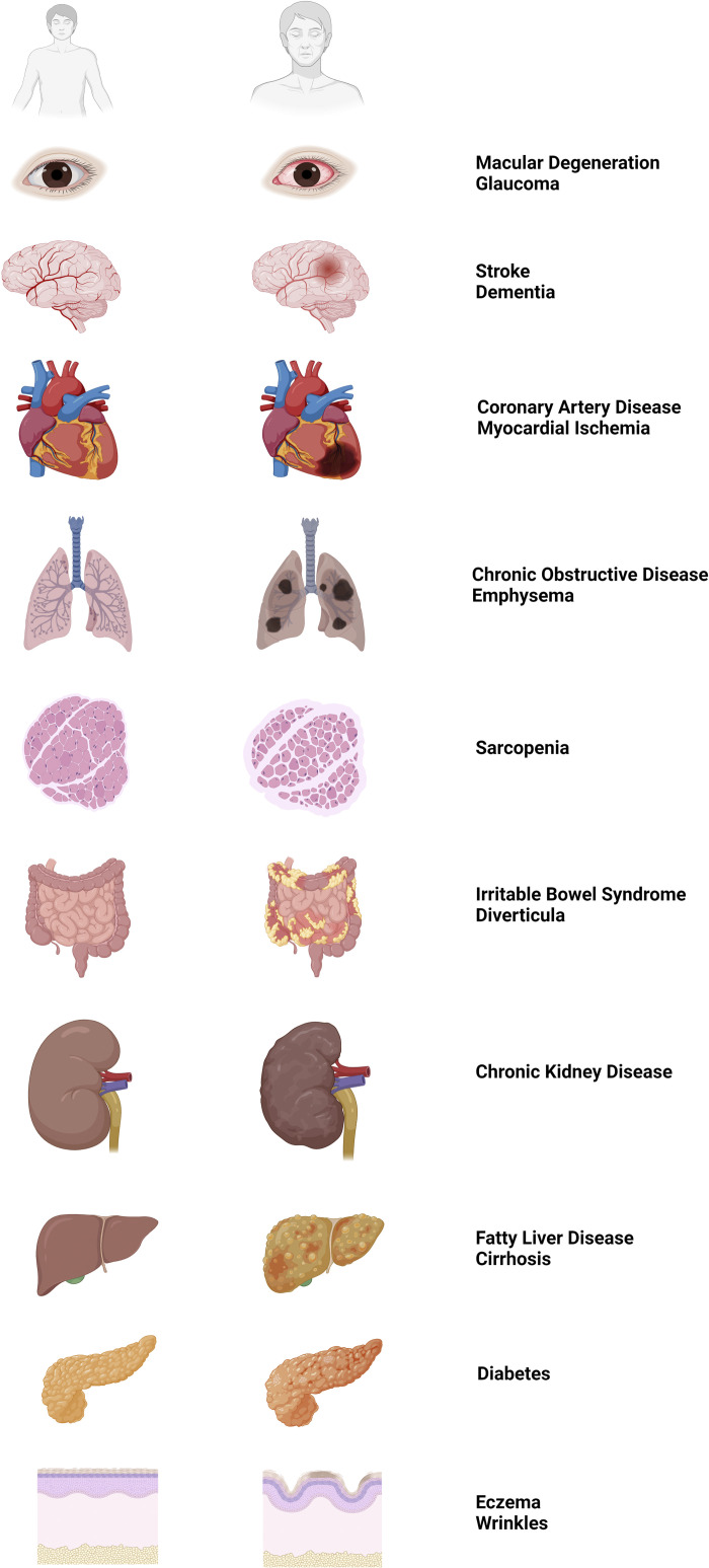
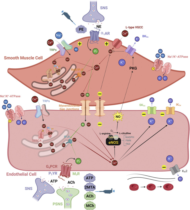
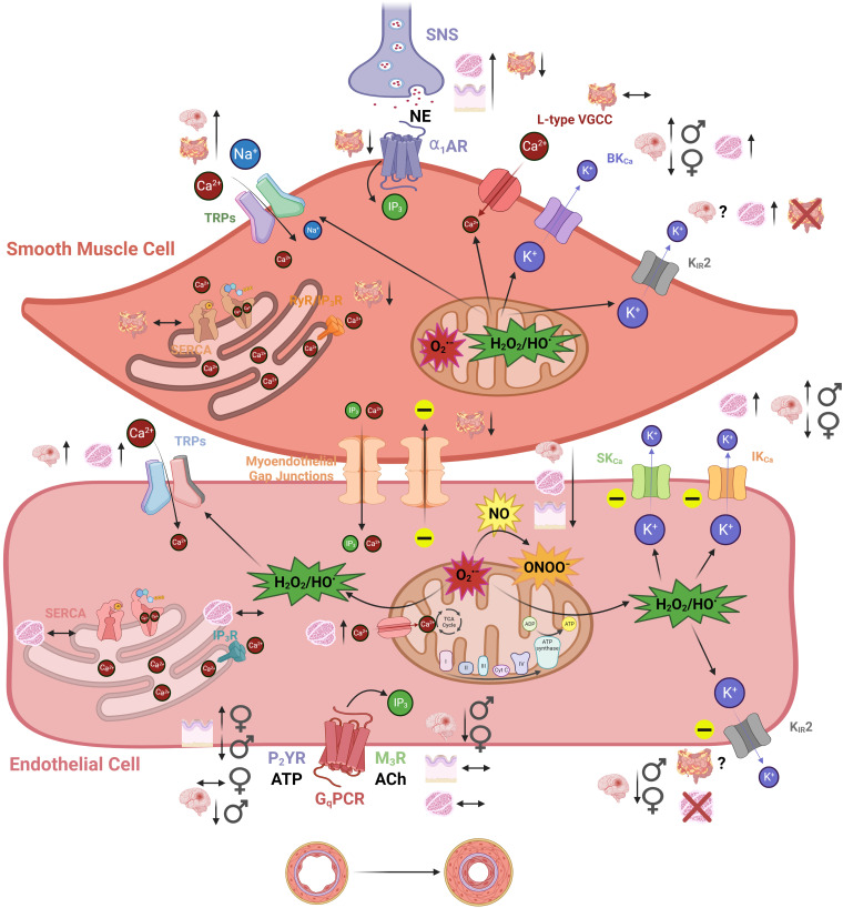
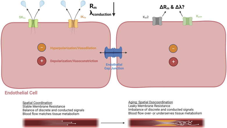
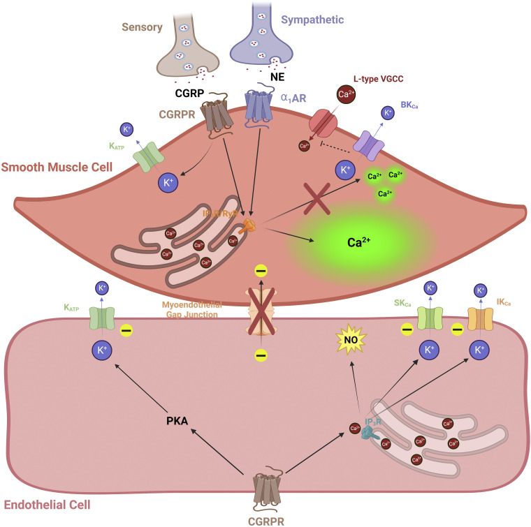
Individuals aged ≥65 yr will comprise ∼20% of the global population by 2030. Cardiovascular disease remains the leading cause of death in the world with age-related endothelial "dysfunction" as a key risk factor. As an organ in and of itself, vascular endothelium courses throughout the mammalian body to coordinate blood flow to all other organs and tissues (e.g., brain, heart, lung, skeletal muscle, gut, kidney, skin) in accord with metabolic demand. In turn, emerging evidence demonstrates that vascular aging and its comorbidities (e.g., neurodegeneration, diabetes, hypertension, kidney disease, heart failure, and cancer) are "channelopathies" in large part. With an emphasis on distinct functional traits and common arrangements across major organs systems, the present literature review encompasses regulation of vascular ion channels that underlie blood flow control throughout the body. The regulation of myoendothelial coupling and local versus conducted signaling are discussed with new perspectives for aging and the development of chronic diseases. Although equipped with an awareness of knowledge gaps in the vascular aging field, a section has been included to encompass general feasibility, role of biological sex, and additional conceptual and experimental considerations (e.g., cell regression and proliferation, gene profile analyses). The ultimate goal is for the reader to see and understand major points of deterioration in vascular function while gaining the ability to think of potential mechanistic and therapeutic strategies to sustain organ perfusion and whole body health with aging.

Keywords: K+ channels; TRP channels; endothelial function; myoendothelial coupling; vascular aging.


Conflict of interest statement

No conflicts of interest, financial or otherwise, are declared by the author.

Figures

Figure 1.
Figure 1.
Aging and the development of chronic diseases among organs throughout the body. Left: cartoon illustrations of healthy organs during young adult age (≈25 yr) as eye, brain, heart, lungs, skeletal muscle, gut, kidney, liver, pancreas, and skin (top to bottom). Middle: corresponding illustrations of diseased organs during old adult age (≥65 yr). Right: list of common diseases that become more prevalent aging as associated with the disease states of each organ illustrated in middle. Images were created with a licensed version of Biorender.com.
Figure 2.
Figure 2.
Classic fundamental anatomy of vascular signaling within resistance arteries: an endothelial perspective. Smooth muscle cells (SMCs) serve the mechanical compliance function of the vascular wall for controlling resistance to blood flow, whereas endothelial cells (ECs) transduce stimuli evoked by signaling molecules carried in the blood within the vascular lumen. The level of [Ca2+]i among cell types in the vascular wall plays a dichotomous role, whereby its global increase in SMCs and ECs generates vasodilation and vasoconstriction, respectively. Either as a cause or effect of increased [Ca2+]i in the vascular wall, vasoconstriction and vasodilation are associated with membrane potential (Vm), indicated as depolarization (“+” sign) and hyperpolarization (“−” sign), respectively. SMC: the primary contributors to elevations in SMC [Ca2+]i are norepinephrine (NE)-stimulated α1-adrenergic receptors (α1ARs) sourced from the sympathetic nervous system (SNS), L-type voltage-gated Ca2+ channels (VGCCs), and Ca2+-permeant transient receptor potential (TRP) channels (e.g., TRPC3, TRPV3, TRPV4). Also, Na+-permeant TRP channels (e.g., TRPM4) can depolarize SMC Vm and activate L-type VGCCs. A distinct event known as Ca2+ “sparks” in SMCs released from endoplasmic reticulum ryanodine receptors (RyRs) will selectively activate high-conductance Ca2+-activated K+ (BKCa) channels to hyperpolarize SMC Vm while deactivating L-type VGCCs. Note that α1ARs can also be activated by the experimental agonist phenylephrine (PE, see pipettor icon). EC: the contributors to increased EC [Ca2+]i are Gq protein-coupled receptors (GqPCRs; purinergic, P2YR and muscarinic, M3R) activated by adenosine triphosphate (ATP) or acetylcholine (ACh) and Ca2+-permeant TRP channels (e.g., TRPV4). Increased EC [Ca2+]i primarily generates nitric oxide (NO) via endothelial NO synthase (eNOS, see main text for more information) and activates small- and intermediate-conductance Ca2+-activated K+ (SKCa and IKCa) channels. In addition to activated SKCa and IKCa channels, hyperpolarization of EC Vm can occur through direct activation of inward-rectifying K+ (KIR) channels via the shear of blood flow or elevated extracellular K+ (6–15 mM). Experimental agonists for P2YRs (2-methylthioadenosine diphosphate or 2MTA) and M3Rs (methacholine or MCh) are also used to mimic actions of the SNS and parasympathetic nervous system (PSNS), respectively (see pipettor icon). Myoendothelial feedback: activation of SMC α1ARs by NE generates inositol-trisphosphate (IP3) to elicit Ca2+ release through IP3 receptors (IP3Rs) in the sarcoplasmic reticulum and thereby produce constriction. When elevated in SMCs for a prolonged period, IP3 and Ca2+ diffuse through myoendothelial gap junctions into the endothelium to activate SKCa and IKCa channels and/or NO production, providing negative feedback to smooth muscle contraction (see broken lines indicating signaling from EC back to SMC). Images were created with a licensed version of Biorender.com.
Figure 3.
Figure 3.
The impact of aging on classical signaling components within the vascular wall of resistance arteries. There are increases (↑), decreases (↓), or stability (↔) among various smooth muscle cell (SMC) and endothelial cell (EC) receptors and ion channels that govern vascular reactivity during old vs. young age. With brain, gut, skeletal muscle, and skin blood vessels most prominently used for aging studies, various vascular types (and biological sex where applicable/known) are illustrated for both SMCs and ECs. Note that KIR2 channels in particular are expressed in both cell types in brain arteries but not skeletal muscle (SMCs only) or gut (ECs only). Mitochondrial reactive oxygen species signaling via superoxide (O2•−), and hydrogen peroxide/peroxyl radicals (H2O2/HO) play more of a role during and old age and impact various Ca2+-permeant pathways (e.g., TRP channels) and K+ channels (e.g., SKCa and IKCa). Although not illustrated, other major sources of O2•− include the nicotinamide adenine dinucleotide phosphate (NADPH) oxidases and uncoupled eNOS (see main text). Note that nitric oxide (NO) bioavailability is decreased during old age because of a greater propensity for uncoupled endothelial NO synthase (eNOS) and the reaction of O2•− with NO form peroxynitrite (ONOO). A primary consequence of altered signaling during aging could be elevated basal constriction and higher vascular resistance as the basis for hypertension. Another outcome may be sustained discrete reactivity to metabolites and neurotransmitters with a decrease in the spatial domain along the vascular wall for conducted signals through endothelial and myoendothelial gap junctions among capillaries, precapillary arterioles, and arteries (see Fig. 4). Images were created with a licensed version of Biorender.com.
Figure 4.
Figure 4.
Conducted vascular signaling and the endothelium as an electrical conduit: leaky ion channels and a restricted spatial domain with aging. In addition to modes of discrete/local changes in vasoreactivity, conducted signaling along the endothelium through gap junctions is crucial for coordinating vascular resistance among arteries, arterioles, and capillaries to match tissue metabolism. With aging, ion channels such as small- and intermediate-conductance Ca2+-activated K+ (SKCa and IKCa) channels become “leaky” because of reactive oxygen species and enhanced Ca2+ entry through transient receptor potential channels (TRPs). Where applicable, changes in modulators of KIR2 (phosphatidylinositol 4,5-bisphosphate or PIP2, cholesterol) and ATP-sensitive K+ channels (KATP) channels (ATP, H2S) with aging may influence optimal distribution of charge (hyperpolarization, depolarization) and corresponding vasoreactivity (vasodilation, vasoconstriction) as well. The length constant of conduction (λ) is favored by a high membrane resistance (Rm) and low-axial resistance as defined by the open-state probability of ion channels and gap junctions, respectively. The consequences of leaky ion channels with aging are a decrease in Rm and thereby a restricted spatial domain for optimally coordinating changes in vascular resistance to blood flow to meet metabolic demand. Images were created with a licensed version of Biorender.com.
Figure 5.
Figure 5.
Novel perivascular nerve interactions to influence Ca2+ and K+ channel activity during aging. A role for afferent (sensory) nerve-signaling mechanisms has recently emerged during aging as a vasodilatory counterbalance to sympathetic (vasoconstrictive) input (209, 211) (see main text). A diminishment in sensory nerve density and signaling [via calcitonin gene-related peptide (CGRP) and its receptor] occurs in tandem with enhanced sympathetic activity [via α1-adrenergic receptor (α1AR)]. Consequently, smooth muscle cell (SMC), Ca2+ “waves” dominate versus “spark” events and thereby enhance cross-bridge interaction of actin and myosin fibers for constriction while inhibiting SMC hyperpolarization via high-conductance Ca2+-activated K+ channels (BKCa). As shown in mesenteric arteries, myoendothelial coupling is reduced during old age as well, potentially precluding the vasodilatory influence of calcitonin gene-related peptide receptors (CGRPRs) on endothelial cells (ECs) via activated K+ channels (KATP, SKCa, and IKCa). Although not as well known in the context of aging and competition with sympathetic nerves, note that substance P and its receptor are also important for mediating perivascular sensory nerve signals particularly via KCa channels and nitric oxide (NO) production. Images were created with a licensed version of Biorender.com.

Similar articles

Cited by

References

    1. Observations on the Harveian doctrine of the circulation of the blood. Edinb Med Surg J 13: 501–507, 1817. - PMC - PubMed
    1. Vaduganathan M, Mensah GA, Turco JV, Fuster V, Roth GA. The global burden of cardiovascular diseases and risk: a compass for future health. J Am Coll Cardiol 80: 2361–2371, 2022. doi:10.1016/j.jacc.2022.11.005. - DOI - PubMed
    1. Divo MJ, Martinez CH, Mannino DM. Ageing and the epidemiology of multimorbidity. Eur Respir J 44: 1055–1068, 2014. doi:10.1183/09031936.00059814. - DOI - PMC - PubMed
    1. Camici GG, Savarese G, Akhmedov A, Lüscher TF. Molecular mechanism of endothelial and vascular aging: implications for cardiovascular disease. Eur Heart J 36: 3392–3403, 2015. doi:10.1093/eurheartj/ehv587. - DOI - PubMed
    1. Bloom SI, Islam MT, Lesniewski LA, Donato AJ. Mechanisms and consequences of endothelial cell senescence. Nat Rev Cardiol 20: 38–51, 2023. doi:10.1038/s41569-022-00739-0. - DOI - PMC - PubMed

Publication types

LinkOut - more resources