. 2023 Aug 7;8(8):401.
doi: 10.3390/tropicalmed8080401.

Nanoformulation-Based 1,2,3-Triazole Sulfonamides for Anti- Toxoplasma In Vitro Study

Affiliations

Nanoformulation-Based 1,2,3-Triazole Sulfonamides for Anti- Toxoplasma In Vitro Study

Fadwa M Arafa et al. Trop Med Infect Dis. .

Abstract

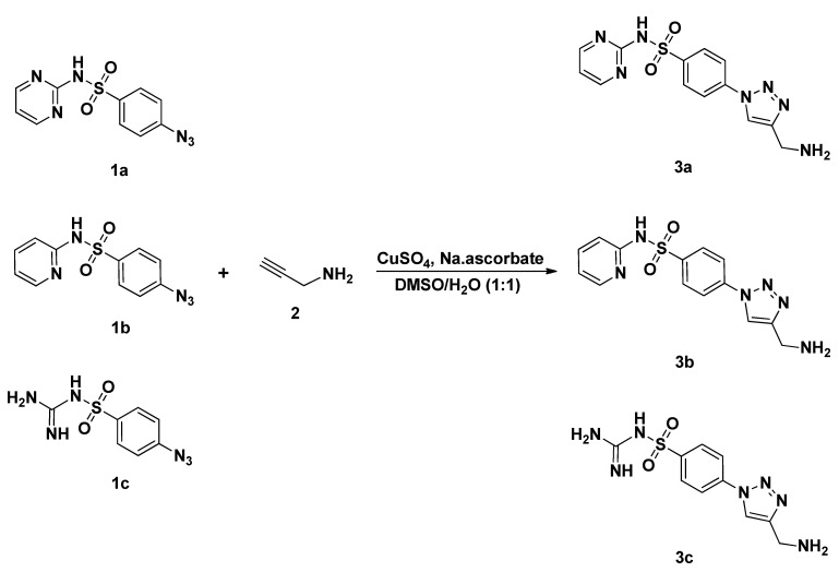
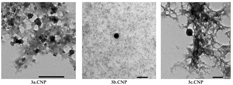
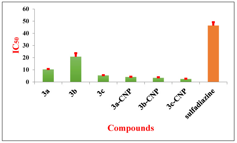
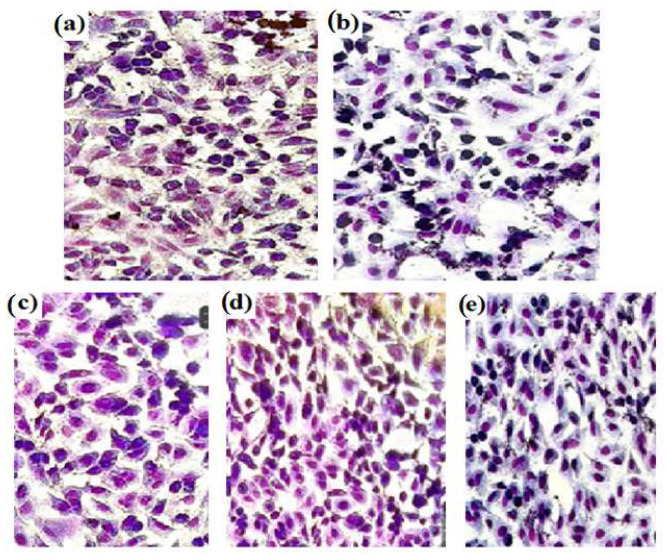
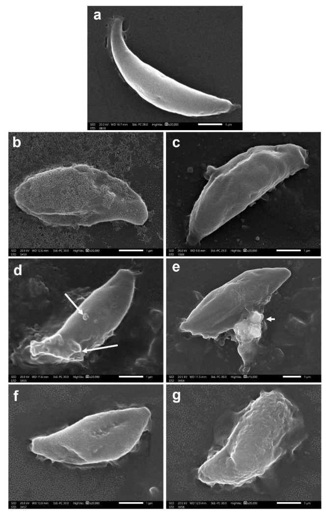
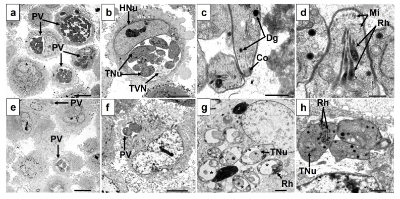
Toxoplasma gondii is deemed a successful parasite worldwide with a wide range of hosts. Currently, a combination of pyrimethamine and sulfadiazine serves as the first-line treatment; however, these drugs have serious adverse effects. Therefore, it is imperative to focus on new therapies that produce the desired effect with the lowest possible dose. The designation and synthesis of sulfonamide-1,2,3-triazole hybrids (3a-c) were performed to create hybrid frameworks. The newly synthesized compounds were loaded on chitosan nanoparticles (CNPs) to form nanoformulations (3a.CNP, 3b.CNP, 3c.CNP) for further in vitro investigation as an anti-Toxoplasma treatment. The current study demonstrated that all examined compounds were active against T. gondii in vitro relative to the control drug, sulfadiazine. 3c.CNP showed the best impact against T. gondii with the lowest IC50 value of 3.64 µg/mL. Using light microscopy, it was found that Vero cells treated with the three nanoformulae showed remarkable morphological improvement, and tachyzoites were rarely seen in the treated cells. Moreover, scanning and transmission electron microscopic studies confirmed the efficacy of the prepared nanoformulae on the parasites. All of them caused parasite ultrastructural damage and altered morphology, suggesting a cytopathic effect and hence confirming their promising anti-Toxoplasma activity.

Keywords: 1,2,3-triazole; Toxoplasma gondii; chitosan nanoparticles; in vitro studies; sulfonamides.


Conflict of interest statement

The authors declare no conflict of interest.

Figures

Scheme 1
Scheme 1
Click synthesis of sulfonamide-1,2,3-triazole molecular conjugates 3a–c.
Figure 1
Figure 1
Transmission electron microscope study of 3a–c.CNP.
Figure 2
Figure 2
The IC50 concentrations (μg/mL) of 3a–c, their nanoformulae (3a–c.CNP), and sulfadiazine against T. gondii tachyzoites.
Figure 3
Figure 3
Morphology of Vero cells after 24 h of exposure to 50 μg/mL of (a) RPMI medium, (b) sulfadiazine, (c) 3a.CNP, (d) 3b.CNP and (e) 3c.CNP. Stained using Giemsa stain (×200).
Figure 4
Figure 4
Morphology of Vero cells infected with T. gondii after 24 h of exposure to (a) RPMI medium: the tachyzoites of T. gondii can be seen inside the inoculated Vero cells (red arrow), as well as adhered to the glass coverslip (black arrow); and 50 μg/mL each of (b) sulfadiazine: the tachyzoites can be seen inside (red arrow) and outside (black arrow) treated Vero cells, (c) 3a.CNP, (d) 3b.CNP, and (e) 3c.CNP: T. gondii tachyzoites were rarely seen in the infected cells; however, some vacuoles, representing degenerated parasites, were noticed (black arrows). Stained using Giemsa stain (×1000).
Figure 5
Figure 5
SEM of T. gondii tachyzoites (a) Regular, non-treated tachyzoite with tapered anterior end and rounded posterior end (×20,000). (b,c) Tachyzoites treated with 3a.CNP showing rough outer surface with multiple depressions and deep furrows (×20,000). (d,e) Tachyzoites treated with 3b.CNP showing multiple surface blebs (arrows) and loss of surface integrity with leakage of cytoplasmic contents (arrowhead) (×20,000). (f,g) Tachyzoites treated with 3c.CNP showing multiple surface depressions and completely damaged and disfigured morphology (×20,000).
Figure 6
Figure 6
Transmission electron microscopic (TEM) images of Toxoplasma gondii-infected, non-treated (ad) and 3a.CNP-treated (eh) Vero cells showing: (a) Multiple infected, non-treated cells showing several parasitophorous vacuoles (PV) containing a large number of tachyzoites (×600). (b) A cross-section of an infected cell where the host cell’s nucleus (HNu) and a large PV containing multiple tachyzoites can be seen. Each tachyzoite contained a clearly delineated nucleus (TNu) and multiple organelles and was surrounded by the structures of the tubulovesicular network (TVN) (×1500). (c,d) A longitudinal section of the anterior end of the tachyzoites showing conoid (Co), dense granules (Dg), micronemes (Mi), and thin elongated rhoptry bulbs (Rh) (×8000 and ×12,000, respectively). (e,f) Infected, 3a.CNP-treated cells showed fewer infected cells and PVs with fewer tachyzoites inside them (×600 and ×1500, respectively). (g) A cross-section of an infected, 3a.CNP-treated cell containing multiple vacuolated tachyzoites where the nuclei (TNu) can be seen surrounded by vacuoles (asterisks) containing degraded materials. The rhoptries (Rh) can be seen to be pushed aside by the vacuoles. Destabilized PV membrane with infiltration of the host cell’s cytoplasmic contents within the PV (×2000). (h) Tachyzoite treated with 3a.CNP showing swollen rhoptry bulbs (Rh) and the nucleus (TNu) lost its lining nuclear membrane. Other tachyzoites appeared tethered to each other (white arrow) with no clear line of demarcation between them (×5000).
Figure 7
Figure 7
Transmission electron microscopic (TEM) images of Toxoplasma gondii-infected, 3b.CNP-treated (ac) and 3c.CNP-treated (df) Vero cells showing: (a) Multiple, 3b.CNP-treated tachyzoites appeared extremely vacuolated without nuclei or organelles (×1500). (b) At higher magnification, tachyzoites appeared to have lost their nuclei and all apical complex structures. They had multiple-sized vacuoles (asterisks) containing degraded materials (×5000). (c) Tachyzoites treated with 3b.CNP were tethered to each other (black arrow) (×8000). (d) Multiple intracellular, 3c.CNP-treated tachyzoites within a PV. Some appeared apparently normal, while others had hazy cytoplasmic membranes (black arrows) without nuclei or organelles. The tubulovesicular network (TVN) appeared dark and granular with an absence of the PV membrane (×4000). (e) Tachyzoites treated with 3c.CNP appeared without any nuclei or organelles, and only multiple-sized vacuoles (asterisk) containing materials at different stages of degradation were seen. The tubulovesicular network (TVN) appeared dark and granular (×6000). (f) Tachyzoites treated with 3c.CNP were severely destructed without nuclei or organelles. Nuclear chromatin could be detected in the cytoplasm without any delineating nuclear membrane (black arrow). A triangular structure resembling the anterior end of another superimposed degenerated tachyzoite could be seen (white arrow) (×6000).

References

    1. Dubey J., Lago E., Gennari S., Su C., Jones J. Toxoplasmosis in humans and animals in Brazil: High prevalence, high burden of disease, and epidemiology. Parasitology. 2012;139:1375–1424. doi: 10.1017/S0031182012000765. - DOI - PubMed
    1. Schlundt J., Toyofuku H., Jansen J., Herbst S. Emerging food–borne zoonoses. Rev. Sci. Et Tech.-Off. Int. Des. Epizoot. 2004;23:513–534. doi: 10.20506/rst.23.2.1506. - DOI - PubMed
    1. Esch G.W. Toxoplasmosis of animals and humans. J. Parasitol. 2010;96:940. doi: 10.1645/GE-2605.1. - DOI
    1. Hermes G., Ajioka J.W., Kelly K.A., Mui E., Roberts F., Kasza K., Mayr T., Kirisits M.J., Wollmann R., Ferguson D.J. Neurological and behavioral abnormalities, ventricular dilatation, altered cellular functions, inflammation, and neuronal injury in brains of mice due to common, persistent, parasitic infection. J. Neuroinflamm. 2008;5:48. doi: 10.1186/1742-2094-5-48. - DOI - PMC - PubMed
    1. Park Y.-H., Han J.-H., Nam H.-W. Clinical features of ocular toxoplasmosis in Korean patients. Korean J. Parasitol. 2011;49:167. doi: 10.3347/kjp.2011.49.2.167. - DOI - PMC - PubMed

LinkOut - more resources