

Raman spectroscopy to discriminate laryngeal squamous cell carcinoma from non-cancerous surrounding tissue
- PMID: 37624524
- PMCID: PMC10457228
- DOI: 10.1007/s10103-023-03849-4
Raman spectroscopy to discriminate laryngeal squamous cell carcinoma from non-cancerous surrounding tissue
Abstract
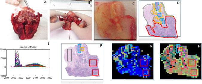
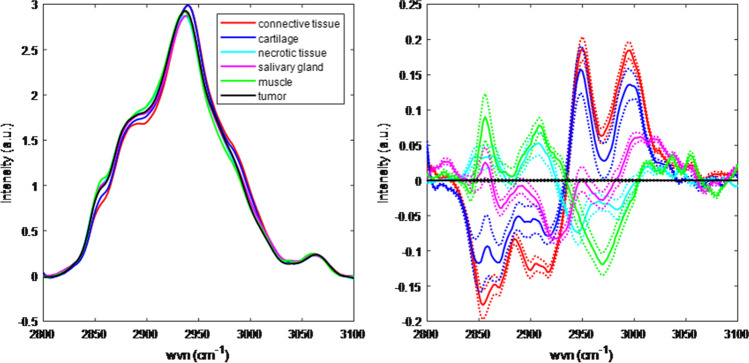
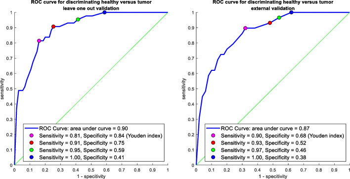
As for many solid cancers, laryngeal cancer is treated surgically, and adequate resection margins are critical for survival. Raman spectroscopy has the capacity to accurately differentiate between cancer and non-cancerous tissue based on their molecular composition, which has been proven in previous work. The aim of this study is to investigate whether Raman spectroscopy can be used to discriminate laryngeal cancer from surrounding non-cancerous tissue. Patients surgically treated for laryngeal cancer were included. Raman mapping experiments were performed ex vivo on resection specimens and correlated to histopathology. Water concentration analysis and CH-stretching region analysis were performed in the high wavenumber range of 2500-4000 cm-1. Thirty-four mapping experiments on 22 resection specimens were used for analysis. Both laryngeal cancer and all non-cancerous tissue structures showed high water concentrations of around 75%. Discriminative information was only found to be present in the CH-stretching region of the Raman spectra of the larynx (discriminative power of 0.87). High wavenumber region Raman spectroscopy can discriminate laryngeal cancer from non-cancerous tissue structures. Contrary to the findings for oral cavity cancer, water concentration is not a discriminating factor for laryngeal cancer.
Keywords: High wavenumber; Larynx; Raman spectroscopy; Squamous cell carcinoma.
© 2023. The Author(s).
Conflict of interest statement
Tom Bakker Schut and Gerwin Puppels are the employees of RiverD International B.V. Senada Koljenović, Gerwin Puppels, Tom Bakker Schut, and Rob Baatenburg de Jong have ownership interest in RiverD International B.V. RiverD International B.V. has supplied the Raman spectrometer that was used in this study.
Figures

References
-
- Helliwell T, Woolgar J (2013) Dataset for histopathology reporting of mucosal malignancies of the larynx. The Royal College of Pathologists. https://www.rcpath.org/static/0d6c0512-e285-40fd-b8a9ee31b13887de/Datase.... Accessed 04–06–2023
-
- Wulff NB, Andersen E, Kristensen CA, Sørensen CH, Charabi B, Homøe P. Prognostic factors for survival after salvage total laryngectomy following radiotherapy or chemoradiation failure: a 10-year retrospective longitudinal study in eastern Denmark. Clin Otolaryngol: Off J ENT-UK: Off J Netherlands Soc Oto-Rhino-Laryngol Cervico-Facial Surg. 2017;42(2):336–346. doi: 10.1111/coa.12726. - DOI - PubMed
-
- Thomas Robbins K, Triantafyllou A, Suárez C, López F, Hunt JL, Strojan P, Williams MD, Braakhuis BJM, de Bree R, Hinni ML, Kowalski LP, Rinaldo A, Rodrigo JP, Vander Poorten V, Nixon IJ, Takes RP, Silver CE, Ferlito A. Surgical margins in head and neck cancer: intra- and postoperative considerations. Auris Nasus Larynx. 2019;46(1):10–17. doi: 10.1016/j.anl.2018.08.011. - DOI - PubMed
-
- Nayanar SK, Krishnanm MA, Mrudula KI, Thavarool PSB, Thiagarajan S. Frozen section evaluation in head and neck oncosurgery: an initial experience in a tertiary cancer center. Frozen section evaluation in head and neck oncosurgery: an initial experience in a tertiary cancer center. Turk Patoloji Dergisi. 2019;35(1):46–51. doi: 10.5146/tjpath.2018.01439. - DOI - PubMed
MeSH terms
Substances
LinkOut - more resources
Full Text Sources
Medical
Research Materials
