. 2023 Aug 25;18(8):e0289770.
doi: 10.1371/journal.pone.0289770. eCollection 2023.

Persistent extrasynaptic hyperdopaminergia in the mouse hippocampus induces plasticity and recognition memory deficits reversed by the atypical antipsychotic sulpiride

Affiliations

Persistent extrasynaptic hyperdopaminergia in the mouse hippocampus induces plasticity and recognition memory deficits reversed by the atypical antipsychotic sulpiride

Jill Rocchetti et al. PLoS One. .

Abstract

Evidence suggests that subcortical hyperdopaminergia alters cognitive function in schizophrenia and antipsychotic drugs (APD) fail at rescuing cognitive deficits in patients. In a previous study, we showed that blocking D2 dopamine receptors (D2R), a core action of APD, led to profound reshaping of mesohippocampal fibers, deficits in synaptic transmission and impairments in learning and memory in the mouse hippocampus (HP). However, it is currently unknown how excessive dopamine affects HP-related cognitive functions, and how APD would impact HP functions in such a state. After verifying the presence of DAT-positive neuronal projections in the ventral (temporal), but not in the dorsal (septal), part of the HP, GBR12935, a blocker of dopamine transporter (DAT), was infused in the CA1 of adult C57Bl/6 mice to produce local hyperdopaminergia. Chronic GBR12935 infusion in temporal CA1 induced a mild learning impairment in the Morris Water Maze and abolished long-term recognition memory in novel-object (NORT) and object-place recognition tasks (OPRT). Deficits were accompanied by a significant decrease in DAT+ mesohippocampal fibers. Intrahippocampal or systemic treatment with sulpiride during GBR infusions improved the NORT deficit but not that of OPRT. In vitro application of GBR on hippocampal slices abolished long-term depression (LTD) of fEPSP in temporal CA1. LTD was rescued by co-application with sulpiride. In conclusion, chronic DAT blockade in temporal CA1 profoundly altered mesohippocampal modulation of hippocampal functions. Contrary to previous observations in normodopaminergic mice, antagonising D2Rs was beneficial for cognitive functions in the context of hippocampal hyperdopaminergia.


Conflict of interest statement

The authors declare no competing interests.

Figures

Fig 1
Fig 1. Topographical organization of DA projections to the hippocampus.
Schematic representations of retrolabeled neuron distribution in ventral (left) and dorsal (right) mesencephalon of C57Bl/6J mice. Table compiling the numbers and proportion of TH and DAT-immunopositive neurons retrogradely labeled in ventral tegmental area (VTA) or substantia nigra pars compacta (SNc) following ventral (n = 10 mice) and dorsal (n = 10 mice) retrobeads bi-lateral administration.
Fig 2
Fig 2. Effect of GBR12935 infusion in the ventral hippocampus (CA1) on spatial and associative Morris water maze (MWM) task.
A) Mean escape latencies after GBR12935 (300nmol/L, .5μL) daily infusion in ventral CA1 (mean latency of two trials, 30 min and 90 min after GBR infusion). There was no significant difference between groups after two-way repeated measure ANOVA. B) Proportion of successful trials per group per day. Posthoc comparison of groups means revealed a significant difference at training day 9 (n = 18, 20; *p < .05) C) Probe testing of spatial memory 24h after the last training session. Both groups spent significantly more time in the target quadrant compared to the others (###p < .001 compared with 25%, ***p < .001 for Quadrant 3 compared with others; no difference between groups). D) Mean escape latencies in the cued/associative version of the MWM. No significant difference between groups (n = 10,10). Bars denote SEM.
Fig 3
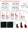
Fig 3. Impairments in recognition memory and mesohippocampal dopaminergic fibers induced in the mouse ventral hippocampus after chronic GBR12935 infusions.
A) Design of the NORT and OPRT performed on cannulated mice between day 1 and day 18 of infusions. A short-term version (1h between habituation and testing) of the OPRT was performed on mice treated for 3 weeks B) NORT scores of mice after 2 or 16 days of GBR (300nmol/L, .5μL) infusion. Visuospatial retention of older object was significantly impaired in GBR-treated mice compared with NaCl-treated mice (n = 9, 9; ###p < .01 compared with .50; *p < .05 between groups) at day 16 but not at day 2 (n = 9, 9; ###p < .01 compared with .50; p = .94 between groups). C) OPRT scores after 4 or 18 days of GBR12935 infusion. Visuospatial retention of older place was preserved in NaCl- and GBR-treated groups at day 4 of treatment (n = 10, 10; ###p < .01 compared with .50; p = .87 between groups) but retention was impaired in GBR-treated animals at day 18 (n = 10, 10; ###p < .01 compared with .50; **p < .01 between groups). D) Short-term OPRT scores after 3 weeks of chronic GBR12935 infusion. Both groups equally displayed significant visuospatial retention of the older place (n = 10, 11; ###p < .01 compared with .50; p = .89 between groups). E) Examples of immunohistolabelling of DAT+ fibers (20X) in NaCl- and GBR-treated mice hippocampal slices (green). Co-staining was performed with Hoescht (blue) F) Quantification of total DAT+ fibers in hippocampi of NaCl- and GBR-treated mice (n = 3, 3). There was significantly fewer DAT+ fibers in all layers of the hippocampus of GBR-treated mice (LMol: **p < .01; Rad: *p < .05; Pyr/Ori: *p < .05 between groups). LMol = lacunosum moleculare, Rad = stratum radiatum, Pyr/Ori = Stratum pyramidale + Stratum oriens. Bars denote SEM.
Fig 4
Fig 4. Effect of D2/D3 antagonist sulpiride on GBR-induced recognition memory impairments.
A) Experimental design of the test. Animals were separated into 4 groups and systemically treated daily with intraperitoneal injection (IP) of saline (.9%) or sulpiride (50mg/kg). 30 min later, animals were intrahippocampally infused with either NaCl or GBR (300nmol/L, .5μL) as previously. Group (3) (sulp local) was treated intrahippocampally with sulpiride (10μmol/L, .5μL) 10 min prior to GBR12935 infusion. After two weeks of repeated daily co-treatment, performances in the NORT and the OPRT were assessed. Results are compared with one-way ANOVA followed by Tukey’s HSD posthoc comparisons of the means. Mean difference with theoretical value of symmetric exploration (.50) was assessed in each group with one-sample Student t test. B) NORT scores after 2 weeks of co-treatment. Local GBR infusion (GBR, n = 26) impaired visuospatial retention of the older object compared to NaCl group (NaCl, n = 21) as previously described (*p < .05 compared with GBR; ###p < .001 compared with .50). Pre-treatment with either sulpiride locally (GBR+sulp local, n = 17; *p < .05 compared with GBR; #p < .05 compared with .50) or sulpiride IP (GBR+sulp IP, n = 19; **p < .01 compared with GBR; ##p < .01 compared with .50) before GBR infusion resulted in restoration of novel-object recognition. C) OPRT scores after 2 weeks of co-treatment. Chronic intrahippocampal GBR infusion impaired spatial recognition of the older place compared to NaCl group ((2) n = 28; ###p < .001 compared with .50; **p < .01 compared with (1)). Pre-treatment with sulpiride locally ((3) n = 18; p = .38 compared with .50; p = .08 compared with (1)) impaired spatial recognition of older place even if significant difference with NaCl group was not fully reached. Pre-treatment with systemic sulpiride ((4) n = 20; p = .89 compared with .50; **p < .01 compared with (1)) resulted in impaired spatial recognition as well. Bars denote SEM.
Fig 5
Fig 5. In vitro acute effect of GBR12935 (30nmol/L) on LTP and LTD in ventral CA1.
A) HFS (3x100pulses/100Hz/20s) induced significant LTP in dorsal CA1 of coronal slices of naïve mice ventral hippocampus (n = 7; ###p < .001 compared with baseline). GBR application (30 min) did not significantly change LTP levels induced by tetanic stimulation (n = 7; #p < .05 compared with baseline; p = .31 between groups). B) Paired-pulse LFS (1200 pairs/1Hz/200ms IPI) induced significant LTD (n = 7; #p < .05 compared with baseline). GBR application (40 min) completely blocked the expression of ppLFS induced LTD at Schaffer collateral-CA1 synapses (n = 7; p = .25 compared with baseline; *p < .05 between groups). C) Co-application of the D1/D5 antagonist SCH23390 (1μmol/L) did not prevent GBR from blocking LTD expression (n = 6; p = .5 compared with baseline). D) Co-application of D2/D3 antagonist sulpiride with GBR allowed the expression of significant LTD (n = 6; ##p < .01 compared with baseline; *p < .05 between groups). Bars are SEM.

Similar articles

Cited by

References

    1. Howes OD, Kapur S. The dopamine hypothesis of schizophrenia: version III—the final common pathway. Schizophr Bull. 2009;35(3):549–62. doi: 10.1093/schbul/sbp006 - DOI - PMC - PubMed
    1. Laruelle M, Abi-Dargham A. Dopamine as the wind of the psychotic fire: new evidence from brain imaging studies. J Psychopharmacol. 1999;13(4):358–71. doi: 10.1177/026988119901300405 - DOI - PubMed
    1. Bussey TJ, Barch DM, Baxter MG. Testing long-term memory in animal models of schizophrenia: suggestions from CNTRICS. Neurosci Biobehav Rev. 2013;37(9 Pt B):2141–8. doi: 10.1016/j.neubiorev.2013.06.005 - DOI - PubMed
    1. Goldman-Rakic PS, Muly EC 3rd, Williams GV. D(1) receptors in prefrontal cells and circuits. Brain research Brain research reviews. 2000;31(2–3):295–301. doi: 10.1016/s0165-0173(99)00045-4 - DOI - PubMed
    1. Eichenbaum H. Hippocampus: cognitive processes and neural representations that underlie declarative memory. Neuron. 2004;44(1):109–20. doi: 10.1016/j.neuron.2004.08.028 - DOI - PubMed

Publication types