. 2023 Aug 25;9(34):eadi4148.
doi: 10.1126/sciadv.adi4148. Epub 2023 Aug 25.

Structural basis of telomeric nucleosome recognition by shelterin factor TRF1

Affiliations

Structural basis of telomeric nucleosome recognition by shelterin factor TRF1

Hongmiao Hu et al. Sci Adv. .

Abstract

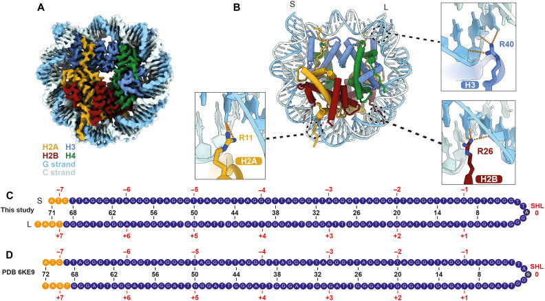
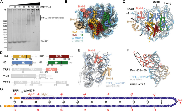
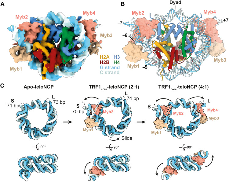
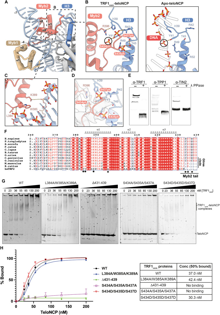
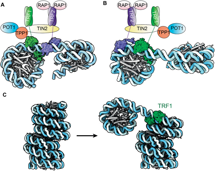
Shelterin and nucleosomes are the key players that organize mammalian chromosome ends into the protective telomere caps. However, how they interact with each other at telomeres remains unknown. We report cryo-electron microscopy structures of a human telomeric nucleosome both unbound and bound to the shelterin factor TRF1. Our structures reveal that TRF1 binds unwrapped nucleosomal DNA ends by engaging both the nucleosomal DNA and the histone octamer. Unexpectedly, TRF1 binding shifts the register of the nucleosomal DNA by 1 bp. We discovered that phosphorylation of the TRF1 C terminus and a noncanomical DNA binding surface on TRF1 are critical for its association with telomeric nucleosomes. These insights into shelterin-chromatin interactions have crucial implications for understanding telomeric chromatin organization and other roles of shelterin at telomeres including replication and transcription.


Figures

Fig. 1.
Fig. 1.. Structure of a human teloNCP.
(A) Cryo-EM reconstruction (2.5 Å) and (B) the atomic model of a human teloNCP. Within the DNA duplex, the G-strand consists of the G-rich TTAGGG repeats. The C-strand consists of the CCCTAA repeats, which are complementary to the TTAGGG repeats. (C) DNA positioning of telomeric sequence observed in the structure shown in (A) and (B). Short (S) and long (L) labels denote the short and long lengths of the DNA on either side of the dyad, respectively. The numbers on top of the sequence (±1, ±2, …, ±7) denote the SHLs relative to the nucleosomal dyad position (SHL 0). The blue nucleotides represent the 23 telomeric repeats. The orange nucleotides represent the nontelomeric sequences resulting from restriction digestion of the DNA construct. The labels and coloring scheme in this figure is used throughout the manuscript. Base-specific interactions between DNA bases and histone H2A, H3, and H2A are shown in the three close-up views. (D) DNA positioning of telomeric sequence in the published teloNCP crystal structure (PDB 6KE9) (16).
Fig. 2.
Fig. 2.. Structure of the 2:1 TRF1core-teloNCP complex.
(A) EMSAs showing titration of TRF1core against teloNCP. Experiments were performed in triplicate. (B) Cryo-EM reconstruction (2.7 Å) and (C) the atomic model of TRF1core-teloNCP complex, respectively. Subunits are colored as labeled. Dyad position and SHLs are indicated in (C). (D) Domain architectures of protein subunits in the complex. Unresolved regions are shown as semi-transparent. (E) Superimposition of the apo-teloNCP (gray) and TRF1core-teloNCP (colored) structures determined in this work to show unwrapping of nucleosomal DNA by TRF1. (F) Comparison of the structures of the Myb domains bound to naked telomeric DNA (PDB 1W0T, gray) (11) and bound to the nucleosomal DNA (colored). (G) Positioning of telomeric DNA sequence in the TRF1core-teloNCP structure. The TRF1 Myb domain binding sites are indicated.
Fig. 3.
Fig. 3.. Structure of the 4:1 TRF1core-teloNCP complex.
(A) Cryo-EM reconstruction (6.7 Å) and (B) the model of 4:1 TRF1core-teloNCP complex, respectively. Subunits are colored as labeled. Dyad position and SHL, which are occupied by the Myb domains of TRF1, are also indicated. (C) Model for the hierarchical assembly of TRF1core on the teloNCP based on the structures of apo-teloNCP and the 2:1 and 4:1 TRF1core-teloNCP complexes determined in this study.
Fig. 4.
Fig. 4.. Noncanonical DNA interaction surface and phosphorylation of TRF1 crucial for nucleosome binding.
(A) Interactions between the two TRF1 Myb domains with teloNCP. Dashed box and circle indicate histone H3-Myb2 and DNA-Myb2 interactions, respectively. (B) Left: Close-up view of the interaction between the phosphorylated C-terminal residues of TRF1 Myb2 domain with histone H3 N-terminal tail in the TRF1core-teloNCP structure. Right: Close-up view of the same region of histone H3 interacting with DNA in the apo-teloNCP structure. Black circles highlight the observation that the phosphate group of S434 occupies the same position as a DNA backbone phosphate. (C) Close-up view showing noncanonical DNA interactions made by helix 1 of Myb2. (D) Cryo-EM density of the phosphorylated C-terminal residues of TRF1 Myb2 domain. (E) Phos-tag gels of the untreated and λ-phosphatase (λ PPase) treated TRF1core samples combined with immunoblotting using TRF1, TPP1, and TIN2 antibodies (α-TRF1, α-TPP1, and α-TIN2). (F) Sequence alignment of TRF1 Myb domains from various mammalian species and human TRF2 Myb domain. The hexagonal dots and stars underneath the sequence denote residues involved in DNA interaction within the noncanonical DNA surface on Myb2 (C) and residues involved in interactions with histone H3 (B), respectively. (G) EMSAs showing titration of purified wild-type (WT) and mutant TRF1core complexes against teloNCP. Experiments were performed in triplicate. (H) Quantification of EMSA experiments shown in (G). In the left, we plotted percentages of unbound teloNCPs as a function of protein concentration in the EMSA reaction of the wild-type and each mutant complex. The right table shows the concentration of each TRF1core complex at which 50% of teloNCP remains unbound as determined from the graphs. Error bars at each concentration point are the SEM obtained from the three replicates.
Fig. 5.
Fig. 5.. Model for the organization of shelterin and nucleosomes on telomeric DNA.
(A and B) Two proposed models for how shelterin and telomeric nucleosomes are organized on telomeric DNA. In both models, TRF1 binds at the junction between nucleosome and linker DNA as shown in our structures. TRF2 binds either the linker DNA (in A) or on the outer DNA gyres (in B). The nucleosome repeat length in these models is 157 bp, based on the published work (5). (C) Remodeling of the columnar telomeric chromatin structure (PDB 7V9K) (17) to accommodate TRF1 binding.

Similar articles

Cited by

References

    1. de Lange T., Shelterin-Mediated Telomere Protection. Annu. Rev. Genet. 52, 223–247 (2018). - PubMed
    1. Lim C. J., Cech T. R., Shaping human telomeres: From shelterin and CST complexes to telomeric chromatin organization. Nat. Rev. Mol. Cell Biol. 22, 283–298 (2021). - PMC - PubMed
    1. Shay J. W., Role of Telomeres and Telomerase in Aging and Cancer. Cancer Discov. 6, 584–593 (2016). - PMC - PubMed
    1. Tommerup H., Dousmanis A., de Lange T., Unusual chromatin in human telomeres. Mol. Cell. Biol. 14, 5777–5785 (1994). - PMC - PubMed
    1. Makarov V. L., Lejnine S., Bedoyan J., Langmore J. P., Nucleosomal Organization of telomere-specific chromatin in rat. Cell 73, 775–787 (1993). - PubMed