. 2023 Nov 9;142(19):1658-1671.
doi: 10.1182/blood.2023020504.

The iron chaperone poly(rC)-binding protein 1 regulates iron efflux through intestinal ferroportin in mice

Affiliations

The iron chaperone poly(rC)-binding protein 1 regulates iron efflux through intestinal ferroportin in mice

Yubo Wang et al. Blood. .

Abstract

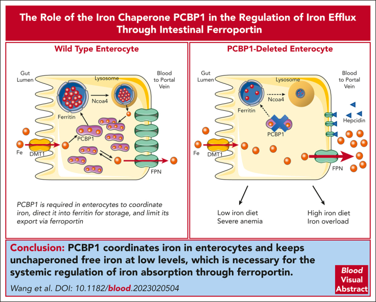
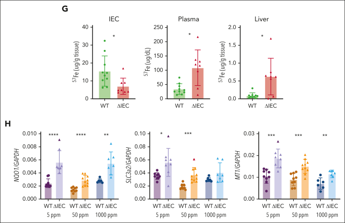
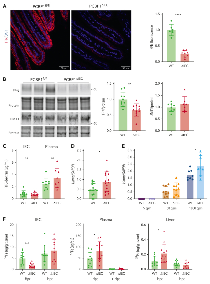
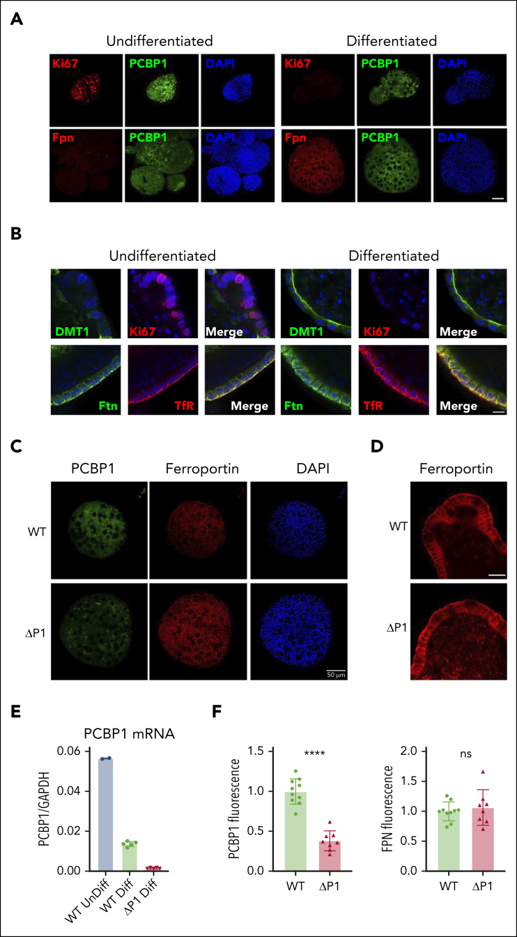
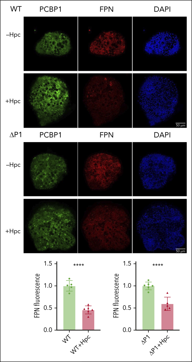
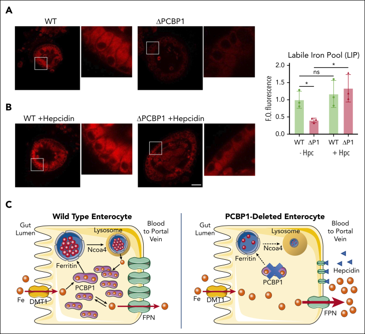
Iron is an essential nutrient required by all cells but used primarily for red blood cell production. Because humans have no effective mechanism for ridding the body of excess iron, the absorption of dietary iron must be precisely regulated. The critical site of regulation is the transfer of iron from the absorptive enterocyte to the portal circulation via the sole iron efflux transporter, ferroportin. Here, we report that poly(rC)-binding protein 1 (PCBP1), the major cytosolic iron chaperone, is necessary for the regulation of iron flux through ferroportin in the intestine of mice. Mice lacking PCBP1 in the intestinal epithelium exhibit low levels of enterocyte iron, poor retention of dietary iron in enterocyte ferritin, and excess efflux of iron through ferroportin. Excess iron efflux occurred despite lower levels of ferroportin protein in enterocytes and upregulation of the iron regulatory hormone hepcidin. PCBP1 deletion and the resulting unregulated dietary iron absorption led to poor growth, severe anemia on a low-iron diet, and liver oxidative stress with iron loading on a high-iron diet. Ex vivo culture of PCBP1-depleted enteroids demonstrated no defects in hepcidin-mediated ferroportin turnover. However, measurement of kinetically labile iron pools in enteroids competent or blocked for iron efflux indicated that PCBP1 functioned to bind and retain cytosolic iron and limit its availability for ferroportin-mediated efflux. Thus, PCBP1 coordinates enterocyte iron and reduces the concentration of unchaperoned "free" iron to a low level that is necessary for hepcidin-mediated regulation of ferroportin activity.


Conflict of interest statement

Conflict-of-interest disclosure: The authors declare no competing financial interests.

The current affiliation for K.D.H. is Eurofins Lancaster Laboratories, Inc, Lancaster, PA.

The current affiliation for M.S.-E. is Laboratory of Cellular and Developmental Biology, National Institutes of Diabetes and Digestive and Kidney Diseases, National Institutes of Health, Bethesda, MD.

Figures

None
Graphical abstract
Figure 1.
Figure 1.
Inducible deletion of PCBP1 specifically in the intestinal epithelium of mouse. (A) Relative levels of PCBP1 mRNA in IECs and livers in PCBP1fl/fl (WT) vs PCBP1ΔIEC (ΔIEC) mice. Weanling mice were fed a tamoxifen diet for 1 month before analysis. mRNA levels from IECs of proximal duodenum were determined by real-time quantitative polymerase chain reaction (qPCR). (B) Detection of PCBP1 in all cells throughout the duodenum of WT mice. Fixed, embedded sections of proximal duodenum analyzed by anti-PCBP1 immunohistochemistry. Brown stain indicates PCBP1. Arrows: epithelial cells. Brackets: duodenal crypts. (C) Specific deletion of PCBP1 in duodenal epithelium of ΔIEC mice. PCBP1 detected by IHC as in panel B, above. Note the absence of brown PCBP1 signal in crypts and epithelial layer with PCBP1 expression in lamina propria and duodenal wall. (D) Absence of PCBP1 protein in immunoblot of IECs from PCBP1ΔIEC mice. IECs were collected and analyzed by immunoblotting for PCBP1 and PCBP2. Representative blot shown on the left, quantitation on the right. (E) Increased villus length and crypt depth in PCBP1ΔIEC mice. Intact, full-length villi and crypts were measured in hematoxylin and eosin–stained sections of duodenum. Mean lengths of structures 1 cm distal to pylorus calculated from each animal; 5 age-matched WT and PCBP1ΔIEC mice were analyzed. Refer to female mice data in supplemental Figure 1.
Figure 2.
Figure 2.
Poor growth and iron depletion in IECs of PCBP1ΔIEC mice. (A) Body weights of WT and PCBP1ΔIEC mice aged 2 and 6 months. Weanling mice fed a tamoxifen diet for 1 month followed by a standard chow diet. (B) Reduced nonheme iron levels in the IECs of PCBP1ΔIEC mice. IECs from 2-month-old mice were collected and analyzed for nonheme iron. (C) Lower body weights of PCBP1ΔIEC mice on defined-iron diets. WT and PCBP1ΔIEC mice were fed 5 ppm, 50 ppm, or 1000 ppm diets for 1 month. Body weight measured at the age 3 months. (D) Reduced nonheme iron levels in the IECs of PCBP1ΔIEC mice on defined-iron diets. IECs from mice in panel C were analyzed for nonheme iron. (E) Lower ferritin and higher NCOA4 levels in IECs of PCBP1ΔIEC vs WT mice. Mice were fed defined-iron diets as in panel C. IECs were collected at 3 months and analyzed by immunoblot for ferritin and NCOA4. A representative blot is shown above with quantitation below. Analyzed mice were male littermates; Female littermates were included as indicated. Refer to data from female mice in supplemental Figure 2.
Figure 3.
Figure 3.
Loss of intestinal PCBP1 disrupts systemic iron homeostasis, causing liver iron loading or anemia. (A) Elevated plasma iron in PCBP1ΔIEC mice. Nonheme iron levels were measured in plasma of WT and PCBP1ΔIEC mice that were fed 5 ppm, 50 ppm, and 1000 ppm iron diets for 1 month. (B) Iron accumulation in the livers of PCBP1ΔIEC mice that were fed a high-iron diet. Nonheme iron measured in liver tissue from WT and PCBP1ΔIEC mice from panel A. (C) Iron accumulation in bone marrow of PCBP1ΔIEC mice. Nonheme iron measured in bone marrow of mice that were fed a tamoxifen diet for 1 month after weaning. (D) More severe anemia in PCBP1ΔIEC vs WT mice on a low-iron diet. Complete blood counts were obtained on mice fed a 5 ppm iron diet for 1 month. Horizontal line indicates values from WT mice that were fed a 50 ppm diet. (E) Impaired maturation of erythroid precursors in PCBP1ΔIEC mice that were fed a low-iron diet. Erythroid precursors of mice that were fed a 5 ppm iron diet analyzed by flow cytometry. Dot plots and gating strategies shown on the left, subgroup quantification shown on the right. Refer to supplemental Figure 4. (F) Increased expression of erythroid regulators ERFE and EPO in PCBP1ΔIEC mice that were fed a low-iron diet. Bone marrow and kidney mRNA was prepared from mice treated as in panel D and analyzed by qPCR. (G) Excess absorption of oral iron through intestine into the circulation in PCBP1ΔIEC mice. Mice were administered 57FeSO4 solution through oral gavage. IEC, plasma, and liver samples were collected at 1 hour and 57Fe levels were measured by ICP-MS. (H) Activation of Nrf2-controlled, oxidative stress response genes in livers of PCBP1ΔIEC mice. Mice were fed defined-iron diets for 1 month and mRNA levels of oxidative stress genes NQO1, SLC3a2, and MT1 were measured in liver tissue by qPCR. Refer to supplemental Figure 3.
Figure 3.
Figure 3.
Loss of intestinal PCBP1 disrupts systemic iron homeostasis, causing liver iron loading or anemia. (A) Elevated plasma iron in PCBP1ΔIEC mice. Nonheme iron levels were measured in plasma of WT and PCBP1ΔIEC mice that were fed 5 ppm, 50 ppm, and 1000 ppm iron diets for 1 month. (B) Iron accumulation in the livers of PCBP1ΔIEC mice that were fed a high-iron diet. Nonheme iron measured in liver tissue from WT and PCBP1ΔIEC mice from panel A. (C) Iron accumulation in bone marrow of PCBP1ΔIEC mice. Nonheme iron measured in bone marrow of mice that were fed a tamoxifen diet for 1 month after weaning. (D) More severe anemia in PCBP1ΔIEC vs WT mice on a low-iron diet. Complete blood counts were obtained on mice fed a 5 ppm iron diet for 1 month. Horizontal line indicates values from WT mice that were fed a 50 ppm diet. (E) Impaired maturation of erythroid precursors in PCBP1ΔIEC mice that were fed a low-iron diet. Erythroid precursors of mice that were fed a 5 ppm iron diet analyzed by flow cytometry. Dot plots and gating strategies shown on the left, subgroup quantification shown on the right. Refer to supplemental Figure 4. (F) Increased expression of erythroid regulators ERFE and EPO in PCBP1ΔIEC mice that were fed a low-iron diet. Bone marrow and kidney mRNA was prepared from mice treated as in panel D and analyzed by qPCR. (G) Excess absorption of oral iron through intestine into the circulation in PCBP1ΔIEC mice. Mice were administered 57FeSO4 solution through oral gavage. IEC, plasma, and liver samples were collected at 1 hour and 57Fe levels were measured by ICP-MS. (H) Activation of Nrf2-controlled, oxidative stress response genes in livers of PCBP1ΔIEC mice. Mice were fed defined-iron diets for 1 month and mRNA levels of oxidative stress genes NQO1, SLC3a2, and MT1 were measured in liver tissue by qPCR. Refer to supplemental Figure 3.
Figure 4.
Figure 4.
Lower ferroportin levels but higher ferroportin activity in PCBP1ΔIEC mice. (A) Lower levels of ferroportin on duodenal epithelium of PCBP1ΔIEC vs WT mice. Fluorescent immunohistochemistry for FPN in proximal duodenum from 2 months old mice. DAPI (4′,6-diamidino-2-phenylindole) stain indicates nuclei. Representative images on the left show quantitative analysis of fluorescence at right. (B) Lower levels of ferroportin in IECs of PCBP1ΔIEC vs WT mice. IECs from 2 months mice analyzed for FPN and DMT1 by immunoblot. Representative blot on the left, quantitation on the right. (C) Intact tight junctions in intestinal epithelium of PCBP1ΔIEC mice. Mice were administered FITC-dextran (4000 MW) through oral gavage. IECs and plasma were collected after 1 hour and FITC-dextran measured. (D) Higher levels of hepcidin mRNA in livers of PCBP1ΔIEC vs WT mice. Hepcidin mRNA levels in liver were measured by qPCR in 2 months mice. (E) Higher levels of hepcidin mRNA in livers of PCBP1ΔIEC mice on a high-iron diet. Mice were fed defined-iron diets for 1 month and liver hepcidin mRNA levels were analyzed. (F) Ferroportin mediates excess iron absorption through intestinal epithelium of PCBP1ΔIEC mice. Mice were injected intraperitoneally with synthetic hepcidin or vehicle followed by oral gavage with 57Fe solution. IECs, plasma, and liver samples were collected after 1 hour and 57Fe levels measured.
Figure 5.
Figure 5.
PCBP1 deletion does not suppress ferroportin expression in differentiated enteroids. (A) Expression of PCBP1 and ferroportin in differentiated WT enteroids. Undifferentiated enteroids on the left, differentiated on the right. Enteroids were fixed and analyzed by indirect immunofluorescence. Proliferation marker Ki67 (red, top panel), FPN (red, bottom panel), PCBP1 (green) and DAPI (blue) are shown. Bar = 50 μm. (B) Expression of key iron homeostatic proteins in differentiated enteroids. Enteroids prepared as in panel A were analyzed for DMT1, Ki67, ferritin (Ftn), and transferrin receptor 1 (TfR) as indicated. DAPI staining in blue. Higher magnification and merged images show distinct subcellular localizations. Bar = 10 μm. (C) Depletion of PCBP1 does not affect FPN expression in the enteroids. PCBP1 depletion was induced with 4-OH tamoxifen in differentiating enteroids from PCBP1fl/fl, TG vil-CreERT2 mice. Indirect immunofluorescence for PCBP1, FPN, and DAPI in WT (no tamoxifen) and ΔPCBP1 (tamoxifen) is shown. Quantitation of fluorescent images is shown below in panel F. (D) No change in basolateral localization of ferroportin in PCBP1-depleted enteroids. Higher magnification images of FPN from central sections in WT and ΔPCBP1 enteroids. Bar = 20 μm. (E) Efficient deletion of PCBP1 in differentiated enteroids. Enteroids treated as in panel C were analyzed by qPCR.
Figure 6.
Figure 6.
Hepcidin-mediated degradation of ferroportin in WT and ΔPCBP1 enteroids. WT and ΔPCBP1 differentiated enteroids were treated with hepcidin for 18 hours and fixed and stained for FPN and PCBP1. Quantitation of FPN signals is shown below.
Figure 7.
Figure 7.
Restoration of labile iron pool in ΔPCBP1 enteroids upon treatment with hepcidin. (A-B) Labile Fe(II) in WT and ΔPCBP1 differentiated enteroids was detected with fluorescent indicator (FerroOrange) without (A) or with (B) hepcidin pretreatment for 1 hour. Central sections of representative enteroids are shown. Images are in pairs with inset at higher magnification on the right. Quantification at right. Enteroids were grown 3 times from individual mice and iron signal from 3 to 7 enteroids in each condition was imaged and measured. Bar = 50 μm. (C) Model of PCBP1-mediated management of cytosolic iron retention and efflux. In WT enterocytes (left), dietary iron imported through DMT1 is rapidly coordinated by PCBP1, which directs excess iron to ferritin for storage and maintains free iron pool at low levels. Lysosomal turnover of ferritin, mediated by NCOA4, occurs when intracellular iron levels are low. Rates of iron efflux through FPN are low and steady. In PCBP1-deleted enterocytes (right), imported iron is not coordinated by PCBP1 and intracellular free iron rapidly rises to high levels. Rates of iron efflux through FPN are high and rapid efflux continues until intracellular free iron falls to low levels. The pool of cytosolic iron buffered by PCBP1 is absent and ferritin storage is low. Iron is initially delivered to portal circulation at a high rate (bolus effect).

Comment in

Similar articles

Cited by

References

    1. Coffey R, Ganz T. Iron homeostasis: an anthropocentric perspective. J Biol Chem. 2017;292(31):12727–12734. - PMC - PubMed
    1. Katsarou A, Pantopoulos K. Basics and principles of cellular and systemic iron homeostasis. Mol Aspects Med. 2020;75 - PubMed
    1. Pasricha SR, Tye-Din J, Muckenthaler MU, Swinkels DW. Iron deficiency. Lancet. 2021;397(10270):233–248. - PubMed
    1. Yanatori I, Kishi F. DMT1 and iron transport. Free Radic Biol Med. 2019;133:55–63. - PubMed
    1. Gunshin H, Mackenzie B, Berger UV, et al. Cloning and characterization of a mammalian proton-coupled metal-ion transporter. Nature. 1997;388(6641):482–488. - PubMed

Publication types