. 2023 Aug 4;8(86):eadg0539.
doi: 10.1126/sciimmunol.adg0539. Epub 2023 Aug 25.

PD-1 blockade increases the self-renewal of stem-like CD8 T cells to compensate for their accelerated differentiation into effectors

Affiliations

PD-1 blockade increases the self-renewal of stem-like CD8 T cells to compensate for their accelerated differentiation into effectors

Amanda L Gill et al. Sci Immunol. .

Abstract

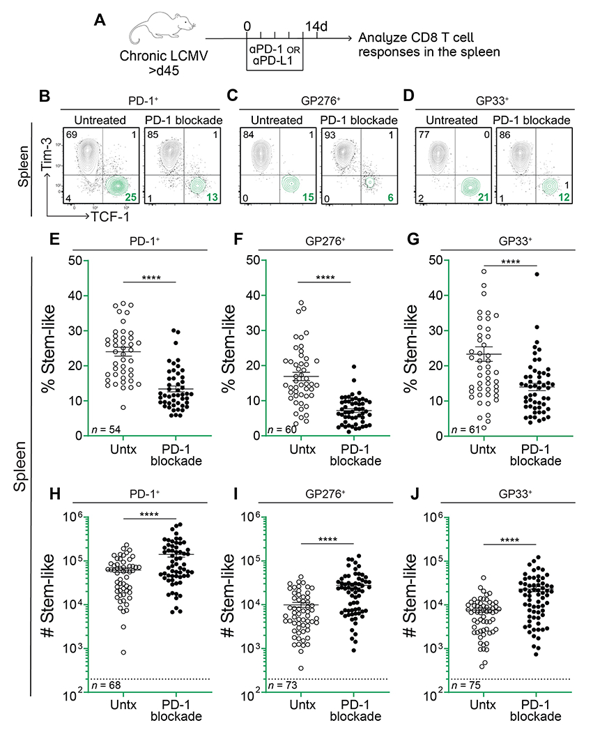
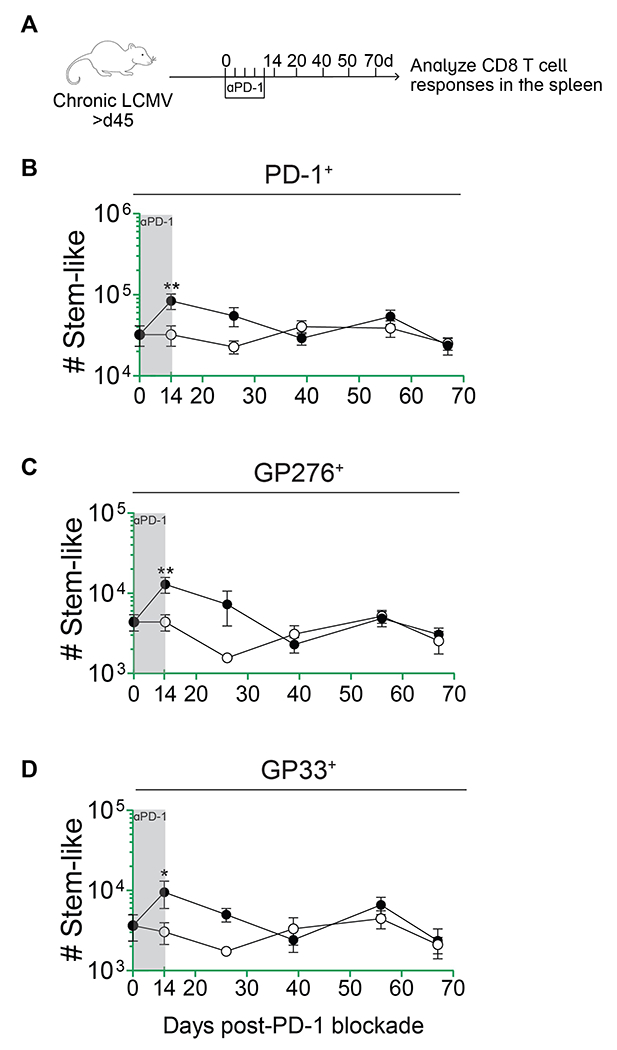
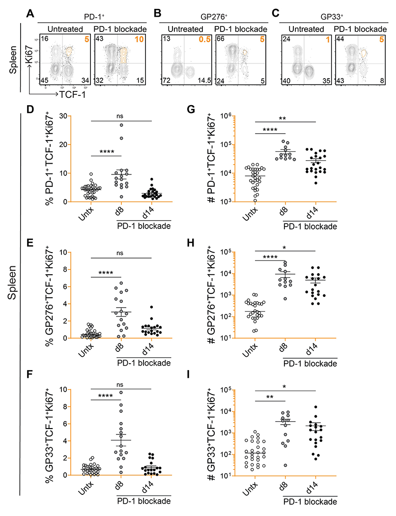
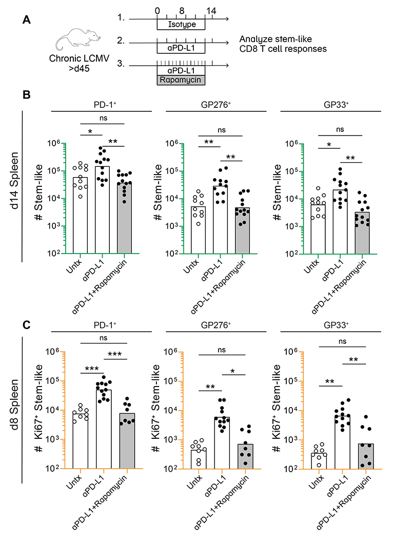
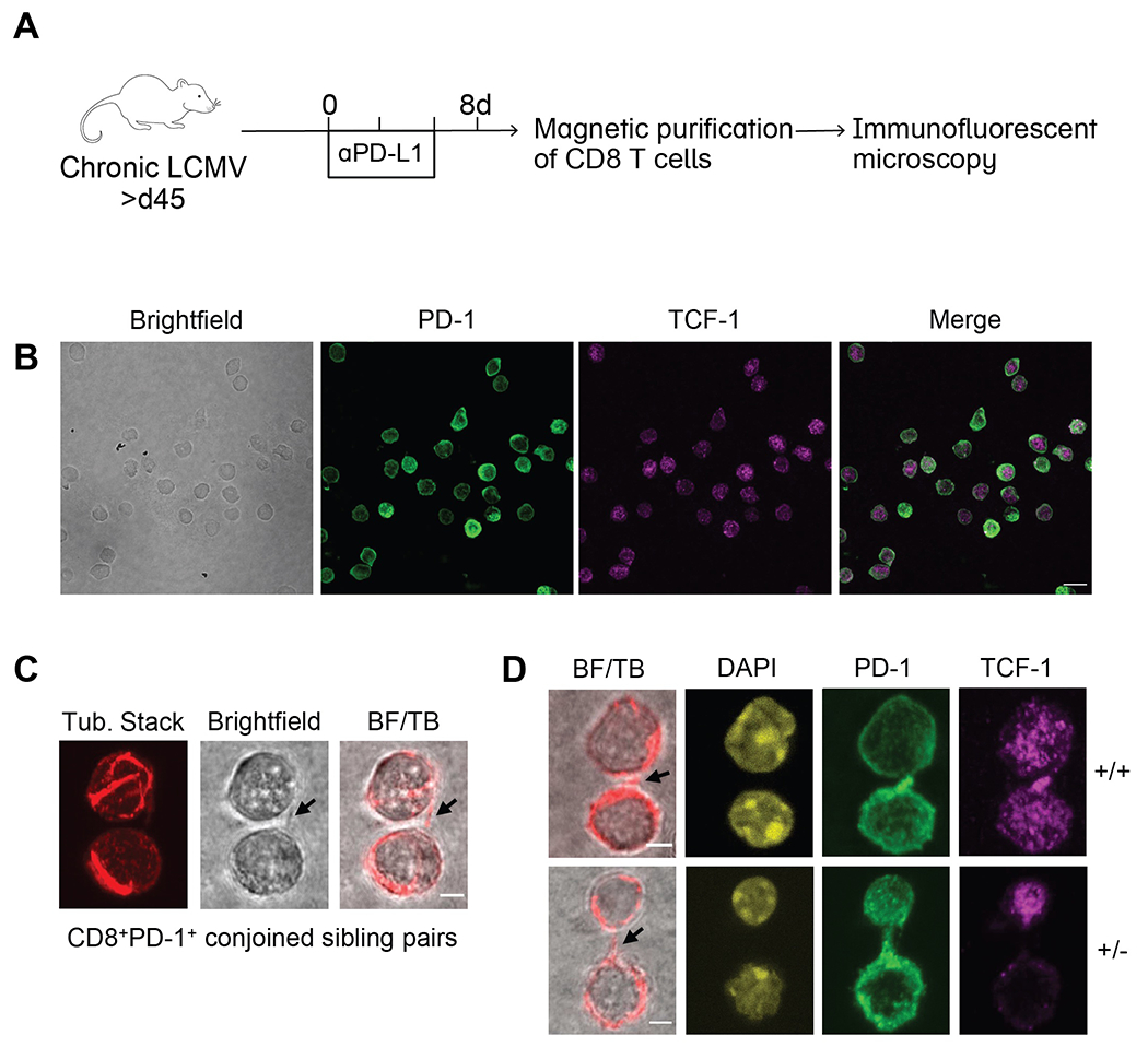
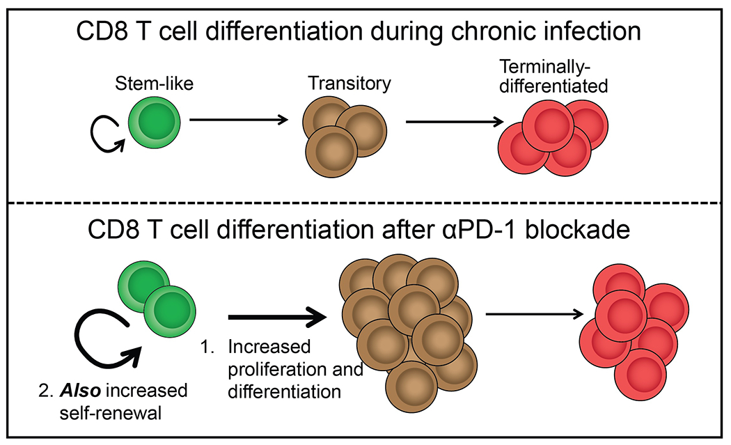
PD-1+TCF-1+ stem-like CD8 T cells act as critical resource cells for maintaining T cell immunity in chronic viral infections and cancer. In addition, they provide the proliferative burst of effector CD8 T cells after programmed death protein 1 (PD-1)-directed immunotherapy. However, it is not known whether checkpoint blockade diminishes the number of these stem-like progenitor cells as effector cell differentiation increases. To investigate this, we used the mouse model of chronic lymphocytic choriomeningitis virus (LCMV) infection. Treatment of chronically infected mice with either αPD-1 or αPD-L1 antibody not only increased effector cell differentiation from the virus-specific stem-like CD8 T cells but also increased their proliferation so their numbers were maintained. The increased self-renewal of LCMV-specific stem-like CD8 T cells was mTOR dependent. We used microscopy to understand the division of these progenitor cells and found that after PD-1 blockade, an individual dividing cell could give rise to a differentiated TCF-1- daughter cell alongside a self-renewing TCF-1+ sister cell. This asymmetric division helped to preserve the number of stem-like cells. Moreover, we found that the PD-1+TCF-1+ stem-like CD8 T cells retained their transcriptional program and their in vivo functionality in terms of responding to viral infection and to repeat PD-1 blockade. Together, our results demonstrate that PD-1 blockade does not deplete the stem-like population despite increasing effector differentiation. These findings have implications for PD-1-directed immunotherapy in humans.


Conflict of interest statement

Competing interests: RA and GJF hold patents on the PD-1 pathway. GJF has served on advisory boards for Roche, Bristol-Myers-Squibb, iTeos, NextPoint, IgM, Jubilant, GV20, IOME, Santa Ana Bio, Simcere of America, and Geode. The other authors declare that they have no competing interests.

Figures

Fig. 1.
Fig. 1.. PD-1 blockade decreases the frequency of virus-specific stem-like CD8 T cells but their numbers increase.
(A) LCMV chronically infected mice were treated with αPD-1 or αPD-L1 every 3 days for 2 weeks. CD8 T cell responses were analyzed on day 14. (B-D) Representative flow plots showing expression of TCF-1 and Tim-3 on total PD-1+, GP276+, and GP33+ CD8 T cells with and without αPD-1/L1 treatment. (E-G) Frequency of TCF-1+Tim-3 stem-like cells among the LCMV-specific CD8 T cell populations indicated above each plot. (H-J) Absolute number of stem-like cells among the LCMV-specific populations indicated above each plot. Dotted lines indicate the limit of detection. The number of mice in each plot is shown in the bottom left corner and values are from 12 independent experiments. Individual data points represent individual mice; bars represent mean ± SEM. Statistical significance in (E-J) was determined using unpaired two-tailed t tests; *, P ≤ 0.05; **, P ≤ 0.01; ***, P ≤ 0.001; ****; P ≤ 0.0001; ns, non-significant.
Fig. 2.
Fig. 2.. Virus-specific stem-like CD8 T cells are maintained up to 8 weeks following initial PD-1 blockade.
(A) LCMV chronically infected mice were treated with αPD-1 every 3 days for 2 weeks. Following treatment cessation, CD8 T cell responses were analyzed at weeks 2, 4, 6, 8, and 10. (B-D) Absolute number of stem-like cells among the LCMV-specific populations indicated above each plot. Data points represent means from 3 pooled experiments ± SEM; n=3-5 mice per group per timepoint; *, P ≤ 0.05; **, P ≤ 0.01; ***, P ≤ 0.001; ****; P ≤ 0.0001; ns, non-significant.
Fig. 3.
Fig. 3.. Increased proliferation and self-renewal of virus-specific stem-like CD8 T cells after PD-1 blockade.
(A-C) Representative flow plots showing expression of TCF-1 and Ki67 on total PD-1+, GP276+, and GP33+ CD8 T cells on day 8 post-PD-1 blockade. (D-I) Frequency and absolute number of stem-like PD-1+, GP276+, or GP33+ CD8 T cells that were proliferating (Ki67+) on days 8 and 14 post-blockade. Individual data points represent individual mice; bars represent mean ± SEM; 4-7 independent experiments per group, n=3-5 mice per experiment. Statistical significance (D-I) was calculated using one-way ANOVA with Tukey’s test for multiple comparisons; *, P ≤ 0.05; **, P ≤ 0.01; ***, P ≤ 0.001; ****; P ≤ 0.0001; ns, non-significant.
Fig. 4.
Fig. 4.. mTOR signaling regulates proliferation of virus-specific stem-like CD8 T cells after PD-1 therapy.
(A) Rapamycin was administered to mice daily in combination with αPD-L1 for 8 or 14 days. Control mice received sham treatment during the same time period. (B) Absolute number of stem-like CD8 T cells in the spleen among total PD-1+, GP276+, and GP33+ CD8 T cells following 2 week treatment with isotype, αPD-L1, or αPD-L1 in combination with rapamycin (C) Absolute number of proliferating (Ki67+) stem-like CD8 T cells among the total LCMV-specific populations indicated above each plot on day 8 post-treatment. (B) and (C) each show values from 3 independent experiments, n=3-5 mice per group, per experiment. Individual data points represent individual mice, and bars indicate group means. Statistical significance (B and C) was calculated using one-way ANOVA with Tukey’s test for multiple comparisons; *, P ≤ 0.05; **, P ≤ 0.01; ***, P ≤ 0.001; ****; P ≤ 0.0001; ns, non-significant.
Fig. 5.
Fig. 5.. Asymmetric division can sustain the stem-like CD8 T cell population.
(A) LCMV chronically infected mice were treated with or without αPD-L1 every 3 days. CD8 T cells were isolated from spleens by magnetic purification on day 8 and immunofluorescence confocal microscopy was performed. (B) Representative photos of singlet CD8 T cells following αPD-L1 treatment were captured on 60X lens with 2X scanning zoom (0.1μm/pixel). Merged fluorescence of PD-1 and TCF-1 are shown. Scale bar is 10μm. (C) Conjoined CD8+PD-1+ sibling pairs were captured on 60X lens with 4X scanning zoom (0.05 μm/pixel). Representative conjoined sibling cell pair illustrating bridge between sibling cells marked by arrows in (l-to-r) brightfield and merge of brightfield with single z-slice tubulin (BF/TB). (D) CD8+PD-1+ sibling pairs from αPD-L1-treated mice illustrating TCF-1 concordant (+/+; top) and TCF-1 discordant (+/−; bottom) divisions. (−/−) sibling pairs were also observed but are not shown. Representative images displaying (l-to-r), brightfield merge with single z-slice tubulin (bridges marked by arrows), DAPI, PD-1, and TCF-1 staining (n=14 doublets imaged per group). Scale bars are 2.5μm. (B) and (C) each show representative images from a total of 3 independent experiments.
Fig. 6.
Fig. 6.. Stem-like CD8 T cells that have received cycle 1 PD-1 therapy can robustly proliferate in response to viral challenge.
(A) Stem-like (PD-1+Tim-3CD73+) and Tim-3+ (PD-1+Tim-3+CD73) CD8 T cells were each sorted from isotype or cycle 1 αPD-1-treated CD45.2+ donor mice. These subsets were transferred into naïve CD45.1+ recipients that were challenged with LCMV Clone 13 (2 × 10⁶ pfu, i.v.) the next day. Donor CD8 T cell responses were analyzed on day 14 post-challenge. (B) Expansion of donor cells among PBMC at day 14 post-challenge. Dotted line indicates the limit of detection based on 5% estimated take of donor cells within the recipient. (C) Frequency of donor (CD45.2+) and recipient (CD45.1+) CD8 T cells in the spleen. Treatment of the donor cells prior to transfer is shown above each plot. Frequency (D-F) or absolute number (G-I) of donor CD8 T cells within the spleen, liver, or lung. (J) Phenotype of donor CD8 T cells based on Tim-3 and TCF-1 expression. Colored arrows indicate the direction of differentiation. Two independent transfer experiments were performed, each with n=2-4 mice per group. In all plots, individual data points represent individual mice; bars represent mean ± SEM. Two-way Statistical significance was determined by ANOVA with Sidak’s test for multiple comparisons (B) or one-way ANOVA with Tukey’s test for multiple comparisons (D-I); *, P ≤ 0.05; **, P ≤ 0.01; ***, P ≤ 0.001; ****; P ≤ 0.0001; ns, non-significant.
Fig. 7.
Fig. 7.. Stem-like CD8 T cells that have received cycle 1 PD-1 therapy can respond to an additional cycle.
(A) Stem-like CD8 T cells were sorted from cycle 1 αPD-1-treated CD45.2+ donor mice. These cells were transferred into infection-matched CD45.1+ recipients that were then treated with an additional round of αPD-1. Donor CD8 T cell responses were analyzed on day 14. (B-E) Absolute number of donor (CD45.2+) CD8 T cells within the PBMC, spleen, liver, or lung at day 14 post-secondary PD-1 blockade. (F) Frequency of donor (CD45.2+) and recipient (CD45.1+) CD8 T cells as a proportion of transferred cells following the recipient’s treatment (αPD-1 or isotype). (G) Frequency of donor CD8 T cells following the recipient’s treatment (H) Phenotype of the donor CD8 T cell population after additional treatment, based on Tim-3 and TCF-1 expression. Colored arrow indicates the direction of differentiation. (I) Absolute number of Tim-3+TCF-1 and Tim-3TCF-1+ donor CD8 T cells after additional treatment. Two independent transfer experiments were performed, each with n=2-4 mice per group. In all plots, individual data points represent individual mice; bars represent mean ± SEM. Statistical significance in (B-E, G, and I) was determined using unpaired two-tailed t tests; *, P ≤ 0.05; **, P ≤ 0.01; ***, P ≤ 0.001; ****; P ≤ 0.0001; ns, non-significant.
Fig. 8.
Fig. 8.. CD8 T cell differentiation during chronic infection and after PD-1 blockade.
At steady state during chronic infection, stem-like CD8 T cells undergo slow self-renewal and steadily turn over into transitory effector CD8 T cells. These cells sustain the antiviral response, but gradually become exhausted. Following PD-1 blockade, stem-like CD8 T cells undergo a proliferative burst to increase the pool of effector cells, but they also increase their self-renewal, ensuring that they are not depleted.

References

    1. Brummelman J, Mazza EMC, Alvisi G, Colombo FS, Grilli A, Mikulak J, Mavilio D, Alloisio M, Ferrari F, Lopci E, Novellis P, Veronesi G, Lugli E, High-dimensional single cell analysis identifies stem-like cytotoxic CD8+ T cells infiltrating human tumors. Journal of Experimental Medicine. 215, 2520–2535 (2018). - PMC - PubMed
    1. Eberhardt CS, Kissick HT, Patel MR, Cardenas MA, Prokhnevska N, Obeng RC, Nasti TH, Griffith CC, Im SJ, Wang X, Shin DM, Carrington M, Chen ZG, Sidney J, Sette A, Saba NF, Wieland A, Ahmed R, Functional HPV-specific PD-1+ stem-like CD8 T cells in head and neck cancer. Nature. 597, 279–284 (2021). - PMC - PubMed
    1. He R, Hou S, Liu C, Zhang A, Bai Q, Han M, Yang Y, Wei G, Shen T, Yang X, Xu L, Chen X, Hao Y, Wang P, Zhu C, Ou J, Liang H, Ni T, Zhang X, Zhou X, Deng K, Chen Y, Luo Y, Xu J, Qi H, Wu Y, Ye L, Follicular CXCR5-expressing CD8+ T cells curtail chronic viral infection. Nature. 537, 412–416 (2016). - PubMed
    1. Leong YA, Chen Y, Ong HS, Wu D, Man K, Deleage C, Minnich M, Meckiff BJ, Wei Y, Hou Z, Zotos D, Fenix KA, Atnerkar A, Preston S, Chipman JG, Beilman GJ, Allison CC, Sun L, Wang P, Xu J, Toe JG, Lu HK, Tao Y, Palendira U, Dent AL, Landay AL, Pellegrini M, Comerford I, McColl SR, Schacker TW, Long HM, Estes JD, Busslinger M, Belz GT, Lewin SR, Kallies A, Yu D, CXCR5+ follicular cytotoxic T cells control viral infection in B cell follicles. Nat Immunol. 17, 1187–1196 (2016). - PubMed
    1. Jansen CS, Prokhnevska N, Master VA, Sanda MG, Carlisle JW, Bilen MA, Cardenas M, Wilkinson S, Lake R, Sowalsky AG, Valanparambil RM, Hudson WH, McGuire D, Melnick K, Khan AI, Kim K, Chang YM, Kim A, Filson CP, Alemozaffar M, Osunkoya AO, Mullane P, Ellis C, Akondy R, Im SJ, Kamphorst AO, Reyes A, Liu Y, Kissick H, An intra-tumoral niche maintains and differentiates stem-like CD8 T cells. Nature. 576, 465–470 (2019). - PMC - PubMed

Publication types

Substances