. 2023 Sep 7;30(9):1217-1234.e7.
doi: 10.1016/j.stem.2023.07.016. Epub 2023 Aug 24.

Durable alveolar engraftment of PSC-derived lung epithelial cells into immunocompetent mice

Affiliations

Durable alveolar engraftment of PSC-derived lung epithelial cells into immunocompetent mice

Michael J Herriges et al. Cell Stem Cell. .

Abstract

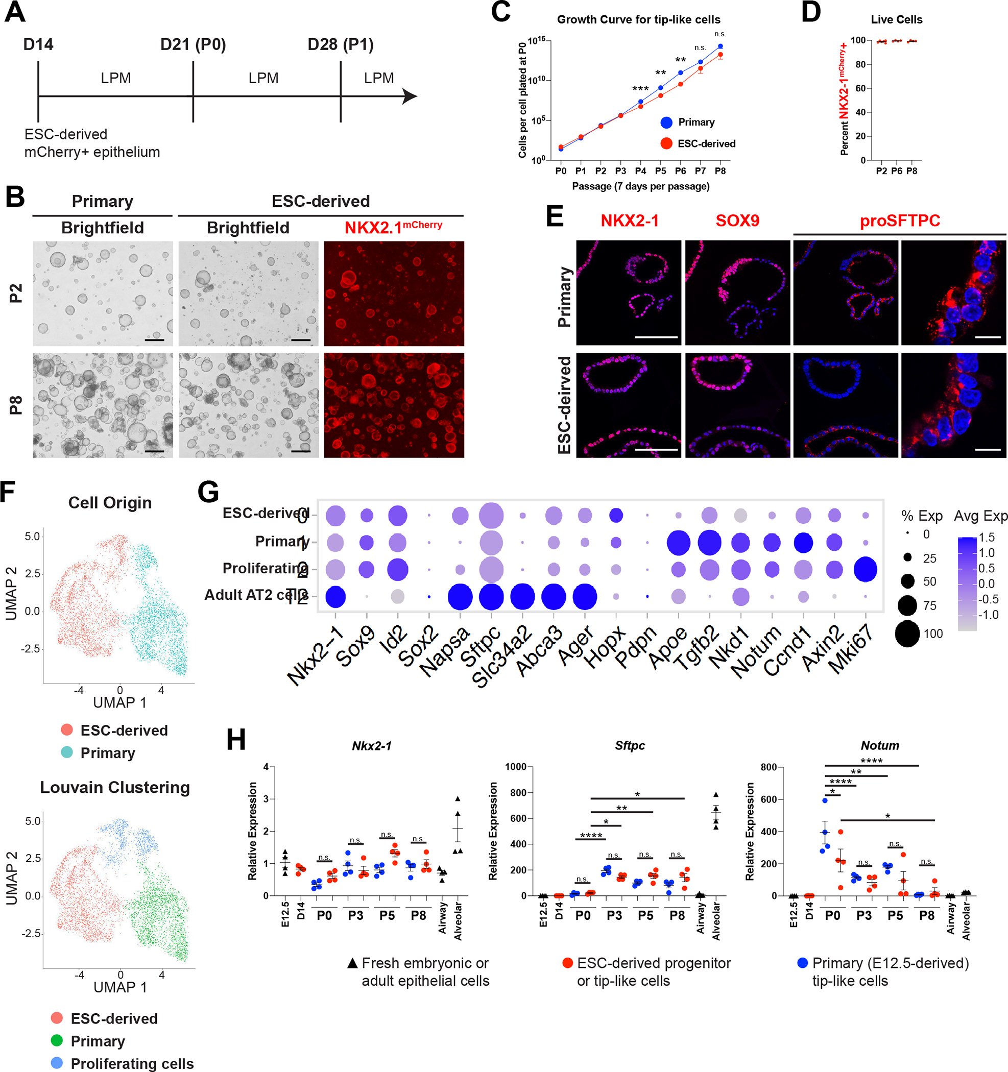
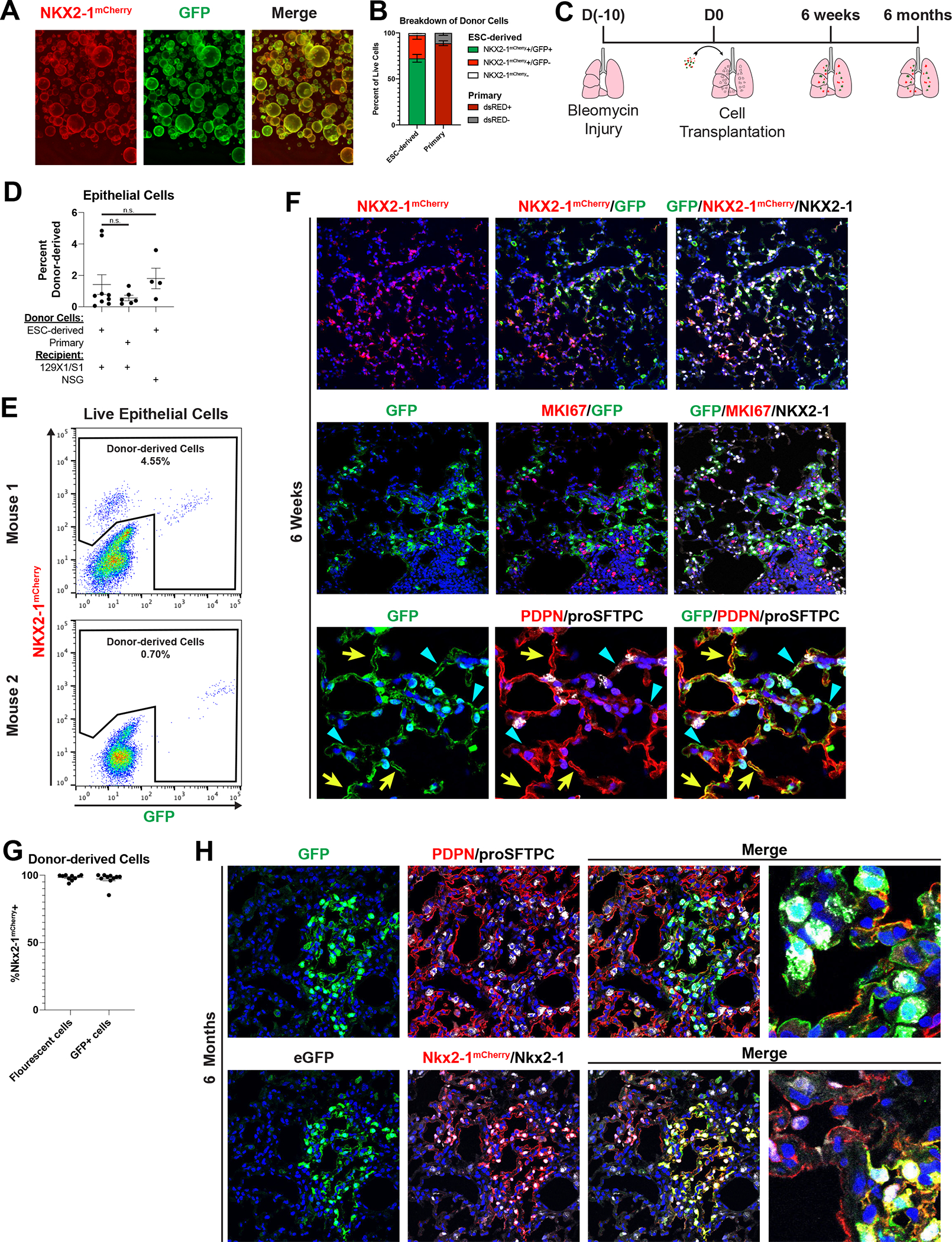
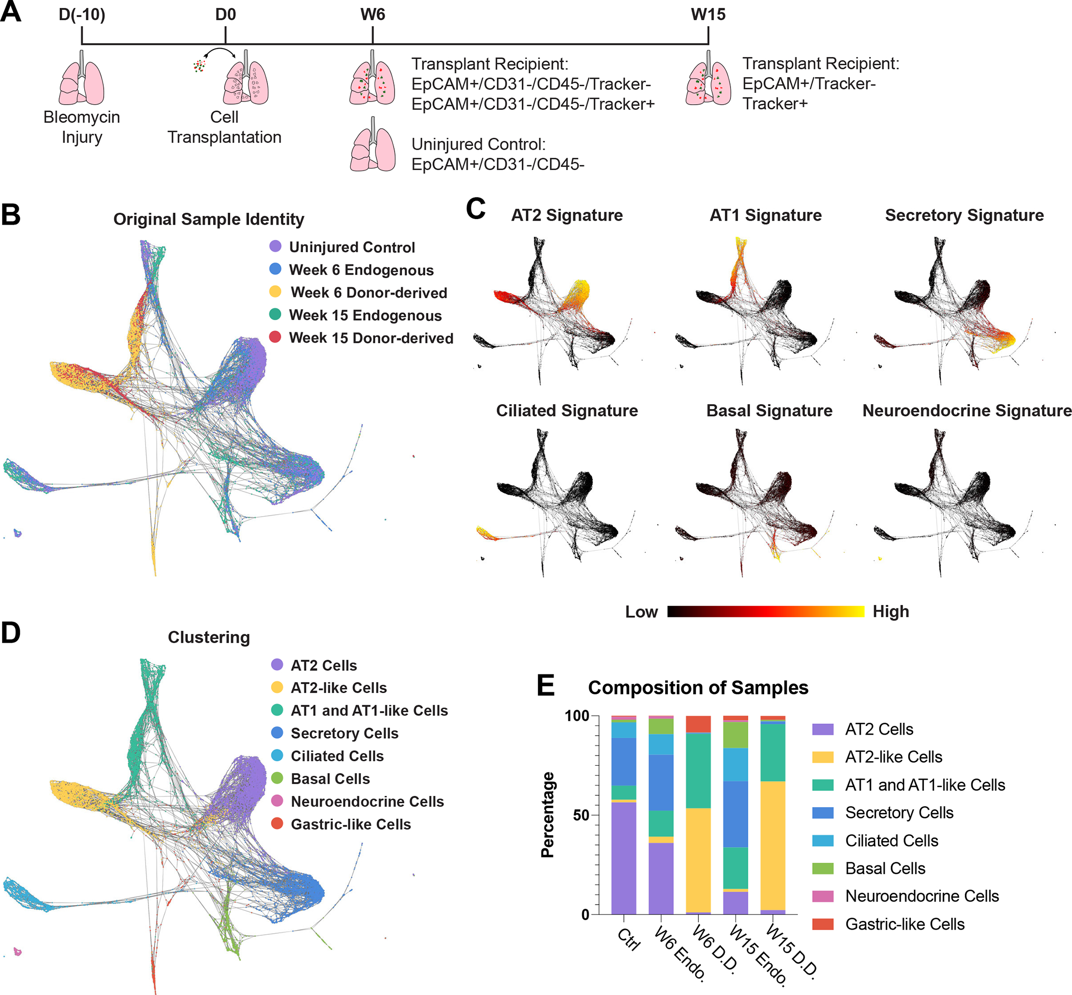
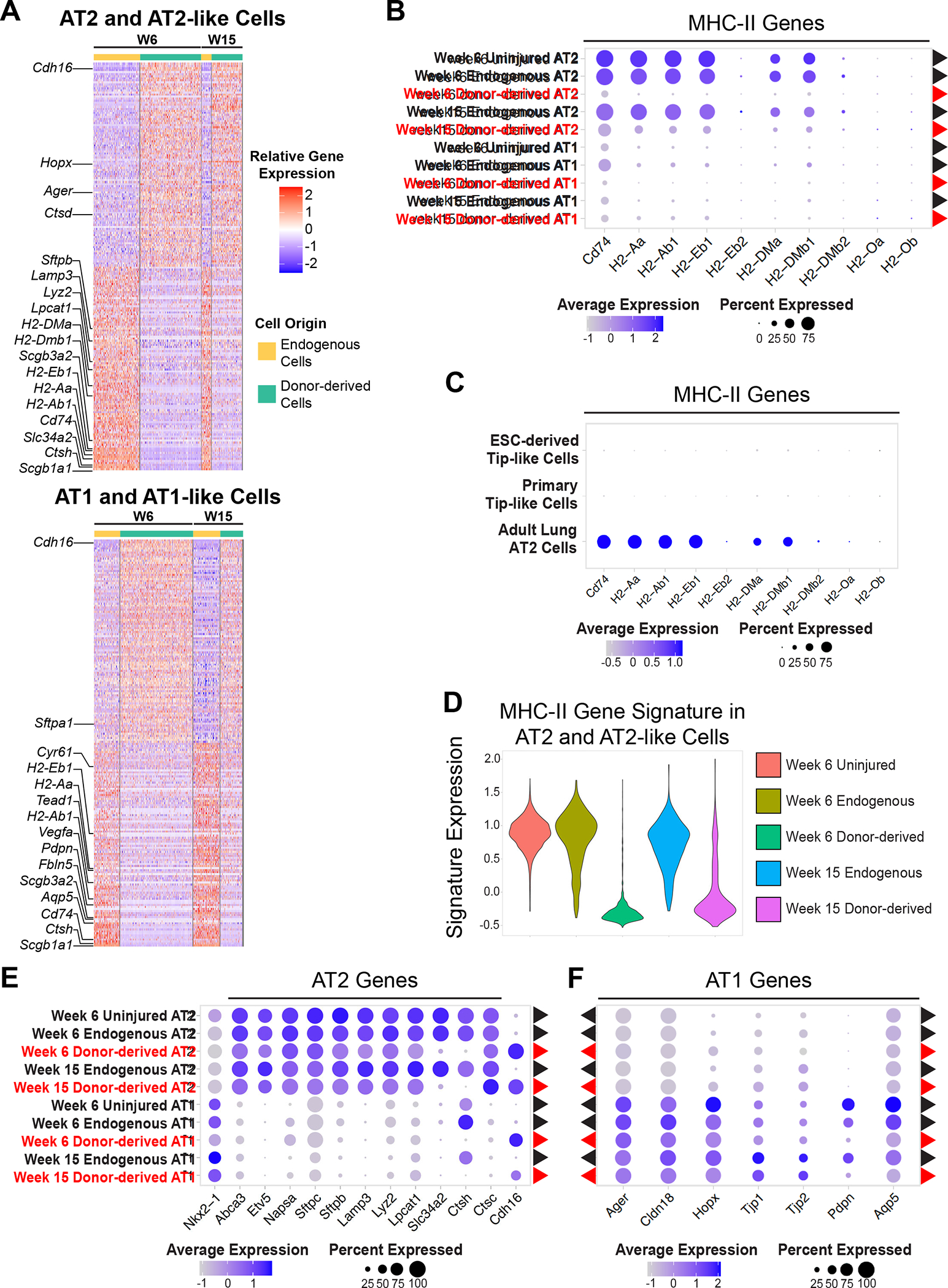
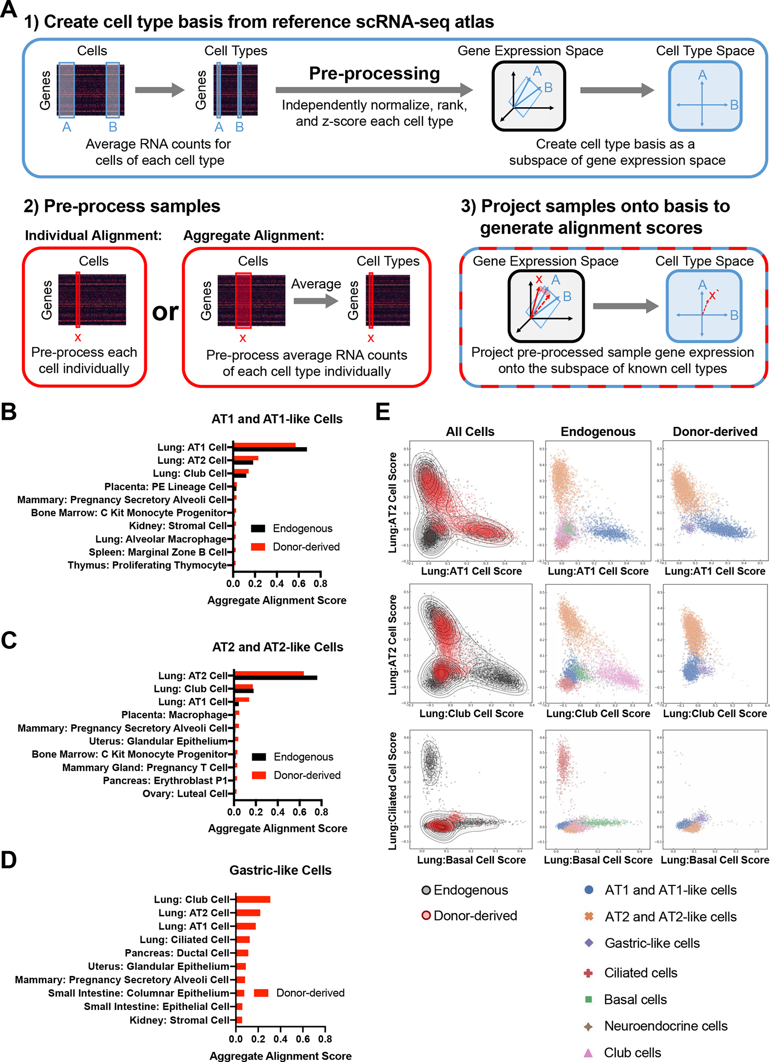
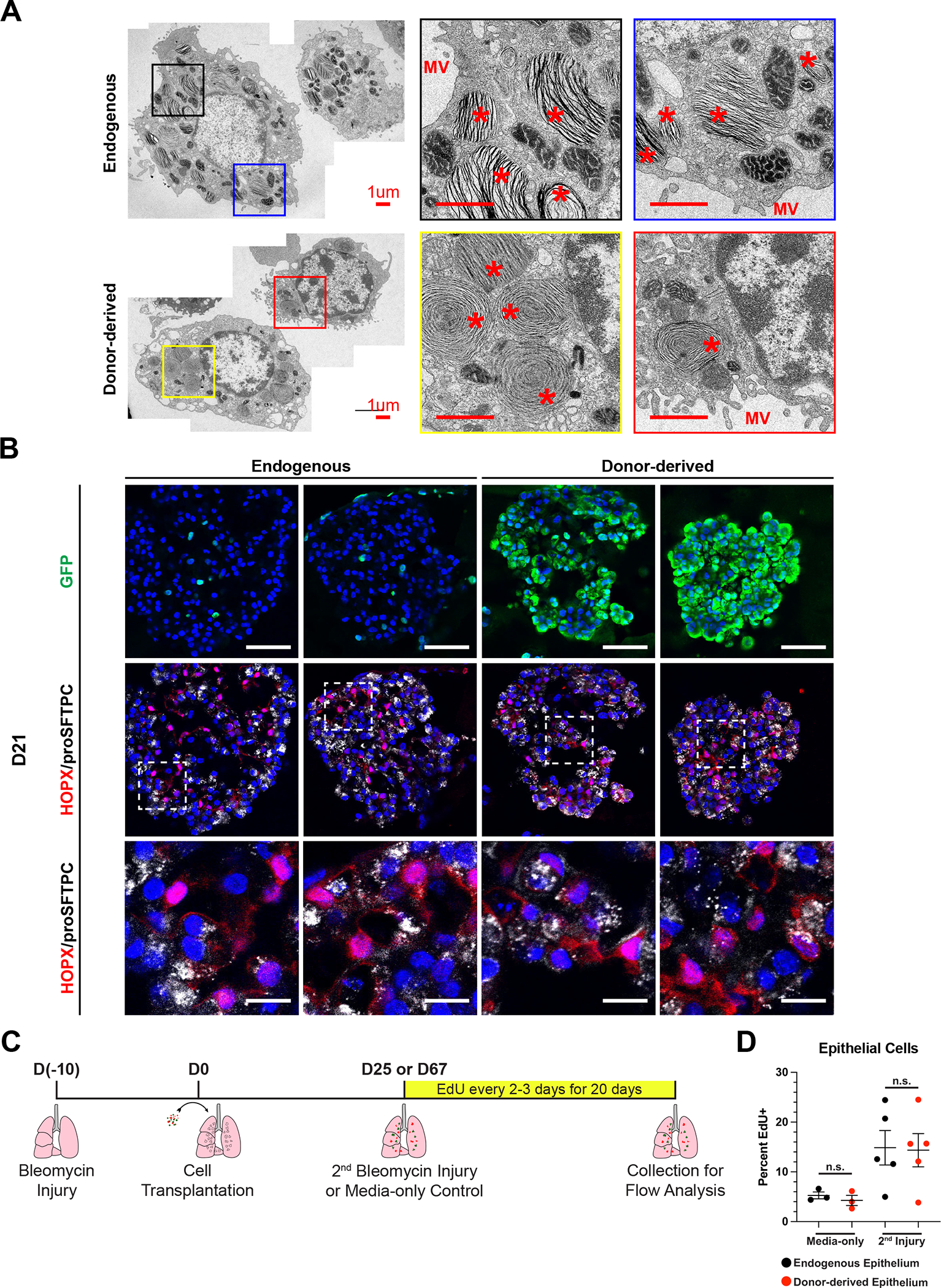
Durable reconstitution of the distal lung epithelium with pluripotent stem cell (PSC) derivatives, if realized, would represent a promising therapy for diseases that result from alveolar damage. Here, we differentiate murine PSCs into self-renewing lung epithelial progenitors able to engraft into the injured distal lung epithelium of immunocompetent, syngeneic mouse recipients. After transplantation, these progenitors mature in the distal lung, assuming the molecular phenotypes of alveolar type 2 (AT2) and type 1 (AT1) cells. After months in vivo, donor-derived cells retain their mature phenotypes, as characterized by single-cell RNA sequencing (scRNA-seq), histologic profiling, and functional assessment that demonstrates continued capacity of the engrafted cells to proliferate and differentiate. These results indicate durable reconstitution of the distal lung's facultative progenitor and differentiated epithelial cell compartments with PSC-derived cells, thus establishing a novel model for pulmonary cell therapy that can be utilized to better understand the mechanisms and utility of engraftment.

Keywords: alveolar epithelium; bleomycin injury; directed differentiation; distal lung bud tip cells; lung; lung progenitors; pluripotent stem cells; stem cells.


Conflict of interest statement

Declaration of interests The authors declare no competing interests.

Figures

Figure 1:
Figure 1:. Modified Lung Specification Protocol Leads to Increased Yield of Lung Progenitors
(A) Schematic of WB and WBRF lung specification protocols for directed differentiation of mouse ESCs carrying an Nkx2–1mCherry reporter. (B) Fluorescent microcopy images of day 8 and 13 of WB and WBRF lung specification protocols. Scale bars are 200um. (C) Quantification of the percent of live cells that are NKX2–1mCherry+/EpCAM+ double positive lung epithelial progenitors on day 8 or day 14 depending on the inclusion of RA and/or FGF10. n.s. not significant, **** p < 0.0001 by one-way ANOVA. n=4 biological replicates. Error bars = mean +/− SEM. (D) FACS plots on day 14 of WB and WBRF lung specification protocols. (E) Quantification of the percent of epithelial cells that are NKX2–1mCherry+ and the yield of NKX2–1mCherry+/EpCAM+ cells per well of a 6-well plate on day 13–14 of lung specification protocols. **** p < 0.0001 by unpaired, two-tailed Student’s t-test. n=15, 22, 8, 15 biological replicates. Error bars = mean +/− SEM. (F) UMAP plot for scRNA-seq of Epcam+ sorted day 13 cells from WBRF lung specification protocol. Plot displays three clusters identified by Louvain clustering. (G) UMAP plot displaying presence or absence of detectable Nkx2–1 in these same day 13 cells. (H) Expression of pulmonary and liver genes in annotated clusters of scRNA-seq data set. See also Figure S1 and Tables S1, S2
Figure 2:
Figure 2:. ESC-derived Tip-like Cells are Morphologically and Transcriptionally Similar to Cultured Primary Tip-like Cells
(A) Schematic of differentiation and passaging of ESC-derived tip-like cells in lung progenitor medium (LPM) following cell sorting of Nkx2–1mCherry+ cells on day 14. (B) Fluorescent microcopy images of primary and ESC-derived tip-like cells at P2 and P8. Scale bars are 500um. (C) Quantification of cell proliferation for primary and ESC-derived tip-like cells across nine passages. n.s. not significant, ** p<0.01, *** p<0.001 by unpaired, two-tailed Student’s t-test. n= 4 biological replicates. Error bars= mean +/− SEM (D) Assessment of NKX2–1mCherry expression throughout passaging of ESC-derived tip-like cells. n= 4 biological replicates. Error bars = mean +/− SEM. (E) Immunofluorescence microscopy for NKX2–1, SOX9, and proSFTPC in primary and ESC-derived tip-like cells. Nuclei stained with Hoechst, scale bars are 100um (first three columns) or 10um (rightmost column). (F) UMAP plot for scRNA-seq of primary and ESC-derived tip-like cells. Top plot distinguishes cells by sample origin; bottom plot displays Louvain clusters. (G) Expression of genes, including those identified as differently expressed between primary and ESC-derived tip-like cells. Cells from clusters annotated in panel F are compared against primary adult AT2 cells collected and sequenced at the same time. (H) Analysis of gene expression by RT-qPCR. Primary and ESC-derived tip-like cells from multiple passages are compared against lung epithelial progenitors from day 14 of the WBRF protocol (D14) and freshly sorted lung epithelial cells from embryonic (E12.5) and adult (Airway and Alveolar) mouse lungs. Reference gating for primary controls can be found in supplemental figure 2C,D. n.s. not significant, * p<0.05, ** p<0.01, **** p<0.0001 by one-way ANOVA. n= 4 biological replicates. Error bars = mean +/− SEM. See also Figures S1, S2 and Tables S1, S2.
Figure 3:
Figure 3:. Transplanted ESC-derived Tip-like Cells Give Rise to AT2-like and AT1-like Cells that Persist for At Least Six Months Post-transplantation in Immunocompetent Mice
(A) Image of ESC-derived tip-like cells, carrying an Nkx2–1mCherry reporter (red), labeled with lentiviral GFP (green). Scale bar is 500um. (B) Percentage of NKX2–1mcherry+/GFP+, NKX2–1mcherry+/GFP−, and NKX2–1mcherry− cells ESC-derived tip-like cells prior to transplantation (n=5 distinct lines). Also shown is the average dsRed+ percentage of cultured primary cells similarly labeled with lentiviral dsRed (n=3 technical replicates of the same line. Note: primary cells do not have an Nkx2–1 reporter. Error bars = mean +/− SEM (C) Schematic for transplantation of cells into bleomycin injured immunocompetent lungs with subsequent histological or flow analysis. (D) Flow cytometry quantitation of the percent of live epithelial (EpCAM+/CD45−/CD31−) cells that are donor-derived after transplantation of ESC-derived or primary tip-like cells based on flow analysis of whole recipient lungs. Recipient mice were either immunocompetent 129X1/S1 mice (6 weeks post-transplantation) or immunocompromised NSG mice (9 weeks post-transplantation). n.s. not significant by unpaired, two-tailed Student’s t-test. n= 9, 6, 4 biological replicates. Error bars = mean +/− SEM. (E) FACS plots identifying donor-derived cells within all live lung epithelial cells using mCherry and GFP expression. The two plots come from transplantations using different donor lines with different levels of GFP silencing. Few GFP+/mCherry- cells were detectable in these samples. (F) Immunofluorescence confocal microscopy of lung tissue sections showing donor-derived cell clusters at 6 weeks post-transplantation, assessing markers of donor trackers, lung lineages, and proliferation. White arrowheads indicate cuboidal proSFTPC+/GFP+ cells, yellow arrows indicate thin PDPN+/GFP+ cells, and blue triangles indicate thin PDPN−/GFP+ cells. Nuclei stained with Hoechst, scale bars are 100um. (G) The percent of all fluorescent (GFP+ or mCherry+) or GFP+ donor-derived cells that express NKX2–1mCherry at 6 weeks post-transplantation as determined by flow cytometry (for lungs with donor-derived cells accounting for >0.5% of assessed epithelium). n= 10 biological replicates. Error bars = mean +/− SEM. (H) Histology of donor-derived cell clusters at 6 months post-transplantation. Nuclei stained with Hoechst, scale bars are 50um (leftmost panels) or 12.5um (rightmost panels). Lower panels indicate some mCherry+ cell clusters are GFP−, presumed due to lentiviral silencing before or after transplantation. See also Figures S3, S4.
Figure 4:
Figure 4:. Single Cell Transcriptomic Profiling of Donor-derived and Endogenous Lung Epithelium
(A) Schematic for generation and collection of samples for scRNA-seq. (B) SPRING plot of epithelial cells characterized by scRNA-seq labeled by sample origin. (C) Expression of lung epithelial cell signatures. Gene sets comprising each signature can be found in Supplementary Table 3. (D) Cell-type annotation of clusters based on supervised Louvain clustering and expression of lung epithelial cell signatures. (E) Composition of each sample based on clusters identified in figure 4D. See also Figure S5 and Table S3.
Figure 5:
Figure 5:. Donor-derived Cells Express Lower Levels of Select MHC-II Components and Maturation Markers
(A) Row-normalized heatmap of the 100 most up-regulated and 100 most down-regulated genes (with adj. p-value <0.05, ordered by logFC) between donor-derived and endogenous cells for both AT2-like and AT1-like cells. Annotated genes are associated with lung epithelial lineages or MHC-II. (B) Expression of MHC-II genes in donor-derived (red) and endogenous (black) cells. (C) Expression of MHC-II genes in ESC-derived and primary tip-like cells compared to adult AT2 cells captured in the same experiment. (D) Violin plots of an MHC-II gene signature composed of genes listed in figure 5B. (E) Expression of AT2 genes in donor-derived (red) and endogenous (black) cells. (F) Expression of AT1 genes in donor-derived (red) and endogenous (black) cells. See also Tables S4–S7.
Figure 6:
Figure 6:. Global Transcriptomic Comparison of Endogenous and Donor-derived Lung Epithelial Cells using scTOP
(A) Schematic of scTOP (Single-Cell Type Order Parameters). (B) The top ten aggregate alignment scores for donor-derived AT1-like cells and the corresponding scores for endogenous AT1 cells. All reference cell types are from adult mice (Mouse Cell Atlas or Control sample as delineated in Figure 4) . (C) The top ten aggregate alignment scores for donor-derived AT2-like cells and the corresponding scores for endogenous AT2 cells. All reference cell types are from adult mice. (D) The top ten aggregate alignment scores for donor-derived gastric-like cells. All reference cell types are from adult mice. (E) Individual alignment scores for all donor-derived and endogenous epithelial cells against the indicated reference cells. Each cell is annotated (by color and shape shown in the key below the graphs) based on sample type or cell type as determined in Figure 4.
Figure 7:
Figure 7:. Functional Assessment of Donor-derived AT2-like Cells
(A) Representative transmission electron micrographs of GFP- endogenous AT2 cells and GFP+ donor-derived AT2-like cells from the same mouse. Scale bars are 1.0 um. * = lamellar body and MV = microvilli. (B) Representative immunofluorescence confocal microscopy of GFP/HOPX/proSFTPC expression in cultured mouse lung alveolospheres, comparing endogenous or donor-derived epithelial cells co-cultured with PDGFRanGFP+ primary lung fibroblasts. PDGFRa-GFP is nuclear, while donor-derived cells have a cytoplasmic GFP. Nuclei stained with Hoechst, scale bars are 50um (top row) or 12.5 um (bottom row). (C) Schematic for EdU labeling of transplant recipient mice following a second bleomycin injury or media-only control. (D) Percent EdU labeling of endogenous and donor-derived cells in ESC-derived tip-like cell recipients for the first 20 days following media-only delivery or a secondary bleomycin injury. Lobes were pruned down to regions containing GFP+ cells, or similar regions in no transplant controls, prior to digestion into single cell suspension. n.s. = not significant by unpaired, two-tailed Student’s t-test. n= 3,5 biological replicates. Error bars = mean +/− SEM.

Similar articles

Cited by

References

    1. Thomas ED, Lochte HL, Lu WC, and Ferrebee JW (1957). Intravenous Infusion of Bone Marrow in Patients Receiving Radiation and Chemotherapy. New Engl J Medicine 257, 491–496. 10.1056/nejm195709122571102. - DOI - PubMed
    1. O’Connor NicholasE., Mulliken JohnB., Banks-Schlegel S, Kehinde O, and Green H (1981). GRAFTING OF BURNS WITH CULTURED EPITHELIUM PREPARED FROM AUTOLOGOUS EPIDERMAL CELLS. Lancet 317, 75–78. 10.1016/s0140-6736(81)90006-4. - DOI - PubMed
    1. Rama P, Matuska S, Paganoni G, Spinelli A, Luca MD, and Pellegrini G (2010). Limbal Stem-Cell Therapy and Long-Term Corneal Regeneration. New Engl J Medicine 363, 147–155. 10.1056/nejmoa0905955. - DOI - PubMed
    1. Schwartz SD, Regillo CD, Lam BL, Eliott D, Rosenfeld PJ, Gregori NZ, Hubschman J-P, Davis JL, Heilwell G, Spirn M, et al. (2015). Human embryonic stem cell-derived retinal pigment epithelium in patients with age-related macular degeneration and Stargardt’s macular dystrophy: follow-up of two open-label phase 1/2 studies. Lancet 385, 509–516. 10.1016/s0140-6736(14)61376-3. - DOI - PubMed
    1. Xuan K, Li B, Guo H, Sun W, Kou X, He X, Zhang Y, Sun J, Liu A, Liao L, et al. (2018). Deciduous autologous tooth stem cells regenerate dental pulp after implantation into injured teeth. Sci Transl Med 10. 10.1126/scitranslmed.aaf3227. - DOI - PubMed

Publication types