. 2023 Nov;29(11):1803-1817.
doi: 10.1261/rna.079738.123. Epub 2023 Aug 25.

Biochemical characterization of mRNA capping enzyme from Faustovirus

Affiliations

Biochemical characterization of mRNA capping enzyme from Faustovirus

S Hong Chan et al. RNA. 2023 Nov.

Abstract

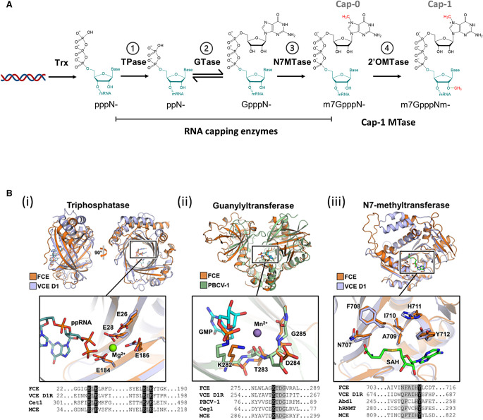
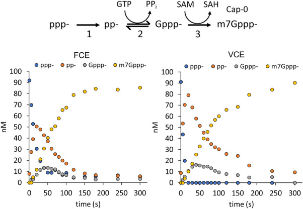
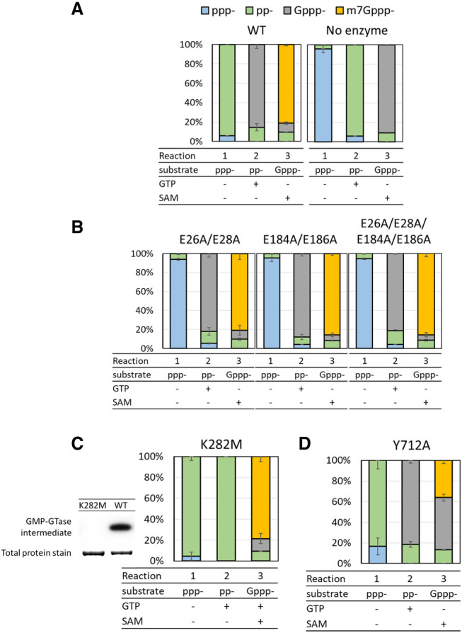
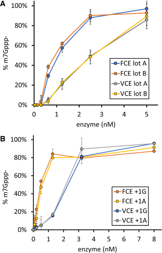
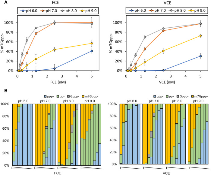
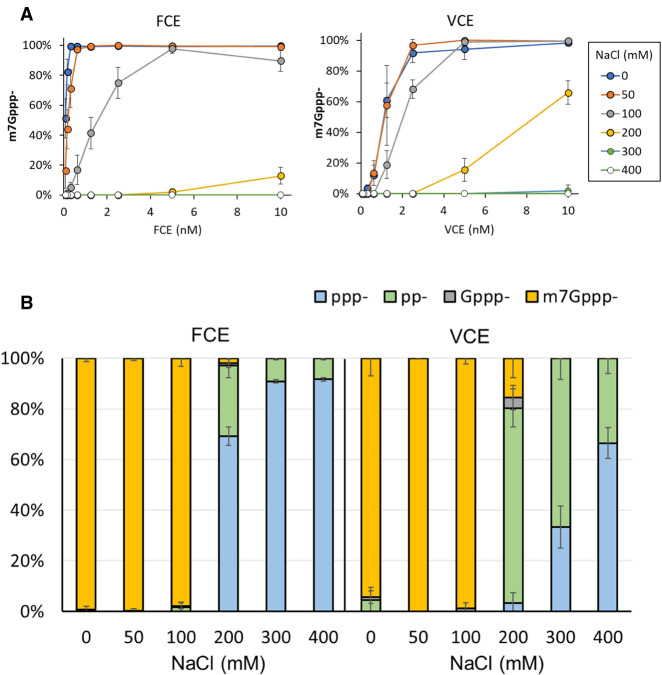
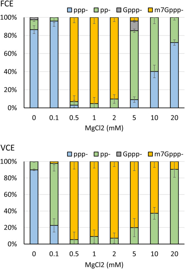
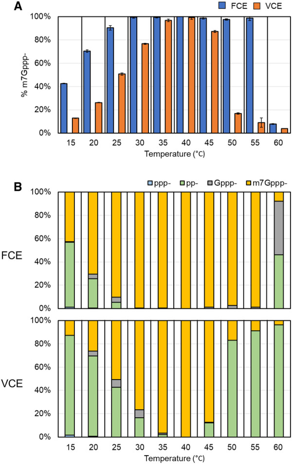
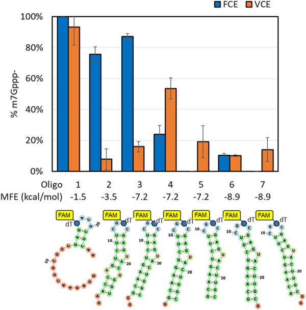
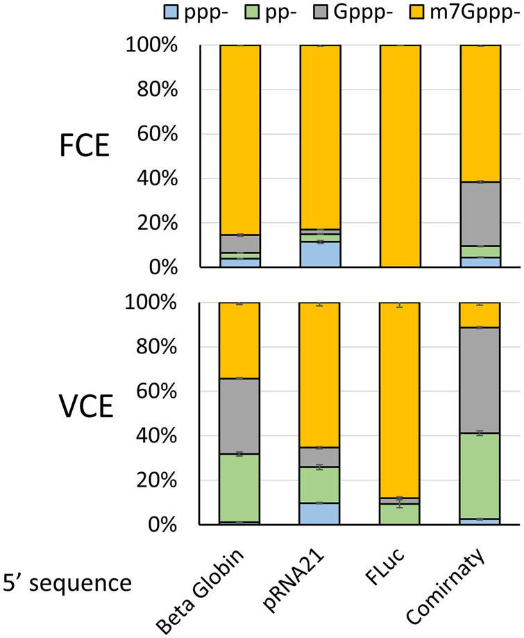
The mammalian mRNA 5' cap structures play important roles in cellular processes such as nuclear export, efficient translation, and evading cellular innate immune surveillance and regulating 5'-mediated mRNA turnover. Hence, installation of the proper 5' cap is crucial in therapeutic applications of synthetic mRNA. The core 5' cap structure, Cap-0, is generated by three sequential enzymatic activities: RNA 5' triphosphatase, RNA guanylyltransferase, and cap N7-guanine methyltransferase. Vaccinia virus RNA capping enzyme (VCE) is a heterodimeric enzyme that has been widely used in synthetic mRNA research and manufacturing. The large subunit of VCE D1R exhibits a modular structure where each of the three structural domains possesses one of the three enzyme activities, whereas the small subunit D12L is required to activate the N7-guanine methyltransferase activity. Here, we report the characterization of a single-subunit RNA capping enzyme from an amoeba giant virus. Faustovirus RNA capping enzyme (FCE) exhibits a modular array of catalytic domains in common with VCE and is highly efficient in generating the Cap-0 structure without an activation subunit. Phylogenetic analysis suggests that FCE and VCE are descended from a common ancestral capping enzyme. We found that compared to VCE, FCE exhibits higher specific activity, higher activity toward RNA containing secondary structures and a free 5' end, and a broader temperature range, properties favorable for synthetic mRNA manufacturing workflows.

Keywords: Faustovirus; RNA capping enzymes; RNA caps; mRNA synthesis; vaccinia capping enzyme.


Figures

FIGURE 1.
FIGURE 1.
(A) Enzymatic reactions involved in RNA 5′ capping. First, the RNA triphosphatase (TPase) hydrolyzes the 5′ triphosphate group (pppN−) to form a 5′ diphosphate (ppN−) (Reaction 1). Then, an RNA 5′ guanylyltransferase adds a GMP group to the 5′ diphosphate to form an unmethylated G-capped structure (GpppN−) through a reversible mechanism (Reaction 2). This is followed by an RNA cap guanosine N7-methyltransferase activity that adds a methyl group to the N7 position of the G cap and forms the Cap-0 structure (m7GpppN−) (Reaction 3). In metazoans, a fourth enzyme RNA cap 2′-O-methyltransferase (2′-OMTase) further adds a methyl group to the 2′-O position of the initiating nucleotide of the transcript, forming the Cap-1 structure (m7GpppNm−) (Reaction 4). (B) Catalytic domains of FCE. (Top panels) Individual domains of the AlphaFold2 prediction of FCE were aligned to published atomic structures. Insets show the alignment of highly conserved residues labeled with FCE residue numbering. (Bottom panels) Amino acid residues of the motifs were aligned with indicated enzymes. Cet1, Ceg1, and Abd1 are the TPase, GTase, and N7MTase of Saccharomyces cerevisiae, respectively. hRNMT is the human RNA cap N7MTase. MCE refers to Acanthamoeba polyphaga mimivirus capping enzyme. Conserved residues are highlighted in gray. FCE residues that are mutated in this study and the corresponding residues in orthologous enzymes are highlighted in black. (i) The predicted structure of FCE's TPase domain is aligned to the VCE TPase domain-ppRNA complex extracted from the CryoEM cotranscriptional capping complex (PDB ID 6RIE). The inset shows the alignment of the conserved ELE–ELE motif. (ii) The predicted structure of FCE's GTase domain is aligned to the X-ray crystal structure of Paramecium bursaria chlorella virus (PBCV-1) GTase with covalent GMP intermediate (PDB ID 1CKN). Inset shows the phosphamide covalent linkage between the catalytic Lys of PBCV-1 GTase and GMP. (iii) The predicted FCE N7MTase domain structure is aligned to the X-ray crystal structure of the N7MTase domain of VCE D1 subunit in complex with co-product SAH (PDB ID 4CKB). Inset shows the conserved SAM-binding site.
FIGURE 2.
FIGURE 2.
FCE and VCE generate the m7Gppp− (Cap-0) structure through the same pathway. Under identical conditions (1× RNA capping buffer, 0.5 mM GTP, 0.1 mM SAM, 1 mM DTT, and 5 nM enzyme at 25°C), FCE and VCE underwent the same reaction pathway to generate the m7Gppp− cap from 5′ triphosphate. As the concentration of 5′ ppp-RNA (blue circles) decreased over time, the concentration of the TPase product pp− (orange circles) increased initially and decreased over time, followed by a similar trend with a smaller amplitude for the GTase product Gppp− (gray circles) and an increase in N7MTase product m7Gppp− (yellow circles).
FIGURE 3.
FIGURE 3.
Catalytic amino acid residues of FCE. (A) Under standard conditions (see Materials and Methods), 2.5 nM of FCE WT enzyme converted ≥90% of ppp-RNA into pp-RNA in the absence of GTP and SAM (Reaction 1), ∼90% pp− into Gppp− in the presence of GTP (Reaction 2) and ∼80% of Gppp− into m7Gppp− in the presence of SAM (Reaction 3). (B) Mutations of the Glu residues of the ELE motifs knocked out TPase activity but did not affect GTase or N7MTase activities. Mutants E26A/E28A, E184A/E186A, and E26A/E28A/E184A/E186A exhibited the same trend. (C) Mutation of Lys282 of the conserved KTDG motif to Met eliminated the formation of the GMP-GTase covalent intermediate (left panel). The mutant K282M exhibited undetectable GTase activity (Reaction 2) but exhibited high level of TPase (Reaction 1) and N7MTase activity (Reaction 3). (D) Mutation of Tyr712 in the conserved SAM-binding motif to Ala significantly decreased the N7MTase activity (Reaction 3) compared to the WT FCE enzyme but not TPase (Reaction 1) or GTase activity (Reaction 2).
FIGURE 4.
FIGURE 4.
(A) FCE exhibits higher specific activity than VCE. RNA capping reactions were performed using decreasing concentrations of two separate preparations of both FCE and VCE under standard conditions at 37°C for 30 min. FCE achieved 50% capping at ∼1 nM enzyme concentration, while VCE achieved the same level of capping at ∼2.5 nM enzyme concentration. (B) FCE and VCE do not discriminate between RNA initiating with guanosine or adenosine. RNA capping reactions were performed on 5′ triphosphate RNA oligonucleotides initiating with a guanosine (+1G) or an adenosine (+1A) under standard conditions at 37°C for 30 min. FCE and VCE exhibited similar level of capping activity on both +1G and +1A RNA. Data points are the average value of triplicated experiments. Error bars represent standard deviations.
FIGURE 5.
FIGURE 5.
Effect of pH on capping activities. (A) FCE and VCE exhibit the highest capping activity at pH 7.0 and pH 8.0. (B) Effect of pH on component enzyme activities. Both enzymes exhibit similar responses outside of optimal pH values. At pH 6.0, the accumulation of ppp− substrate indicates that the TPase activity of both enzymes is negatively affected. At pH 9.0, the accumulation of pp− product indicates that the forward GTase activity of both enzymes is negatively affected. Data points are the average value of triplicated experiments. Error bars represent standard deviations.
FIGURE 6.
FIGURE 6.
Effect of NaCl concentration on capping activities. (A) FCE and VCE exhibit the highest capping activity in zero or 50 mM NaCl. At enzyme concentrations of 5 nM or higher, both enzymes exhibit high capping activity in 100 mM NaCl. (B) Differential effect of NaCl on FCE and VCE. In 200 mM NaCl, the TPase activity of FCE decreased significantly whereas the GTase, but not TPase, activity of VCE was affected. The TPase activity of VCE started to be negatively impacted by 300 mM of NaCl. Data points are the average value of triplicated experiments. Error bars represent standard deviations.
FIGURE 7.
FIGURE 7.
Differential effect of MgCl2 concentration on capping activities. FCE and VCE exhibit the highest capping activity in 0.5–2 mM MgCl2, but individual activities of the two enzymes are differentially affected outside of the optimal MgCl2 concentration range. At 0.1 mM MgCl2, the TPase activity of FCE is impeded whereas in VCE the GTase activity was affected. For FCE, the GTase activity was negatively impacted at 5 mM MgCl2 with TPase starting to be affected at 10 mM MgCl2. For VCE, the GTase activity started to be affected at 5 mM MgCl2 as indicated by pp-RNA accumulation. However, the TPase activity of VCE was not affected up to 20 mM MgCl2 due to the lack of ppp− accumulation. In each experiment, 5 nM of enzymes and 0.5 mM of RNA substrate were used. Data points are the average value of triplicated experiments. Error bars represent standard deviations.
FIGURE 8.
FIGURE 8.
RNA capping activity of FCE and VCE across a temperature range. RNA capping assays were performed on 0.5 µM of a 20 nt poly(A) synthetic oligonucleotide with a 3′ FAM group using 5 nM of FCE or VCE under standard conditions (see Materials and Methods). (A) FCE generated more m7Gppp-cap at all temperatures tested. Data points are the average of triplicated reactions. Error bars represent standard deviations. (B) A breakdown of the intermediate products across the temperature range after 30 min.
FIGURE 9.
FIGURE 9.
RNA capping activity of FCE and VCE on structured model RNA. Synthetic oligonucleotides are designed to adopt hairpin structures with different folding energies and availability of the 5′ triphosphate. All RNA oligos contain a FAM-dT for capillary electrophoresis readout and their theoretical minimum free energies (MFEs) of unfolding are listed. Capping reactions were carried out on 0.5 µM of the RNA oligos using 2 nM of FCE or VCE at 37°C under standard conditions (see Materials and Methods). Data points are the average of triplicated reactions. Error bars represent standard deviations.
FIGURE 10.
FIGURE 10.
RNA capping activity of FCE and VCE on FLuc transcript (1.8 kb) containing a 5′sequence derived from the first 50 nt of human β globin variant 1, synthetic 5′ UTR of pRNA21, synthetic 5′ UTR of pFLuc, and Comirnaty. FCE or VCE (20 nM) was used to cap 3.5 µM of the in vitro transcripts for 1 h at 37°C. Following capping reactions, mRNA capping was measured using targeted RNase H cleavage and LC–MS. Data points are the average of triplicated experiments. Error bars represent standard deviations.
FIGURE 11.
FIGURE 11.
(A) The CheckV database of complete viral genomes was searched for viruses which contain a cap M7MTase domain using hmmsearch and the PFAM profile PF03291. The 83 identified viruses were clustered by proteome similarity using the vContact2 program. The program was run using DIAMOND (Buchfink et al. 2021) for the all-vs-all protein alignment, and the figure was prepared by excluding the reference virus database. (B) A subset of the 83 capping enzymes was selected to represent the diversity of the data set, aligned and used to construct a phylogenetic tree. The tree is built from an MAFFT alignment of the N7MTase domains; for viruses which contain separate genes from TPase, GTase and N7MTase domains, only the N7MTase protein was used. The viruses were mapped to the clustering in (A) (color dots), showing that the clustering based on the whole proteome is consistent with the phylogeny of the cap N7MTase genes. On the right, putative TPase (TP), GTase (GT), and N7MTase (MT) domains, as well as homologs of the regulatory subunit (D12L) of VCE are shown as white hexagons. Domains attached by solid lines are found within one gene.

Similar articles

Cited by

References

    1. Ahola T, Kääriäinen L, Pan J, Xiang N, Tien P, Ahola T, Guo D. 1995. Reaction in alphavirus mRNA capping: formation of a covalent complex of nonstructural protein nsP1 with 7-methyl-GMP. Proc Natl Acad Sci 92: 507–511. 10.1073/pnas.92.2.507 - DOI - PMC - PubMed
    1. Allen MJ, Forster T, Schroeder DC, Hall M, Roy D, Ghazal P, Wilson WH. 2006. Locus-specific gene expression pattern suggests a unique propagation strategy for a giant algal virus. J Virol 80: 7699–7705. 10.1128/JVI.00491-06 - DOI - PMC - PubMed
    1. Barbosa E, Moss B. 1978. mRNA(nucleoside-2′-)-methyltransferase from vaccinia virus. Purification and physical properties. J Biol Chem 253: 7692–7697. 10.1016/S0021-9258(17)34425-3 - DOI - PubMed
    1. Benarroch D, Smith P, Shuman S. 2008. Characterization of a trifunctional mimivirus mRNA capping enzyme and crystal structure of the RNA triphosphatase domain. Structure 16: 501–512. 10.1016/j.str.2008.01.009 - DOI - PubMed
    1. Beverly M, Dell A, Parmar P, Houghton L. 2016. Label-free analysis of mRNA capping efficiency using RNase H probes and LC-MS. Anal Bioanal Chem 408: 5021–5030. 10.1007/s00216-016-9605-x - DOI - PubMed

Publication types

LinkOut - more resources