

New insight into protein glycosylation in the development of Alzheimer's disease
- PMID: 37626031
- PMCID: PMC10457297
- DOI: 10.1038/s41420-023-01617-5
New insight into protein glycosylation in the development of Alzheimer's disease
Abstract
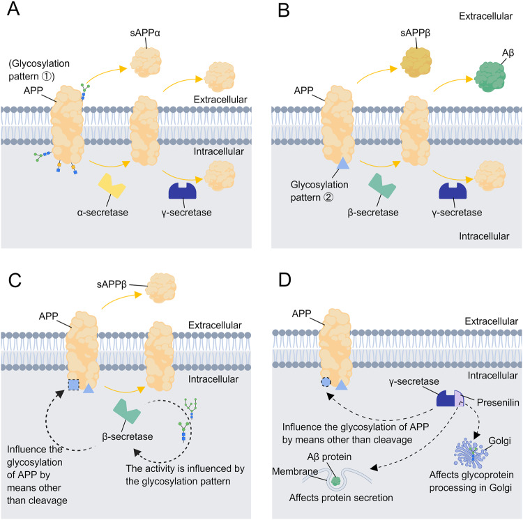
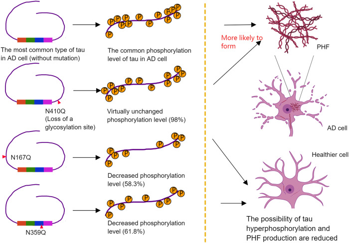
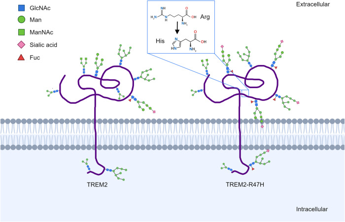
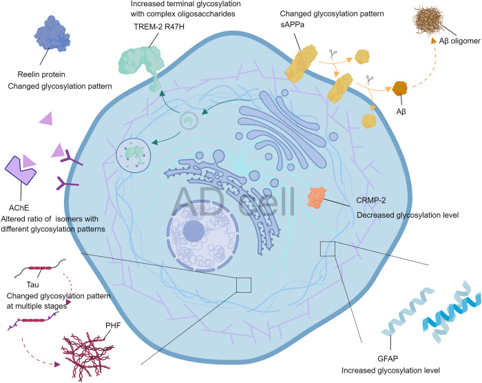
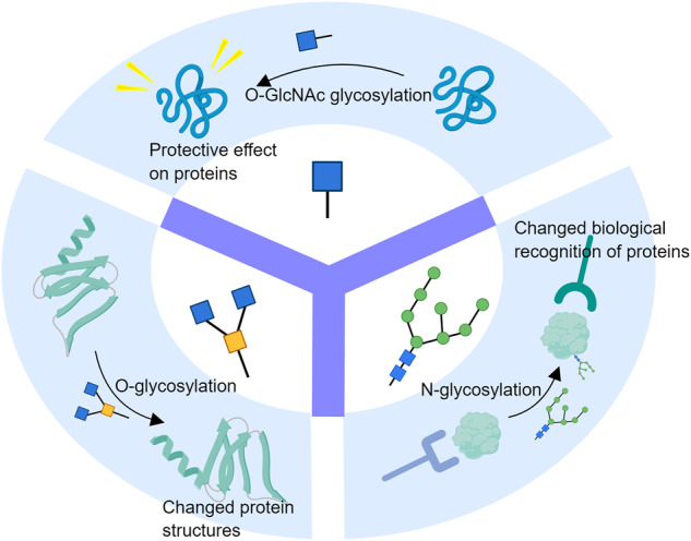
Alzheimer's disease (AD) is a chronic neurodegenerative disease that seriously endangers the physical and mental health of patients, however, there are still no effective drugs or methods to cure this disease up to now. Protein glycosylation is the most common modifications of the translated proteins in eukaryotic cells. Recently many researches disclosed that aberrant glycosylation happens in some important AD-related proteins, such as APP, Tau, Reelin and CRMP-2, etc, suggesting a close link between abnormal protein glycosylation and AD. Because of its complexity and diversity, glycosylation is thus considered a completely new entry point for understanding the precise cause of AD. This review comprehensively summarized the currently discovered changes in protein glycosylation patterns in AD, and especially introduced the latest progress on the mechanism of protein glycosylation affecting the progression of AD and the potential application of protein glycosylation in AD detection and treatment, thereby providing a wide range of opportunities for uncovering the pathogenesis of AD and promoting the translation of glycosylation research into future clinical applications.
© 2023. Cell Death Differentiation Association (ADMC).
Conflict of interest statement
The authors declare no competing interests.
Figures


Similar articles
-
Implications of Glycosylation in Alzheimer's Disease.Front Neurosci. 2021 Jan 13;14:625348. doi: 10.3389/fnins.2020.625348. eCollection 2020. Front Neurosci. 2021. PMID: 33519371 Free PMC article. Review.
-
N-glycan and Alzheimer's disease.Biochim Biophys Acta Gen Subj. 2017 Oct;1861(10):2447-2454. doi: 10.1016/j.bbagen.2017.04.012. Epub 2017 Apr 29. Biochim Biophys Acta Gen Subj. 2017. PMID: 28465241 Review.
-
Early Stage Glycosylation Biomarkers in Alzheimer's Disease.Medicines (Basel). 2019 Sep 3;6(3):92. doi: 10.3390/medicines6030092. Medicines (Basel). 2019. PMID: 31484367 Free PMC article. Review.
-
Clinical relevance of biomarkers, new therapeutic approaches, and role of post-translational modifications in the pathogenesis of Alzheimer's disease.Front Aging Neurosci. 2022 Sep 7;14:977411. doi: 10.3389/fnagi.2022.977411. eCollection 2022. Front Aging Neurosci. 2022. PMID: 36158539 Free PMC article. Review.
-
Glycation vs. glycosylation: a tale of two different chemistries and biology in Alzheimer's disease.Glycoconj J. 2016 Aug;33(4):487-97. doi: 10.1007/s10719-016-9690-2. Epub 2016 Jun 21. Glycoconj J. 2016. PMID: 27325408 Review.
Cited by
-
Recent Advances in Mass Spectrometry-Based Studies of Post-Translational Modifications in Alzheimer's Disease.Mol Cell Proteomics. 2025 Jul;24(7):101003. doi: 10.1016/j.mcpro.2025.101003. Epub 2025 May 29. Mol Cell Proteomics. 2025. PMID: 40449795 Free PMC article. Review.
-
Role of Tau Protein in Neurodegenerative Diseases and Development of Its Targeted Drugs: A Literature Review.Molecules. 2024 Jun 13;29(12):2812. doi: 10.3390/molecules29122812. Molecules. 2024. PMID: 38930877 Free PMC article. Review.
-
Glycosylation in aging and neurodegenerative diseases.Acta Biochim Biophys Sin (Shanghai). 2024 Aug 15;56(8):1208-1220. doi: 10.3724/abbs.2024136. Acta Biochim Biophys Sin (Shanghai). 2024. PMID: 39225075 Free PMC article. Review.
-
Intracellular accumulation of amyloid-ß is a marker of selective neuronal vulnerability in Alzheimer's disease.Nat Commun. 2025 Jun 4;16(1):5189. doi: 10.1038/s41467-025-60328-w. Nat Commun. 2025. PMID: 40467545 Free PMC article.
-
Reelin Signaling in Neurodevelopmental Disorders and Neurodegenerative Diseases.Brain Sci. 2023 Oct 19;13(10):1479. doi: 10.3390/brainsci13101479. Brain Sci. 2023. PMID: 37891846 Free PMC article. Review.
References
Publication types
Grants and funding
LinkOut - more resources
Full Text Sources
Miscellaneous
