. 2023 Aug 26;14(8):562.
doi: 10.1038/s41419-023-06074-7.

Targeted deletion of von-Hippel-Lindau in the proximal tubule conditions the kidney against early diabetic kidney disease

Affiliations

Targeted deletion of von-Hippel-Lindau in the proximal tubule conditions the kidney against early diabetic kidney disease

Madlen Kunke et al. Cell Death Dis. .

Abstract

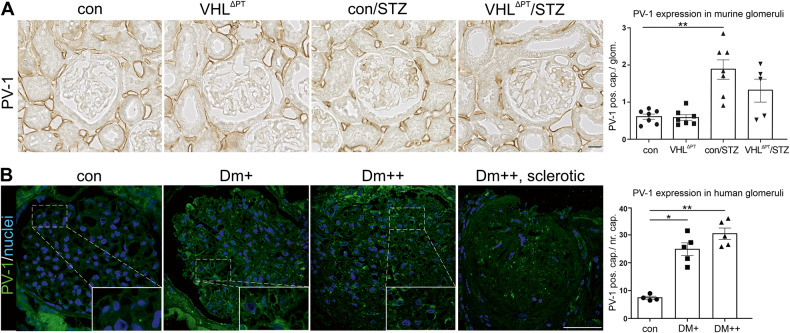
Diabetic kidney disease (DKD) is the leading cause of end-stage renal disease. Glomerular hyperfiltration and albuminuria subject the proximal tubule (PT) to a subsequent elevation of workload, growth, and hypoxia. Hypoxia plays an ambiguous role in the development and progression of DKD and shall be clarified in our study. PT-von-Hippel-Lindau (Vhl)-deleted mouse model in combination with streptozotocin (STZ)-induced type I diabetes mellitus (DM) was phenotyped. In contrary to PT-Vhl-deleted STZ-induced type 1 DM mice, proteinuria and glomerular hyperfiltration occurred in diabetic control mice the latter due to higher nitric oxide synthase 1 and sodium and glucose transporter expression. PT Vhl deletion and DKD share common alterations in gene expression profiles, including glomerular and tubular morphology, and tubular transport and metabolism. Compared to diabetic control mice, the most significantly altered in PT Vhl-deleted STZ-induced type 1 DM mice were Ldc-1, regulating cellular oxygen consumption rate, and Zbtb16, inhibiting autophagy. Alignment of altered genes in heat maps uncovered that Vhl deletion prior to STZ-induced DM preconditioned the kidney against DKD. HIF-1α stabilization leading to histone modification and chromatin remodeling resets most genes altered upon DKD towards the control level. These data demonstrate that PT HIF-1α stabilization is a hallmark of early DKD and that targeting hypoxia prior to the onset of type 1 DM normalizes renal cell homeostasis and prevents DKD development.


Conflict of interest statement

The authors declare no competing interests.

Figures

Fig. 1
Fig. 1. Deletion of Vhl expression in early proximal tubule segments (S1/2) and renal function after induction of type 1 diabetes mellitus.
A Scheme illustrating deletion of the von-Hippel-Lindau (Vhl) gene under the sodium-glucose co-transporter 2 (Sglt2) promoter in S1 and S2 segments of the proximal tubule and time-line of the experiment. B Semi-quantitative analysis of Vhl mRNA expression derived from Exon 1 in S1/2 segments of control, VHLΔPT, con/STZ, and VHLΔPT/STZ. Arithmetic means ± SEM of n = 6 per group; *P < 0.05. C STED images of cilia from proximal tubules obtained from control and VHLΔPT and morphometric analysis of cilia length. BBM, brush border membrane. Arithmetic means ± SEM of n = 7 per group. Scale bar = 2 µm. Nonparametric Mann–Whitney-U-test. D Western blot of renal nuclear HIF-1α abundance and densitometric evaluation of control, VHLΔPT, con/STZ, and VHLΔPT/STZ with histone H3 as reference. Arithmetic means ± SEM of n = 5–6 per group; *P < 0.05, **P < 0.01, ***P < 0.001. EH Renal function data: blood glucose (E), glomerular filtration rate (GFR) determined by FITC-sinistrin plasma kinetic measurements (F), proteinuria of 24 h urine collection (G), and proximal tubular albumin uptake (H) of control, VHLΔPT, con/STZ, and VHLΔPT/STZ. Arithmetic means ± SEM of n = 6–7 per group; *P < 0.05, **P < 0.01, ***P < 0.001. BG Each point represents an individual mouse. B, D, EG Nonparametric Kruskal–Wallis with Dunn’s post-test.
Fig. 2
Fig. 2. Sodium and glucose transporter of the proximal and distal tubule and nitric oxide synthase 1.
(Left) Representative images of immunohistochemical staining of sodium-glucose co-transporter 2 (SGLT2), glucose transporter-1 (GLUT1), sodium hydrogen exchanger-3 (NHE3), and nitric oxide synthase 1 (NOS1) obtained from control, VHLΔPT, con/STZ and VHLΔPT/STZ. Asterisks indicate macula densa cells located in the pars recta of the distal tubule. Scale bar = 20 µm. (Right) Representative blots and densitometrical evaluation of western blots of SGLT2, GLUT1, NHE3, and semi-quantitative evaluation of NOS1 obtained from control, VHLΔPT, con/STZ, and VHLΔPT/STZ. Flotillin-1 was used as a blot reference. Arithmetic means ± SEM of n = 5–7 per group; *P < 0.05, **P < 0.01, ***P < 0.001. (Right) Each band/point represents an individual mouse. Nonparametric Kruskal–Wallis with Dunn’s post test.
Fig. 3
Fig. 3. Vhl deletion preconditions against STZ-induced DKD development.
A Venn diagram of the number of genes altered comparing VHLΔPT vs. control, con/STZ vs. control, and VHLΔPT + con/STZ + VHL∆PT/STZ vs. control based on filter criteria DESeq P-values < 0.05, fold-change >1.5. B and C Heat maps of genes significantly altered in VHLΔPT & con/STZ & VHLΔPT/STZ vs. control, filter criteria used DESeq P-values < 0.05, fold-change > 1.5. B Altered genes sorted depending on known glomerular function related to podocyte, basement membrane (GBM), mesangial cells (mes), angio/vasculogenesis, and endocytosis. C Altered genes are sorted depending on known tubular function related to cell polarity, cell stress, transport, metabolism, and endocytosis. D Light microscopy of semi-thin sections showing representative glomeruli from each group. Glomeruli are marked by a “G”. The number of capillary profiles is higher upon Vhl deletion and diabetes induction and augmented glomerular size is visible upon diabetes induction. Scale bar = 20 µm. E and F Electron microscopy of ultra-thin sections showing representative images of glomerular filtration barrier (E) and of proximal tubule cells from S1 segments (F). Note, thickening of glomerular basement membrane (GBM) upon Vhl deletion and diabetes induction, foot process effacement of podocytes (Pod) only in the con/STZ group, and augmented epithelial height and tubular basement membrane (TBM) height upon Vhl deletion and STZ-induced diabetes. Scale bar = 500 nm and 2 µm, respectively. Glomerular endothelial cells (Endo), brush border membrane of proximal tubule cells (BBM), and proximal tubular cell (PTC).
Fig. 4
Fig. 4. Reduced glomerular VEGF backflow.
A and B Representative images and semi-quantitative analysis of PV-1 positive capillaries per glomeruli of control, VHLΔPT, con/STZ, and VHLΔPT/STZ (A) and from human biopsies of patients with interstitial nephritis (control), mild DM (DM + ), moderate DM (DM++) and severe DM (DM + ++). B. As control biopsies taken from patients with interstitial nephritis were used. Arithmetic means ± SEM of n = 4–7 per group; *P < 0.05, **P < 0.01. Scale bar = 20 µm (A) and scale bar = 50 µm (B). A and B Each point represents an individual mouse or patient. Nonparametric Kruskal–Wallis with Dunn’s post-test.
Fig. 5
Fig. 5. Proximal tubular Vhl deletion resets all genes altered upon induction of DKD by reduced H3K4me3 histone modification.
A and B Heat maps of significantly commonly altered genes of VHLΔPT + con/STZ + VHLΔPT/STZ (HIF-1α related) vs. control (A) and remaining HIF-1α unrelated genes altered in con/STZ vs. control (B). Filter criteria DESeq P-values < 0.05, fold-change > 1.5. C Gene ontology analysis of VHLΔPT and VHLΔPT/STZ vs. control; top 23 biological processes enriched/depleted in the corresponding comparisons. In addition, processes containing the term “chromatin modification” are presented in green, and “insulin“is shown in orange, if significant. D STRING network analysis of Gene ontology analysis underlying genes, including Ash1l, Ogt, Kmt2a, Uty, Kmt2d, Kmt2c, Ep300, Kat6a, Arid4b, Prkaa2, Kmt2e, Kdm5a, Kdm1b, Arid5b, Baz2a, Huwe1, Atxn7. Hif-1α is identified as a functional interaction partner (red circle). E and F Representative blot, densitometrical evaluation, and immunohistochemical images of H3K4me3 double stained with megalin to mark proximal tubules obtained from control, VHLΔPT, con/STZ and VHLΔPT/STZ with histone H3 as reference. Arithmetic means ± SEM of n = 5–7 per group; *P < 0.05. Scale bar = 50 µm. F G: glomeruli. Each point represents an individual mouse. Nonparametric Kruskal–Wallis with Dunn’s post-test.
Fig. 6
Fig. 6. Significantly altered genes between con/STZ and VHLΔPT/STZ.
A Heat map of genes significantly changed between con/STZ and VHLΔPT/ STZ based on filter criteria DESeq P-values < 0.05, fold-change > 1.5. B and C Analysis of Ldc-1 mRNA per cell (B) and western blot of nuclear fractions and densitometrical analysis of ZBTB16 with histone H3 as reference (C). Arithmetic means ± SEM of n = 5–7 per group; **P < 0.01. Each point or band represents an individual mouse. Nonparametric Kruskal–Wallis with Dunn’s post test. D Cellular oxygen consumption rate (OCR) of opossum kidney cells (OKC) treated with 1 mM isopentylamine leading to strongly increased OCR. Arithmetic means ± SEM, n = 5–6 independent experiments; **P < 0.01. Nonparametric Mann–Whitney-U-test. E Green fluorescence protein (GFP) and ZBTB16 western blot and immunohistochemical staining confirming the generation of control (GFP overexpressing) and ZBTB16 overexpressing OKC line. Scale bar = 10 µm. F Western blot of LC3 protein and densitometrical evaluation of LC3II. Arithmetic means ± SEM, n = 6 independent experiments; *P < 0.05. Nonparametric Kruskal–Wallis with Dunn’s post test. G Western blot and densitometrical evaluation of p62 (SQSTM1). Arithmetic means ± SEM, n = 8 independent experiments; **P < 0.01. Nonparametric Mann–Whitney-U-test. (H) Autophagic flux (ratio between LC3II levels in the presence/ absence of bafilomycin A1, BafA1) of control (GFP overexpressing) and ZBTB16 overexpressing OKC. Arithmetic means ± SEM, n = 8 independent experiments. Nonparametric Mann–Whitney-U-test. EH Αctin was used as a reference.
Fig. 7
Fig. 7. Proximal tubular Vhl deletion preconditions against diabetic kidney disease.
Scheme illustrating glomerular and tubular alterations including glomerular hyperfiltration, proteinuria, foot process effacement, basement membrane thickening, tubular transport, PT cell growth, and tubular HIF-1α stabilization in DKD. Vhl-induced deletion preconditions against DKD mediated by high long-term HIF-1α stabilization, Klotho and glucocorticoid receptor (Nr3c1) expression, and low H3K4me3 modification and thereby normalizing GFR, tubular transport, metabolism, gene expression profile, and ameliorating kidney morphology. T1DM type 1 diabetes mellitus, GFR glomerular filtration rate, PT proximal tubule, PTC proximal tubule cell.

References

    1. Thomas MC, Brownlee M, Susztak K, Sharma K, Jandeleit-Dahm KA, Zoungas S, et al. Diabetic kidney disease. Nat Rev Dis Prim. 2015;1:15018. doi: 10.1038/nrdp.2015.18. - DOI - PMC - PubMed
    1. Kimmelstiel P, Wilson C. Intercapillary lesions in the glomeruli of the kidney. Am J Pathol. 1936;12:83–987. - PMC - PubMed
    1. Hasegawa K, Wakino S, Simic P, Sakamaki Y, Minakuchi H, Fujimura K, et al. Renal tubular Sirt1 attenuates diabetic albuminuria by epigenetically suppressing Claudin-1 overexpression in podocytes. Nat Med. 2013;19:1496–504. doi: 10.1038/nm.3363. - DOI - PMC - PubMed
    1. Haraguchi R, Kohara Y, Matsubayashi K, Kitazawa R, Kitazawa S. New insights into the pathogenesis of diabetic nephropathy: proximal renal tubules are primary target of oxidative stress in diabetic kidney. Acta Histochem Cytochem. 2020;53:21–31. doi: 10.1267/ahc.20008. - DOI - PMC - PubMed
    1. Mosenzon O, Wiviott SD, Cahn A, Rozenberg A, Yanuv I, Goodrich EL, et al. Effects of dapagliflozin on development and progression of kidney disease in patients with type 2 diabetes: an analysis from the DECLARE-TIMI 58 randomised trial. Lancet Diabetes Endocrinol. 2019;7:606–17. doi: 10.1016/S2213-8587(19)30180-9. - DOI - PubMed

Publication types

MeSH terms