. 2023 Aug 25;13(1):13942.
doi: 10.1038/s41598-023-39344-7.

Maintenance of pig brain function under extracorporeal pulsatile circulatory control (EPCC)

Affiliations

Maintenance of pig brain function under extracorporeal pulsatile circulatory control (EPCC)

Muhammed Shariff et al. Sci Rep. .

Abstract

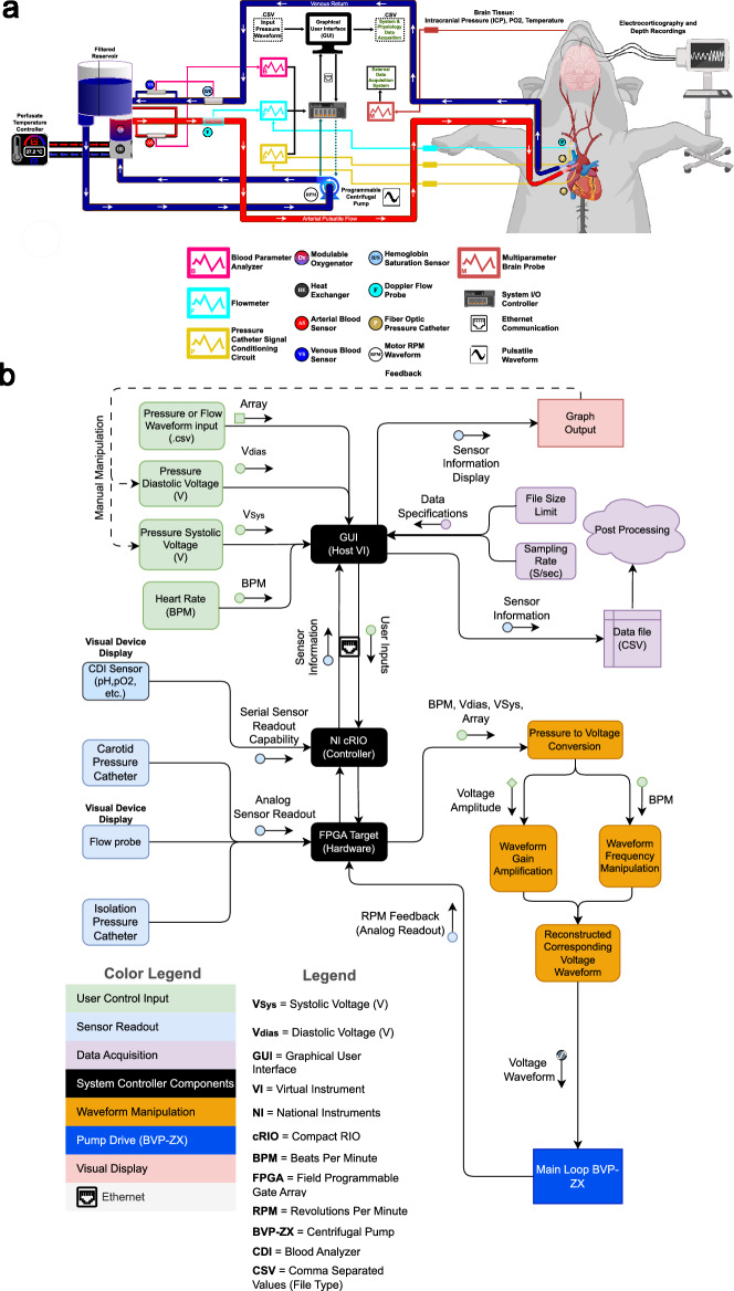
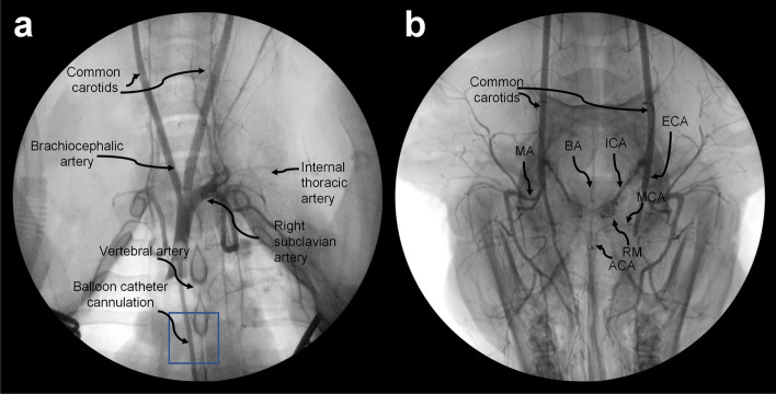
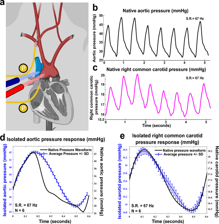
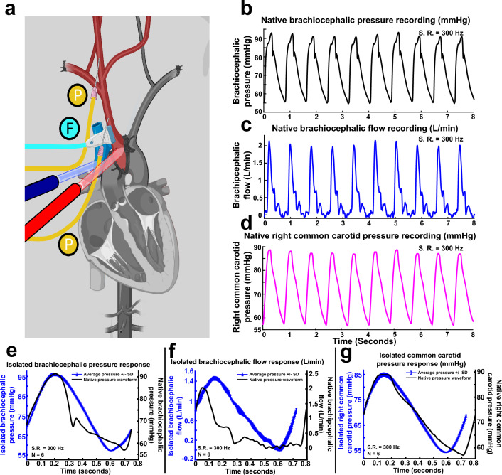
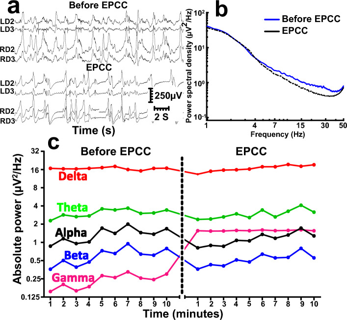
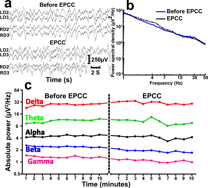
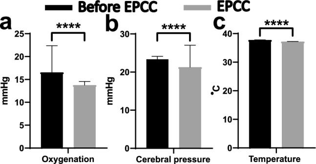
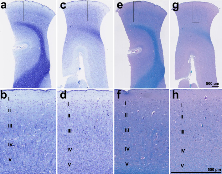
Selective vascular access to the brain is desirable in metabolic tracer, pharmacological and other studies aimed to characterize neural properties in isolation from somatic influences from chest, abdomen or limbs. However, current methods for artificial control of cerebral circulation can abolish pulsatility-dependent vascular signaling or neural network phenomena such as the electrocorticogram even while preserving individual neuronal activity. Thus, we set out to mechanically render cerebral hemodynamics fully regulable to replicate or modify native pig brain perfusion. To this end, blood flow to the head was surgically separated from the systemic circulation and full extracorporeal pulsatile circulatory control (EPCC) was delivered via a modified aorta or brachiocephalic artery. This control relied on a computerized algorithm that maintained, for several hours, blood pressure, flow and pulsatility at near-native values individually measured before EPCC. Continuous electrocorticography and brain depth electrode recordings were used to evaluate brain activity relative to the standard offered by awake human electrocorticography. Under EPCC, this activity remained unaltered or minimally perturbed compared to the native circulation state, as did cerebral oxygenation, pressure, temperature and microscopic structure. Thus, our approach enables the study of neural activity and its circulatory manipulation in independence of most of the rest of the organism.


Conflict of interest statement

The authors declare no competing interests.

Figures

Figure 1
Figure 1
Schematic overview of EPCC and control mechanism. (a) Mechanical constitutents of EPCC in relation to blood flow and exchange. Arterial or oxygen-rich and venous or oxygen-depleted blood are represented in red and blue, respectively. (b) Representation of the computerized control elements. Relationships between numeric data sources, manipulations and outputs.
Figure 2
Figure 2
Arterial angiograms of chest and head. Thoracic angiogram obtained via brachiocephalic cannulation (a) and extension of the radiographic field to the head (b) to illustrate the arteries derived from the carotids. The position of the balloon catheter is indicated in (a) with a box. The usual craniocaudal orientation is reversed in (b), with the snout pointing toward the inferior part of the image. MA: mandibular artery; BA: basilar artery; ICA: internal carotid artery; ECA: external carotid; MCA: middle cerebral artery; ACA: anterior cerebral artery; RM: rete mirabile.
Figure 3
Figure 3
EPCC pressure relative to native perfusion pressure following aortic isolation (subject 1). (a) Vascular structure and aortic isolation. Red: arteries; blue: veins. P (yellow) indicates pressure measurement locations. (b) Native aortic pressure. (c) Native right common carotid pressure. (d)–(e) Comparative waveforms of 6 averaged recordings under EPCC (designated here as isolated condition) with native waveforms used as input for the aortic pressure and for the right common carotid pressure, respectively. All recordings were acquired at 67 samples per s. S.R.: sampling rate. In (d)–(e) the left vertical axis is colored in blue, as is the arterial pressure measured under EPCC.
Figure 4
Figure 4
EPCC pressure relative to native perfusion pressure following brachiocephalic aortic isolation (subject 2). (a) Vascular structure and brachiocephalic isolation. Red: arteries; blue: veins. P (yellow) indicates pressure measurement locations. (b) Native brachiocephalic pressure. (c) Native Doppler-measured brachiocephalic flow. (d) Native right common carotid pressure recordings. (e)–(g) Comparative waveform analysis of 6 average recordings under EPCC (designated here as isolated condition) and sampled native waveforms used as input for the brachiocephalic pressure, brachiocephalic flow and right common carotid pressure, respectively. All recordings were sampled at 300 samples per s. S.R.: sampling rate. In (e)–(g) the left vertical axis is colored in blue, as is the arterial pressure measured under EPCC.
Figure 5
Figure 5
Comparison of depth neurophysiological activity before and after aortic EPCC. Depth recordings from subject 1 before and after EPCC. (a) Depth activity from left (L) and right (R) depth (D) electrodes corresponding to subcortical brain regions spanning from the subcortical white matter (LD2 and RD2) to the dorsal striatum (LD3 and RD3) before and following 4 h of EPCC. (b) Absolute power spectral density of native (blue spectra), post-EPCC (black dotted spectra) LD2 depth recordings in subject 1. (c) Absolute power spectra of delta (red), theta (green), alpha (black), beta (blue), and gamma (magenta) activity defined as in standard electroencephalography and measured in LD2 using 10-min epochs under native perfusion and under EPCC. Electrical noise (50 Hz) interfered with the accurate measurement of gamma frequency after EPCC.
Figure 6
Figure 6
Comparison of depth neurophysiological activity before and after brachiocephalic EPCC. Depth recordings from subject 2 before and following 4 h of EPCC. (a) Depth activity from left (L) and right (R) depth (D) electrodes corresponding to subcortical brain regions spanning from the subcortical white matter (LD2 and RD2) to the dorsal striatum (LD3 and RD3) before and following EPCC. (b) Absolute power spectral density of native (blue spectra), post-EPCC (black dotted spectra) LD2 depth recordings in subject 1. (c) Absolute power spectra of delta (red), theta (green), alpha (black), beta (blue), and gamma (magenta) activity defined as in standard electroencephalography and measured in LD2 using 10-min epochs under native perfusion and under EPCC.
Figure 7
Figure 7
Physical characteristics of cerebral tissue after EPCC. Brain probe measurements of frontal lobe cerebral oxygenation (a) (mmHg of oxygen), barometric pressure (b) (mmHg) and temperature (c) (°C) in subject 2. Black bars represent average and SD of measurements obtained in the native, pre-EPCC state for 10 min with a sampling rate of 1 Hz. Gray bars indicate averaged values of 10 min epochs under EPCC, measured over 5 h. **** represents a significance of p < 0.0001, unpaired t-test.
Figure 8
Figure 8
Microscopic structure of the cerebral cortex after EPCC. A and B: Nissl/luxol fast blue-stained cortical structure of subject 1 after EPCC. C and D: Nissl/luxol fast blue-stained cortical structure of subject 2 after EPCC. E and F: luxol fast blue/periodic acid Schiff-stained cortex of subject 1. G and H. luxol fast blue/periodic acid Schiff-stained cortex of subject 2. Section thickness: 5 µm, photographs obtained with 2.5× and 10× objectives.

Similar articles

Cited by

References

    1. Antognini JF, Kien ND. A method for preferential delivery of volatile anesthetics to the in situ goat brain. Anesthesiology. 1994;80:1148–1154. doi: 10.1097/00000542-199405000-00024. - DOI - PubMed
    1. Marin-Valencia I, Good LB, Ma Q, Malloy CR, Pascual JM. Heptanoate as a neural fuel: Energetic and neurotransmitter precursors in normal and glucose transporter I-deficient (G1D) brain. J. Cereb. Blood Flow Metab. 2013;33:175–182. doi: 10.1038/jcbfm.2012.151. - DOI - PMC - PubMed
    1. White RJ, Albin MS, Verdura J. Isolation of the monkey brain: In vitro preparation and maintenance. Science. 1963;141:1060–1061. doi: 10.1126/science.141.3585.1060. - DOI - PubMed
    1. White RJ, Albin MS, Verdura J. Preservation of viability in the isolated monkey brain utilizing a mechanical extracorporeal circulation. Nature. 1964;202:1082–1083. doi: 10.1038/2021082a0. - DOI - PubMed
    1. Gilboe DD, Cotanch WW, Glover MB. Extracorporeal perfusion of the isolated head of a dog. Nature. 1964;202:399–400. doi: 10.1038/202399a0. - DOI - PubMed

Publication types