

Screening and identification of genes related to ferroptosis in keratoconus
- PMID: 37626095
- PMCID: PMC10457308
- DOI: 10.1038/s41598-023-41194-2
Screening and identification of genes related to ferroptosis in keratoconus
Abstract
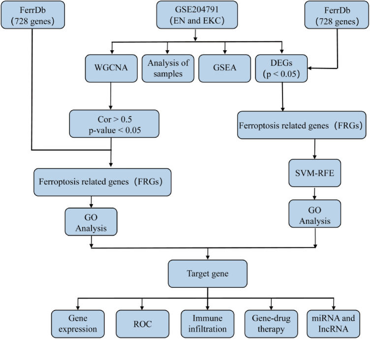
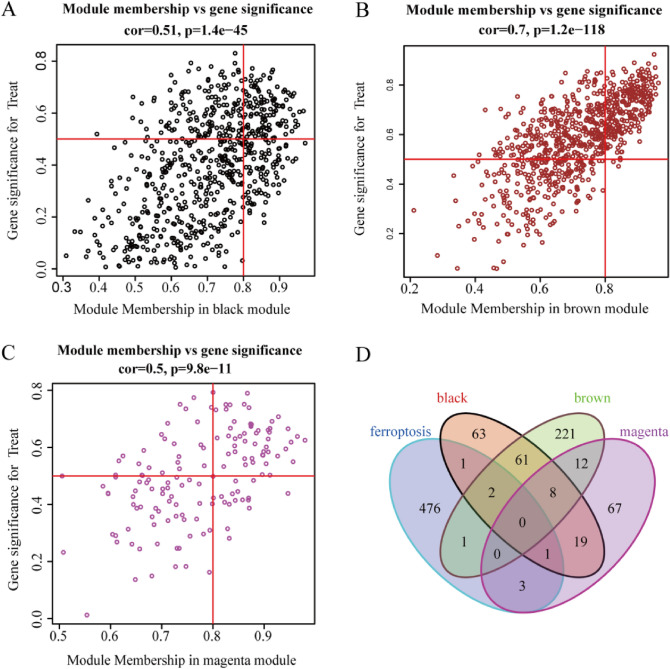
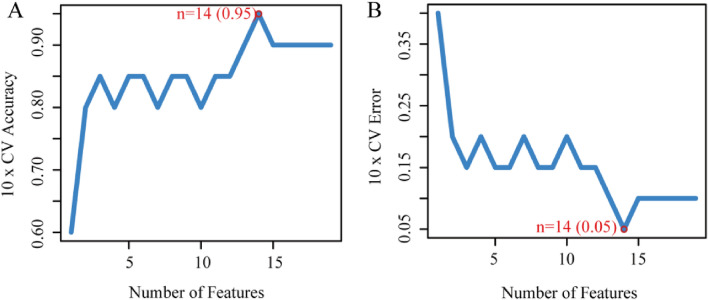
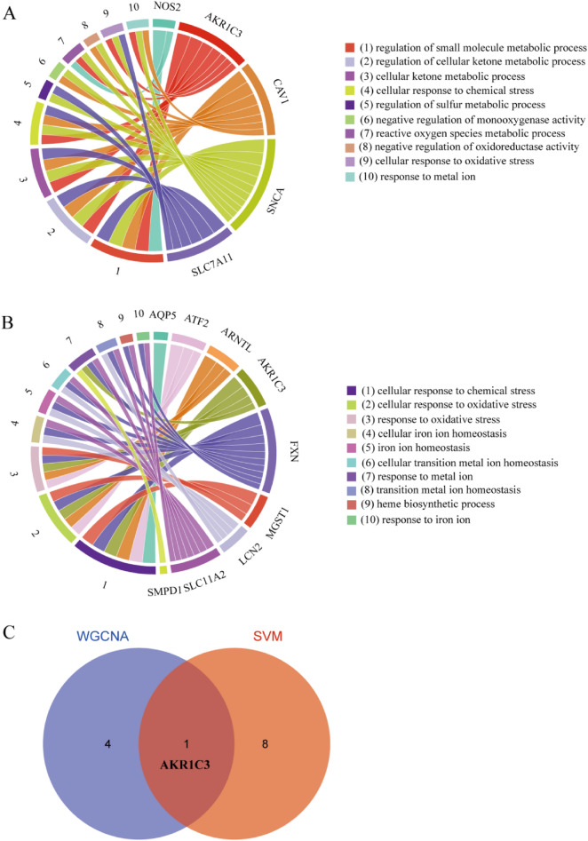
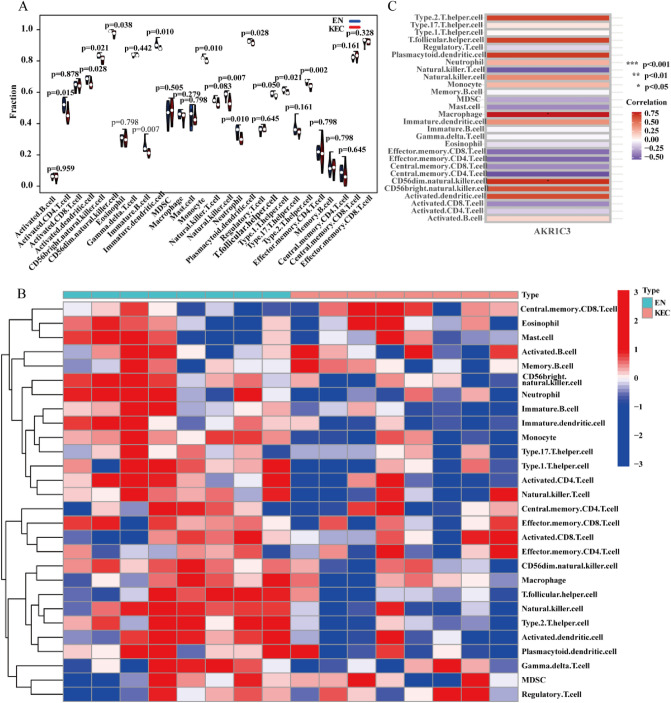
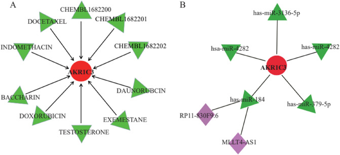
Corneal keratoconus (KC) is a dilated (ectatic) corneal disease characterized by a central thinning of the cornea, which causes protrusion into a conical shape that seriously affects vision. However, due to the complex etiology of keratoconus, its entire mechanism remains unclear and there is no mechanism-directed treatment method. Ferroptosis is a novel programmed cell death mechanism related to lipid peroxidation, stress, and amino acid metabolism, which plays a crucial role in various diseases. This study aimed to explore the relationship between keratoconus and ferroptosis, to provide new insights into the mechanism of keratoconus development, and potential treatment options based on further elucidation of this mechanism. The corresponding mRNA microarray expression matrix data of KC patients were obtained from GEO database (GSE204791). Weighted co-expression network analysis (WGCNA) and support vector machine recursive feature elimination (SVM-RFE) were selected to screen hub genes, which were overlapped with ferroptosis genes (FRGs) from FerrDb. GO and GSEA were performed to analyze differential pathways, ssGSEA was used to determine immune status, and then, feasible drugs were predicted by gene-drug network. Additionally, we predicted the miRNA and IncRNA of hub genes to identify the underlying mechanism of disease so as to predict treatment for the disease. The epithelial transcriptome from keratoconus tissue mRNA microarray data (GSE204791) was extracted for the main analysis, including eight epithelial cells and eight epithelial control cells. The differential genes that were overlapped by WGCAN, SVM-RFE and FRGs were mainly related to oxidative stress, immune regulation, cellular inflammation, and metal ion transport. Through further analysis, aldo-keto reductase family 1 member C3 (AKR1C3) was selected, and negatively correlated with mature CD56 natural killer (NK) cells and macrophages. Then, gene-drug interaction network analysis and miRNA prediction were performed through the website. It was concluded that four immune-related drugs (INDOMETHACIN, DAUNORUBICIN, DOXORUBICIN, DOCETAXEL) and a miRNA (has-miR-184) were screened to predict potential drugs and targets for disease treatment. To our knowledge, this was the first report of KC being associated with ferroptosis and prompted search for differential genes to predict drug targets of gene immunotherapy. Our findings provided insight and a solid basis for the analysis and treatment of KC.
© 2023. Springer Nature Limited.
Conflict of interest statement
The authors declare no competing interests.
Figures


Similar articles
-
Identification and Verification of Ferroptosis-Related Genes in Keratoconus Using Bioinformatics Analysis.J Inflamm Res. 2024 Apr 20;17:2383-2397. doi: 10.2147/JIR.S455337. eCollection 2024. J Inflamm Res. 2024. PMID: 38660574 Free PMC article.
-
Identification of ferroptosis-related genes in male mice with sepsis-induced acute lung injury based on transcriptome sequencing.BMC Pulm Med. 2023 Apr 20;23(1):133. doi: 10.1186/s12890-023-02361-3. BMC Pulm Med. 2023. PMID: 37081490 Free PMC article.
-
A novel combined oxidative stress and extracellular matrix related predictive gene signature for keratoconus.Biochem Biophys Res Commun. 2025 Jan;742:151144. doi: 10.1016/j.bbrc.2024.151144. Epub 2024 Dec 5. Biochem Biophys Res Commun. 2025. PMID: 39657357
-
Pathogenesis of keratoconus: NRF2-antioxidant, extracellular matrix and cellular dysfunctions.Exp Eye Res. 2022 Jun;219:109062. doi: 10.1016/j.exer.2022.109062. Epub 2022 Apr 3. Exp Eye Res. 2022. PMID: 35385756 Free PMC article. Review.
-
Animal Models for the Study of Keratoconus.Cells. 2023 Nov 22;12(23):2681. doi: 10.3390/cells12232681. Cells. 2023. PMID: 38067109 Free PMC article. Review.
Cited by
-
Mechanical Strain of Corneal Epithelium Influences the Expression of Genes Implicated in Keratoconus.Invest Ophthalmol Vis Sci. 2025 Jan 2;66(1):52. doi: 10.1167/iovs.66.1.52. Invest Ophthalmol Vis Sci. 2025. PMID: 39847367 Free PMC article.
References
Publication types
MeSH terms
Substances
LinkOut - more resources
Full Text Sources
Research Materials
Miscellaneous
