. 2023 Aug 25;21(1):223.
doi: 10.1186/s12964-023-01209-x.

VSIG2 promotes malignant progression of pancreatic ductal adenocarcinoma by enhancing LAMTOR2-mediated mTOR activation

Affiliations

VSIG2 promotes malignant progression of pancreatic ductal adenocarcinoma by enhancing LAMTOR2-mediated mTOR activation

Jichuan Xu et al. Cell Commun Signal. .

Abstract

Background: Pancreatic ductal adenocarcinoma (PDAC) is one of the most intractable malignancies to overcome clinically due to its insidious onset as well as rapid progression. It is urgent to seek new diagnostic markers and therapeutic targets in order to furthest ameliorate the prognosis of patients with PDAC. V-set and immunoglobulin domain containing 2 (VSIG2) belongs to immunoglobulin superfamily (IgSF), which function as coinhibitory molecule to mediate immune evasion of tumors. Nevertheless, the role of VSIG2 in PDAC and related mechanism still keep unclear.

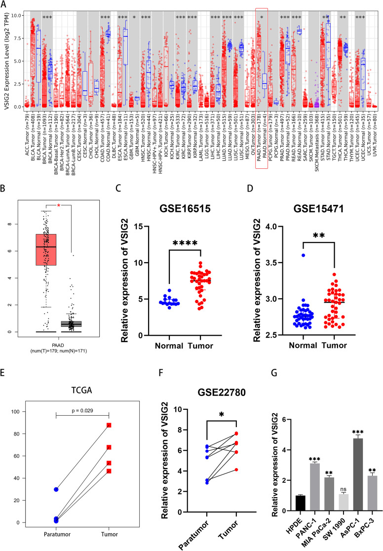
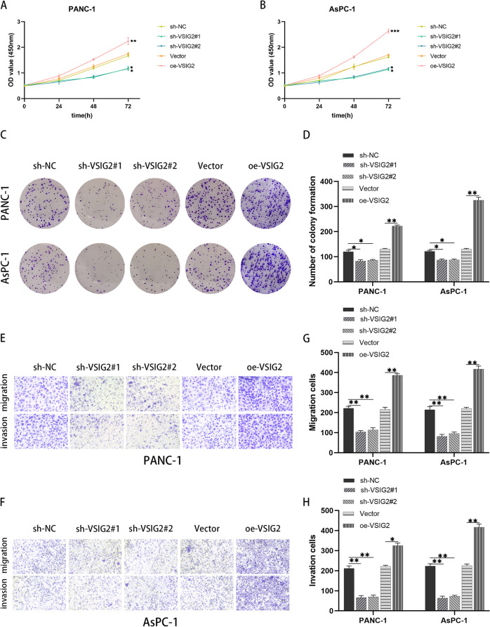
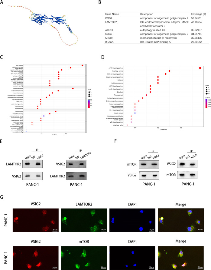
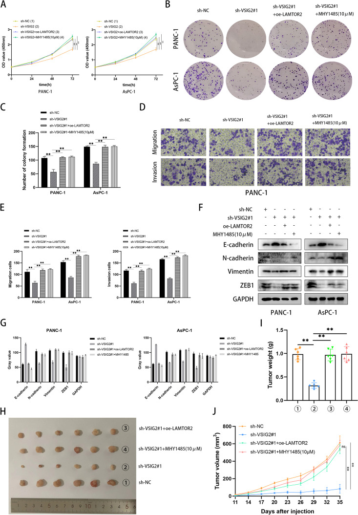
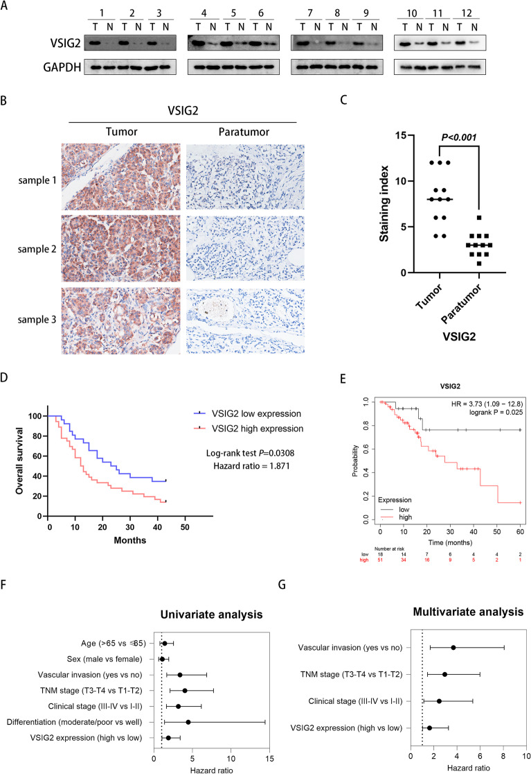
Methods: Different expression of VSIG2 in PDAC tissues and cells were detected by bioinformatic analysis, immunohistochemistry, real-time quantitative PCR as well as western blotting. CCK-8, colony formation, Transwell assay, and scratch experiment were utilized to assess proliferation, invasion and migration properties of PDAC cells. The relationship of VSIG2 with late endosomal/lysosomal adaptor, MAPK and MTOR activator 2 (LAMTOR2) and mechanistic target of rapamycin (mTOR) was identified using mass spectrometry, co-immunoprecipitation and immunofluorescence. GO and KEGG enrichment analysis were performed for further pathway verification using western blotting. Additionally, subcutaneous xenograft tumor model and clinical samples analysis were implemented to further elucidate the oncogenic effect of VSIG2 on PDAC in vivo and clinically.

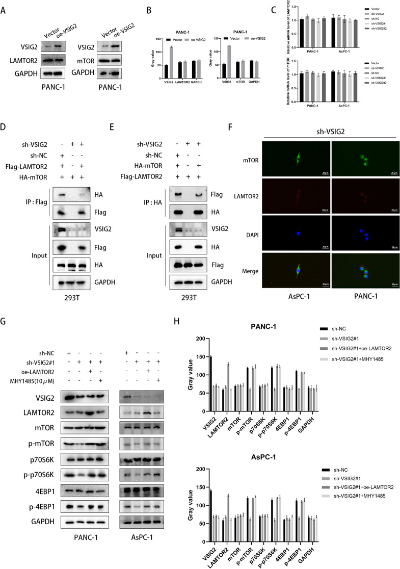
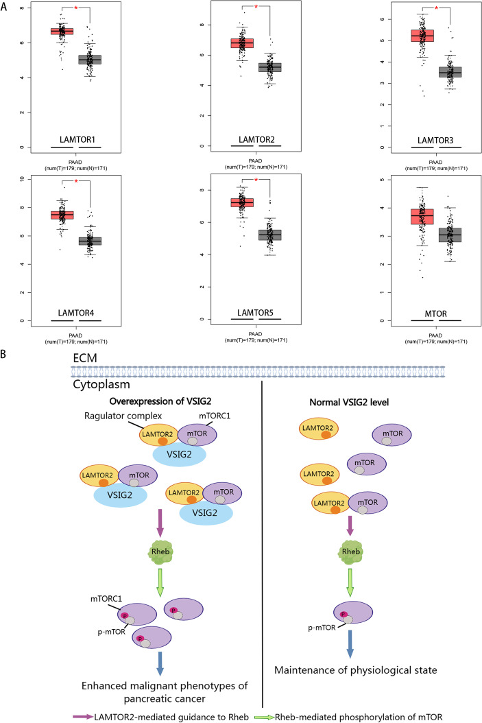
Results: VSIG2 was highly expressed in PDAC tissues and cells. Overexpression of VSIG2 facilitated the proliferation, invasion and migration abilities of PDAC cells, while VSIG2-inhibition exerted opposite effects. Mechanistically, VSIG2 could simultaneously bind to LAMTOR2 and mTOR, thereby enhancing interaction between two molecules, which resulted in elevated phosphorylation-modificatory activation of mTOR and downstream key molecules. Clinically, up-regulation of VSIG2 was positively associated with advanced stage, overall survival and disease-free survival of PDAC patients.

Conclusions: Our study disclosed that VSIG2 was overexpressed in PDAC, which promoted the proliferation, invasion and metastasis. Mechanically, VSIG2 acted as a scaffold to recruit LAMTOR2 and mTOR simultaneously, stabilize the interaction between them, thus enhancing LAMTOR2-mediated mTOR phosphorylated activation. Collectively, VSIG2 could be exploited as a biomarker for diagnosis and prognosis monitor of PDAC in the future, meanwhile, targeting VSIG2 in PDAC management is expected to be a novel strategy. Video Abstract. Video Abstract.

Keywords: Late endosomal/lysosomal adaptor; MAPK and MTOR activator 2 (LAMTOR2); Malignant progression; Mechanistic target of rapamycin (mTOR); Pancreatic ductal adenocarcinoma; V-set and immunoglobulin domain containing 2 (VSIG2).


Conflict of interest statement

The authors declare that they have no competing interests.

Figures

Fig. 1
Fig. 1
VSIG2 was overexpressed in PDAC tissues and cells. A Pan-cancer analysis of VSIG2 in multiple malignancies through TIMER2.0 (http://timer.comp-genomics.org/) database. B, C, D Differential expression analysis of VSIG2 in PDAC and normal pancreatic tissues through GEPIA (http://gepia.cancer-pku.cn/) and GEO (https://www.ncbi.nlm.nih.gov/) databases. E, F Paired sample analysis of VSIG2 expression levels in PDAC and para-cancer tissues through TCGA (https://portal.gdc.cancer.gov/) and GEO (https://www.ncbi.nlm.nih.gov/) databases. G Detection of VSIG2 relative expression in human pancreatic ductal epithelial and five PDAC cell lines by RT-qPCR
Fig. 2
Fig. 2
Effects of VSIG2 on PDAC cell proliferation, invasion and migration. A, B The proliferation ability of PANC-1 and AsPC-1 cells was detected by CCK-8 assay, and the comparison between experimental and control groups was adopted by OD450 value at 72h, *P<0.05, **P<0.01, ***P<0.001. C, D The proliferation ability of VSIG2 knockdown and overexpression groups in PANC-1 and AsPC-1 cells was detected by colony formation assay, *P<0.05, **P<0.01. E, F, G, H The invasion and migration properties of experimental and control groups in PANC-1 and AsPC-1 cells were clarified by Transwell assay, *P<0.05, **P<0.01
Fig. 3
Fig. 3
The role of VSIG2 in cell migration and epithelial-mesenchymal transition (EMT) of PDAC. A, B, C, D The migration ability of PANC-1 and AsPC-1 cells with VSIG2 down-regulation and overexpression was identified by evaluating relative migration rate via scratch assay, *P<0.05, **P<0.01. E, F, G, H The levels of EMT after knocking down and up-regulating VSIG2 expression in PANC-1 and AsPC-1 cells were verified by immunoblotting through assessing proteins expression of EMT related indicators including E-cadherin, N-cadherin, Vimentin, and ZEB1, when GAPDH was used as a loading control
Fig. 4
Fig. 4
VSIG2 interacted with LAMTOR2 and mTOR. A AlphaFold Protein Structure Database (https://alphafold.ebi.ac.uk/) predicted the three-dimensional structure of VSIG2. B The results of VSIG2 mass spectrometry showed the possible interacting proteins, including LAMTOR2 and mTOR. C Proteins in VSIG2 mass spectrometry were analyzed from biological program, cellular condition and molecular function aspects by Gene Ontology (GO). D Kyoto Encyclopedia of Genes and Genomes (KEGG) predicted the related signaling pathway according to the results of VSIG2 mass spectrometry. E The interaction between VSIG2 and LAMTOR2 was testified by endogenic co-immunoprecipitation (Co-IP) in PANC-1 cells. F The interaction between VSIG2 and mTOR was verified by endogenic Co-IP assay in PANC-1 cells. G The interaction of VSIG2 with LAMTOR2 and mTOR and its spatial localization in PANC-1 cells were observed by immunofluorescence
Fig. 5
Fig. 5
VSIG2 promoted the interaction between LAMTOR2 and mTOR, thereby increasing the activation of mTOR signaling pathway via LAMTOR2. A, B Overexpression of VSIG2 detected by immunoblotting in PANC-1 cells had no effect on protein expression of LAMTOR2 and mTOR. GAPDH was used as a loading control. C Knockdown and up-regulation of VSIG2 exerted no influence on mRNA expression of LAMTOR2 and mTOR, which were clarified by RT-qPCR in PANC-1 and AsPC-1 cells. D, E, F The level of interaction between LAMTOR2 and mTOR was testified through knockdown of VSIG2 and transfection of Flag-tagged as well as HA-tagged plasmids in 293T cells via further Co-IP assay and immunofluorescence. GAPDH was used as a loading control. G, H After VSIG2 knockdown, LAMTOR2 overexpression and mTOR agonist MHY1485 were added in PANC-1 and AsPC-1 cells, the expression and activation of mTOR and downstream key molecules were detected by western blots. GAPDH was used as a loading control
Fig. 6
Fig. 6
VSIG2 advanced PDAC progression through LAMTOR2-mediated mTOR activation. A CCK-8 assays were performed to detect the proliferation of PDAC cells regarding to down-regulation of VSIG2 and simultaneous elevation of LAMTOR2 or supplement of MHY1485, **P <0.01. B, C The properties of proliferation about PDAC cells according to different groups were demonstrated using colony formation assays, **P <0.01. D, E Transwell assays were used to certify the ability of migration and invasion about PANC-1 cells regarding to down-regulation of VSIG2 and simultaneous elevation of LAMTOR2 or supplement of MHY1485. F, G The related indicators of EMT were clarified by western blots after VSIG2 knockdown, LAMTOR2 overexpression and addition of mTOR activator MHY1485 in PANC-1 and AsPC-1 cells. GAPDH was utilized as a loading control. The bar charts showed statistical differences connected with migration and invasion PDAC cells, **P<0.01. H, I, J Subcutaneous xenograft tumor model was constructed to ulteriorly prove the conclusion, and tumor diameter was recorded once every three days, while tumor weight was investigated after dissection, **P<0.01
Fig. 7
Fig. 7
Overexpression of VSIG2 was associated with poor prognosis of PDAC patients. A Western blotting was performed on 12 pairs of pancreatic and para-cancerous tissues collected from PDAC patients, when GAPDH was served as a loading control. B 3 pairs of tumor and para-tumor tissues collected from PDAC patients were compared by immunohistochemistry (IHC) assay. C Scatter diagram of staining index about 12 pairs PDAC and adjacent tissue samples using 12-tier IHC scoring. D Overall survival curve was drawn through observing and recording the condition of PDAC patients for 43 months, while all PDAC patients were divided into two groups by VSIG2 expression level. E Disease-free survival probability of PDAC patients with different expression level of VSIG2 was acquired by Kaplan-Meier (https://kmplot.com/analysis/) database. F Forest plot of univariate Cox analysis regarding to multiple parameters. G Forest plot of multivariate Cox analysis regarding to multiple parameters
Fig. 8
Fig. 8
VSIG2 phosphorylated mTORC1, which contained mTOR, through the oncogenic effects of the Ragulator complex containing LAMTOR2. A GEPIA (http://gepia.cancer-pku.cn/) database predicted the component sections of Ragulator complex including LAMTOR1, LAMTOR2, LAMTOR3, LAMTOR4, LAMTOR5 were all highly expressed in PDAC compared to normal tissues, while the expression of MTOR had no difference between PDAC and normal pancreas tissues. B Compared with the expression level of VSIG2 under normal physiological conditions, when the expression of VSIG2 in PDAC cells was upregulated, VSIG2 can act as a scaffold to simultaneously play a recruitment effect on Ragulator and mTORC1, thus enhancing the interaction between mTORC1 and Ragulator complex including LAMTOR2. Subsequently, a large amount of mTORC1 was directed by Ragulator to the small GTPase Rheb, where phosphorylated activation of mTOR was increased and further initiated the malignant progression of PDAC

References

    1. Chrystoja CC, Diamandis EP, Brand R, et al. Pancreatic cancer. Clin Chem. 2013;59(1):41–46. doi: 10.1373/clinchem.2012.196642. - DOI - PubMed
    1. Xu J, Xu J, Liu X, et al. The role of lncRNA-mediated ceRNA regulatory networks in pancreatic cancer. Cell Death Discov. 2022;8(1):287. doi: 10.1038/s41420-022-01061-x. - DOI - PMC - PubMed
    1. Vincent A, Herman J, Schulick R, et al. Pancreatic cancer. Lancet. 2011;378(9791):607–620. doi: 10.1016/s0140-6736(10)62307-0. - DOI - PMC - PubMed
    1. Kelsen DP, Portenoy R, Thaler H, et al. Pain as a predictor of outcome in patients with operable pancreatic carcinoma. Surgery. 1997;122(1):53–59. doi: 10.1016/s0039-6060(97)90264-6. - DOI - PubMed
    1. Saluja A, Maitra A. Pancreatitis and pancreatic cancer. Gastroenterology. 2019;156(7):1937–1940. doi: 10.1053/j.gastro.2019.03.050. - DOI - PubMed

Publication types

MeSH terms