. 2023 Jul 25;11(8):2098.
doi: 10.3390/biomedicines11082098.

Induction of Periodontitis Using Bacterial Strains Isolated from the Human Oral Microbiome in an Experimental Rat Model

Affiliations

Induction of Periodontitis Using Bacterial Strains Isolated from the Human Oral Microbiome in an Experimental Rat Model

Diana Larisa Ancuta et al. Biomedicines. .

Abstract

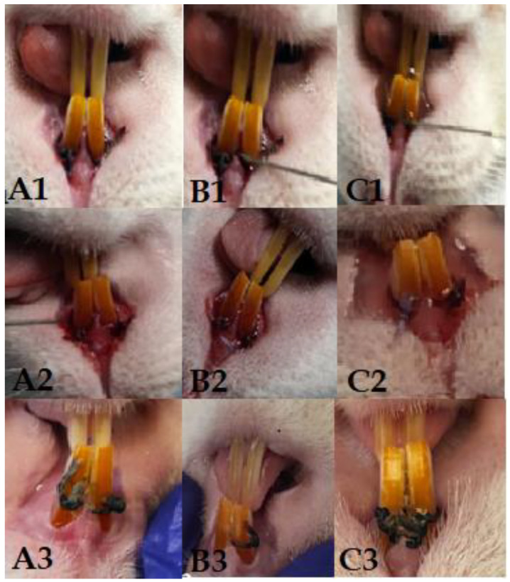
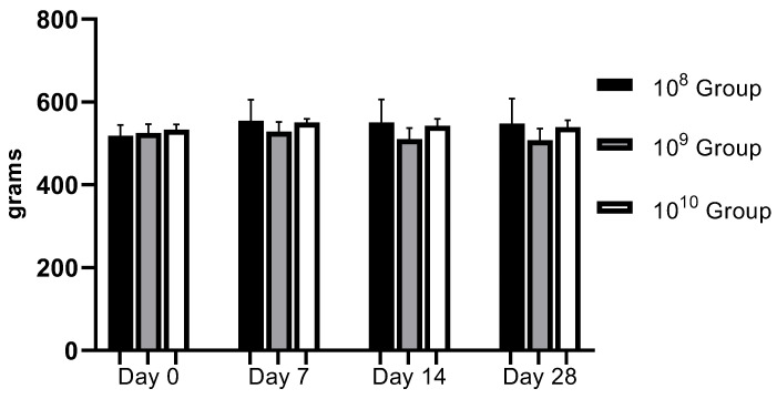
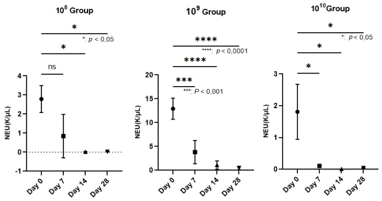
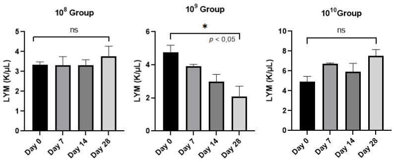
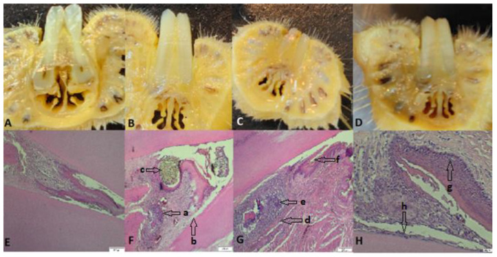
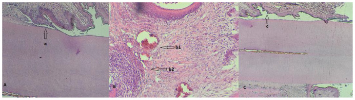
Periodontal disease is that condition resulting in the destruction of periodontal tissues, bone resorption, and tooth loss, the etiology of which is linked to immunological and microbiological factors. The aim of this study was to evaluate the potential trigger of periodontal disease in a rat model using bacterial species incriminated in the pathology of human periodontitis and to establish their optimal concentrations capable of reproducing the disease, with the idea of subsequently developing innovative treatments for the condition. In this study, we included 15 male Wistar rats, aged 20 weeks, which we divided into three groups. In each group, we applied ligatures with gingival retraction wire on the maxillary incisors. The ligature and the gingival sac were contaminated by oral gavage with a mixture of fresh cultures of Aggregatibacter actinomycetemcomitans (A.a), Fusobacterium nucleatum (F.n) and Streptococcus oralis (S.o) in concentrations of 108, 109, and 1010 CFU/mL each for 5 days a week for 4 weeks. During the clinical monitoring period of 28 days, overlapped with the period of oral contamination, we followed the expression of clinical signs specific to periodontitis. We also monitored the evolution of body weight and took weekly samples from the oral cavity for the microbiological identification of the tested bacteria and blood samples for hematological examination. At the end of the study, the animals were euthanized, and the ligated incisors were taken for histopathological analysis. The characteristic symptomatology of periodontal disease was expressed from the first week of the study and was maintained until the end, and we were able to identify the bacteria during each examination. Hematologically, the number of neutrophils decreased dramatically (p < 0.0001) in the case of the 109 group, unlike the other groups, as did the number of lymphocytes. Histopathologically, we identified neutrophilic infiltrate in all groups, as well as the presence of coccobacilli, periodontal tissue hyperplasia, and periodontal lysis. In the 109 group, we also observed pulpal tissue with necrotic bone fragments and pyogranulomatous inflammatory reaction. By corroborating the data, we can conclude that for the development of periodontal disease using A.a, F.n, and S.o, a concentration of 109 or 1010 CFU/mL is required, which must necessarily contaminate a ligature thread applied to the level of the rat's dental pack.

Keywords: Aggregatibacter actinomycetemcomitans; Fusobacterium nucleatum; Streptococcus oralis; ligature; periodontitis; rat.


Conflict of interest statement

The authors declare no conflict of interest.

Figures

Figure 1
Figure 1
Columns (A1A3) represent the 108 group, with clinical aspect at the time of application of the first ligature (A1), after 14 days of contamination (A2), and on day 28 (A3). Columns (B1B3) represent the 109 group, with clinical aspect at the time of application of the first ligature (B1), after 14 days of contamination (B2), and on day 28 (B3). Columns (C1C3) represent the 1010 group, with clinical aspect at the time of application of the first ligature (C1), after 14 days of contamination (C2), and on day 28 (C3).
Figure 2
Figure 2
Evolution of body weight during the study (regardless of the bacterial concentration used, no statistically significant changes were recorded).
Figure 3
Figure 3
Reduction in the number of neutrophils (NEU) on the wall of the PD installation depending on the bacterial concentration used (In group 108, neutrophils decreased starting the 3rd week after contamination; p < 0.05). The most relevant decrease in neutrophils was observed in group 109, where, starting the second week, their number began to decrease more dramatically than in the case of group 108, registering increasingly lower values until the end of the study, when p < 0.0001. In group 1010, the number of neutrophils decreased constantly during the study, with a p value < 0.05.
Figure 4
Figure 4
Evolution of the number of lymphocytes (LYM)/group analyzed from day 0 to day 28.
Figure 5
Figure 5
(AD) Images with cross sections through the collected part; (E) the part collected from a control rat, uncontaminated and without ligature, showing intact alveolar bone and continuous cementum; (F) the part from a rat from group 108, which presents hypertrophy, granulation tissue (a) with nucleated cells that peel off, abundant neutrophils, coccobacilli, inflammation, hyperemia, hyperplasia of the gingival epithelium, and periodontal lysis (b), with a ligature thread present (c); (G) rat teeth from group 109 (granuloma) (d), alveolar bone lysis, pulp tissue with necrotic bone fragments surrounded by pyogranulomatous inflammation, neutrophilic infiltrate (e), abundant granulation tissue, bacteria (coccobacilli), and fodder in the space periodontally invaded with bacteria (f); (H) teeth of rats from group 1010 with hyperplasia (g), an intact alveolodental ligament, abundant granulation tissue, fewer inflammatory cells, bacteria in the alveolar sac, and anucleated desquamation cells on the alveolodental ligament area (h). Sections were stained with H&E. Original magnification: 4×; scale bars = 200 μm.
Figure 6
Figure 6
(A) Detail of a rat tooth from group 108 with a partially desquamated alveolodental ligament (a); (B) strong reaction of hyperemia (b1) and neutrophilic infiltrates (b2) in a rat from group 109; (C) overview highlighting the periodontal sac (c) of a rat from group 1010.

Similar articles

Cited by

References

    1. Bhattarai G., Poudel S.B., Kook S.-H., Lee J.-C. Resveratrol prevents alveolar bone loss in an experimental rat model of periodontitis. Acta Biomater. 2016;29:398–408. doi: 10.1016/j.actbio.2015.10.031. - DOI - PubMed
    1. Okuda K., Kato T., Ishihara K. Involvement of periodontopathic biofilm in vascular diseases. Oral Dis. 2004;10:5–12. doi: 10.1046/j.1354-523X.2003.00979.x. - DOI - PubMed
    1. Graves D.T., Jiang Y., Genco C. Periodontal disease: Bacterial virulence factors, host response and impact on systemic health. Curr. Opin. Infect. Dis. 2000;13:227–232. doi: 10.1097/00001432-200006000-00005. - DOI - PubMed
    1. Kobayashi N., Ishihara K., Sugihara N., Kusumoto M., Yakushiji M., Okuda K. Colonization pattern of periodontal bacteria in Japanese children and their mothers. J. Periodontal. Res. 2008;43:156–161. doi: 10.1111/j.1600-0765.2007.01005.x. - DOI - PubMed
    1. Eke P.I., Dye B.A., Wei L., Slade G.D., Thornton-Evans G.O., Borgnakke W.S., Taylor G.W., Page R.C., Beck J.D., Genco R.J. Update on Prevalence of Periodontitis in Adults in the United States: NHANES 2009 to 2012. J. Periodontol. 2015;86:611–622. doi: 10.1902/jop.2015.140520. - DOI - PMC - PubMed

LinkOut - more resources