. 2023 Aug 3;11(8):2184.
doi: 10.3390/biomedicines11082184.

Immunomorphogenesis in Degenerative Disc Disease: The Role of Proinflammatory Cytokines and Angiogenesis Factors

Affiliations

Immunomorphogenesis in Degenerative Disc Disease: The Role of Proinflammatory Cytokines and Angiogenesis Factors

Natalya G Pravdyuk et al. Biomedicines. .

Abstract

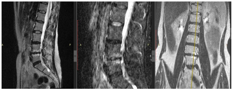
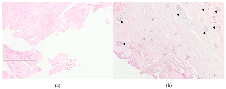
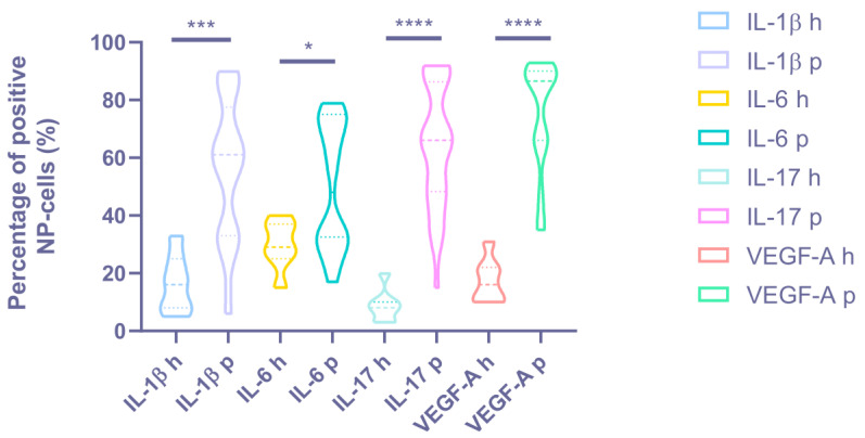
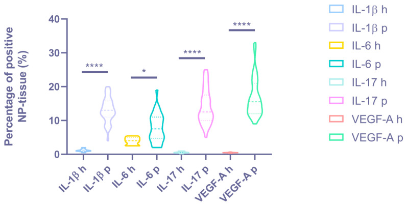
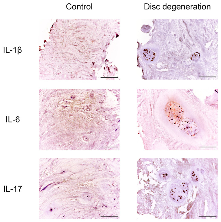
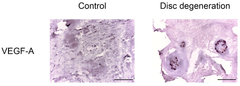
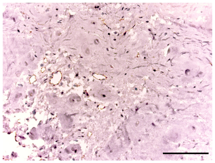
Back pain (BP) due to degenerative disc disease (DDD) is a severe, often disabling condition. The aim of this study was to determine the association between the expression level of proinflammatory cytokines (IL-1β, IL-6, and IL-17), angiogenesis markers (VEGF-A and CD31) in intervertebral disc (IVD) tissue and IVD degeneration in young people with discogenic BP. In patients who underwent discectomy for a disc herniation, a clinical examination, magnetic resonance imaging of the lumbar spine, histological and immunohistochemical analyses of these factors in IVD were performed in comparison with the parameters of healthy group samples (controls). Histology image analysis of IVD fragments of the DDD group detected zones of inflammatory infiltration, combined with vascularization, the presence of granulation tissue and clusters of chondrocytes in the tissue of nucleus pulposus (NP). Statistically significant increased expression of IL-1β, IL-6, IL-17, VEGF-A and CD31 was evident in the samples of the DDD group compared with the controls, that showed a strong correlation with the histological disc degeneration stage. Our results denote an immunoinflammatory potential of chondrocytes and demonstrates their altered morphogenetic properties, also NP cells may trigger the angiogenesis.

Keywords: Angiogenesis markers; CD31; Modic changes; back pain; chondrocyte clusters; degenerative disc disease; immunohistochemistry; proinflammatory cytokines; young age.


Conflict of interest statement

The authors declare no conflict of interest.

Figures

Figure 1
Figure 1
Patient with back pain. Magnetic resonance imaging of the lumbar spine, modes T2-weighted images with fat suppression in sagittal sections (left and center images, respectively), T2-weighted short-tau inversion-recovery image in the coronary section (right image). Right-sided lumbar scoliosis, the 5th stage of degenerative disc disease by Pfirrmann at the L4/5 and 4th stage—at the L5/S1 level, with IVD hernias L4/5 and L5/S1, erosion of the endplates and Modic changes type 1 in the vertebral bodies L4/5 (yellow axis carried out via Modic-1). Modic-1 = the bone marrow edema; Modic-2 = the bone marrow fatty degeneration.
Figure 2
Figure 2
Patient intervertebral disc sample obtained as a result of microdiscectomy in the patient in her 40 s, with a hernia of the L5/S1 vertebrae. Own data, 2020. Hematoxylin–eosin staining. (a). Light microscopy, magnification 100×. Zones of inflammatory infiltration, combined with vascularization and granulation tissue. (b). The sample of the same patient, magnification 100×. Clusters of nucleus pulposus cells (triangles) are a characteristic sign of disc degeneration.
Figure 3
Figure 3
The percentage of positive NP cells in the intervertebral disc tissue of the degenerative disc disease (“p”—patients) and control group (“h”—healthy) discs. * p < 0.05. *** p < 0.001. **** p < 0.0001. NP = nucleus pulposus, IL = interleukin, and VEGF-A = vascular endothelial growth factor A.
Figure 4
Figure 4
The cytokine expression degree in the intervertebral disc matrix of the degenerative disc disease (“p”—patients) and control groups (“h”—healthy). * p < 0.05. **** p < 0.0001. NP = nucleus pulposus, IL = interleukin, and VEGF-A = vascular endothelial growth factor A.
Figure 5
Figure 5
Expression of interleukin (IL)-1β, IL-6, and IL-17 in intervertebral disc tissue. Scale bars: 100 µm.
Figure 6
Figure 6
Vascular endothelial growth factor (VEGF)-A expression in intervertebral disc tissue. Scale bars: 100 µm.
Figure 7
Figure 7
CD31 expression in the intervertebral disc tissue of the patient of the degenerative disc disease group. Scale bar: 100 µm.

References

    1. Ochsmann E.B., Escobar Pinzón C.L., Letzel S., Kraus T., Michaelis M., Muenster E. Prevalence of Diagnosis and Direct Treatment Costs of Back Disorders in 644,773 Children and Youths in Germany. BMC Musculoskelet. Disord. 2010;11:193. doi: 10.1186/1471-2474-11-193. - DOI - PMC - PubMed
    1. Karademir M., Eser O., Karavelioglu E. Adolescent Lumbar Disc Herniation: Impact, Diagnosis, and Treatment. J. Back Musculoskelet. Rehabil. 2017;30:347–352. doi: 10.3233/BMR-160572. - DOI - PubMed
    1. Diamond S., Borenstein D. Chronic Low Back Pain in a Working-Age Adult. Best Pract. Res. Clin. Rheumatol. 2006;20:707–720. doi: 10.1016/j.berh.2006.04.002. - DOI - PubMed
    1. Meucci R.D., Fassa A.G., Paniz V.M., Silva M.C., Wegman D.H. Increase of Chronic Low Back Pain Prevalence in a Medium-Sized City of Southern Brazil. BMC Musculoskelet. Disord. 2013;14:155. doi: 10.1186/1471-2474-14-155. - DOI - PMC - PubMed
    1. Andersson G.B. Epidemiological Features of Chronic Low-Back Pain. Lancet. 1999;354:581–585. doi: 10.1016/S0140-6736(99)01312-4. - DOI - PubMed