

Apocynin, an NADPH Oxidase Enzyme Inhibitor, Prevents Amebic Liver Abscess in Hamster
- PMID: 37626818
- PMCID: PMC10452916
- DOI: 10.3390/biomedicines11082322
Apocynin, an NADPH Oxidase Enzyme Inhibitor, Prevents Amebic Liver Abscess in Hamster
Abstract
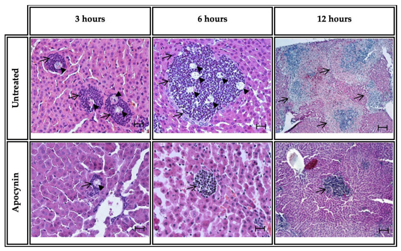
Amebiasis is an intestinal infection caused by Entamoeba histolytica. Amebic liver abscess (ALA) is the most common extraintestinal complication of amebiasis. In animal models of ALA, neutrophils have been shown to be the first cells to come into contact with Entamoeba histolytica during the initial phase of ALA. One of the multiple mechanisms by which neutrophils exhibit amebicidal activity is through reactive oxygen species (ROS) and the enzyme NADPH oxidase (NOX2), which generates and transports electrons to subsequently reduce molecular oxygen into superoxide anion. Previous reports have shown that ROS release in the susceptible animal species (hamster) is mainly stimulated by the pathogen, in turn provoking such an exacerbated inflammatory reaction that it is unable to be controlled and results in the death of the animal model. Apocynin is a natural inhibitor of NADPH oxidase. No information is available on the role of NOX in the evolution of ALA in the hamster, a susceptible model. Our study showed that administration of a selective NADPH oxidase 2 (NOX2) enzyme inhibitor significantly decreases the percentage of ALA, the size of inflammatory foci, the number of neutrophils, and NOX activity indicated by the reduction in superoxide anion (O2-) production. Moreover, in vitro, the apocynin damages amoebae. Our results showed that apocynin administration induces a decrease in the activity of NOX that could favor a decrease in ALA progression.
Keywords: Entamoeba histolytica; NADPH oxidase; NOX2; amebic liver abscess; apocynin; hamster; neutrophil.
Conflict of interest statement
The authors declare no conflict of interest. The funders had no role in the design of the study; in the collection, analyses, or interpretation of data; in the writing of the manuscript; or in the decision to publish the results.
Figures


Similar articles
-
Apocynin prevents GM-CSF-induced-ERK1/2 activation and -neutrophil survival independently of its inhibitory effect on the phagocyte NADPH oxidase NOX2.Biochem Pharmacol. 2020 Jul;177:113950. doi: 10.1016/j.bcp.2020.113950. Epub 2020 Apr 3. Biochem Pharmacol. 2020. PMID: 32251677
-
A review of the proposed role of neutrophils in rodent amebic liver abscess models.Parasite. 2016;23:6. doi: 10.1051/parasite/2016006. Epub 2016 Feb 15. Parasite. 2016. PMID: 26880421 Free PMC article. Review.
-
NOX1 participates in ROS-dependent cell death of colon epithelial Caco2 cells induced by Entamoeba histolytica.Microbes Infect. 2011 Nov;13(12-13):1052-61. doi: 10.1016/j.micinf.2011.06.001. Epub 2011 Jun 30. Microbes Infect. 2011. PMID: 21723410
-
Ethanol-induced erectile dysfunction and increased expression of pro-inflammatory proteins in the rat cavernosal smooth muscle are mediated by NADPH oxidase-derived reactive oxygen species.Eur J Pharmacol. 2017 Jun 5;804:82-93. doi: 10.1016/j.ejphar.2017.03.024. Epub 2017 Mar 15. Eur J Pharmacol. 2017. PMID: 28315342
-
Amebic liver abscess by Entamoeba histolytica.World J Clin Cases. 2022 Dec 26;10(36):13157-13166. doi: 10.12998/wjcc.v10.i36.13157. World J Clin Cases. 2022. PMID: 36683647 Free PMC article. Review.
Cited by
-
Neutrophils versus Protozoan Parasites: Plasmodium, Trichomonas, Leishmania, Trypanosoma, and Entameoba.Microorganisms. 2024 Apr 19;12(4):827. doi: 10.3390/microorganisms12040827. Microorganisms. 2024. PMID: 38674770 Free PMC article. Review.
-
Amebic liver abscess: An update.World J Hepatol. 2024 Mar 27;16(3):316-330. doi: 10.4254/wjh.v16.i3.316. World J Hepatol. 2024. PMID: 38577528 Free PMC article. Review.
References
Grants and funding
- 20221473/research was funded by Sistema de Administración de Programa y Proyectos de investigación (SAPPI-IPN). Multidisciplinar Project for Judith Pacheco-Yépez
- 20221514/research was funded by Sistema de Administración de Programa y Proyectos de investigación (SAPPI-IPN). Multidisciplinary Project for Ivonne Maciel Arciniega-Martínez
- 20221426/research was funded by Sistema de Administración de Programa y Proyectos de investigación (SAPPI-IPN). Multidisciplinary Project for Aldo A. Reséndiz-Albor
- 20230994/Germán Higuera-Martínez received financial support through the scholarship by BEIFI-IPN
- 820039/Germán Higuera-Martínez received scholarship Granted by Consejo Nacional de Ciencia y Tecnología (CONACyT)-Méx
LinkOut - more resources
Full Text Sources
Miscellaneous
