. 2023 Aug 8;12(16):2024.
doi: 10.3390/cells12162024.

Neuronal atg1 Coordinates Autophagy Induction and Physiological Adaptations to Balance mTORC1 Signalling

Affiliations

Neuronal atg1 Coordinates Autophagy Induction and Physiological Adaptations to Balance mTORC1 Signalling

Athanasios Metaxakis et al. Cells. .

Abstract

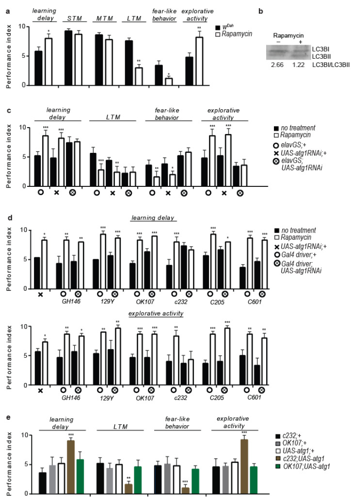
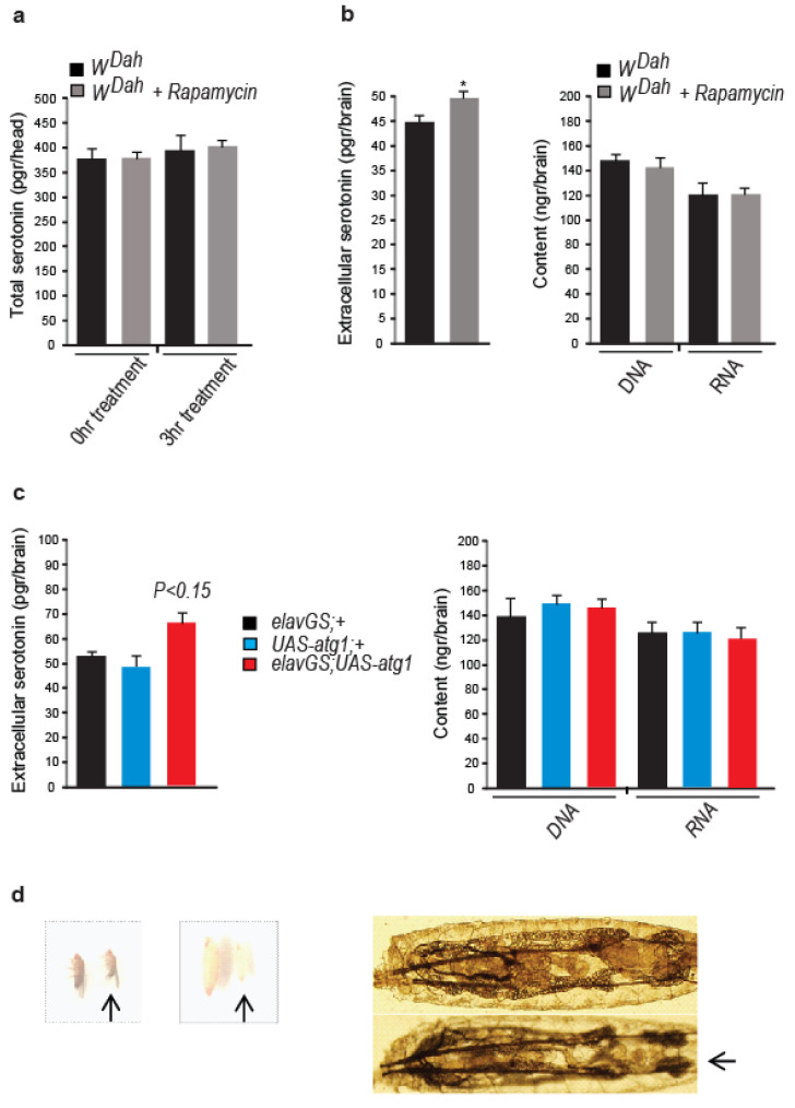
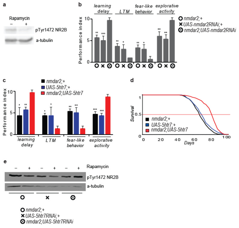
The mTORC1 nutrient-sensing pathway integrates metabolic and endocrine signals into the brain to evoke physiological responses to food deprivation, such as autophagy. Nevertheless, the impact of neuronal mTORC1 activity on neuronal circuits and organismal metabolism remains obscure. Here, we show that mTORC1 inhibition acutely perturbs serotonergic neurotransmission via proteostatic alterations evoked by the autophagy inducer atg1. Neuronal ATG1 alters the intracellular localization of the serotonin transporter, which increases the extracellular serotonin and stimulates the 5HTR7 postsynaptic receptor. 5HTR7 enhances food-searching behaviour and ecdysone-induced catabolism in Drosophila. Along similar lines, the pharmacological inhibition of mTORC1 in zebrafish also stimulates food-searching behaviour via serotonergic activity. These effects occur in parallel with neuronal autophagy induction, irrespective of the autophagic activity and the protein synthesis reduction. In addition, ectopic neuronal atg1 expression enhances catabolism via insulin pathway downregulation, impedes peptidergic secretion, and activates non-cell autonomous cAMP/PKA. The above exert diverse systemic effects on organismal metabolism, development, melanisation, and longevity. We conclude that neuronal atg1 aligns neuronal autophagy induction with distinct physiological modulations, to orchestrate a coordinated physiological response against reduced mTORC1 activity.

Keywords: 5HTR7 receptor; ATG1; ageing; autophagy; behaviour; cAMP/PKA; ecdysone; longevity; mTORC1; metabolism; serotonin transporter.


Conflict of interest statement

The authors declare no conflict of interests.

Figures

Figure 1
Figure 1
Rapamycin treatment alters cognitive and behavioural patterns in flies. (a) Four-day rapamycin treatment decreases the learning ability, LTM formation, and fear-like behaviour, while it increases the exploratory activity and does not alter the STM and MTM formation in 10-day-old WDah flies (n = 5). Individual comparisons by two-tailed Mann–Whitney test. (b) Four-day rapamycin treatment increases autophagy in the heads of 10-day-old WDah flies. (c) Reduced neuronal atg1 expression decreased the LTM and ameliorated the rapamycin effects on behaviour (n = 5). Ten-day-old WDah flies were fed with rapamycin for 4 days. For learning delay: F (5, 24) = 10.79, for LTM: F (5, 24) = 12.50, for fear-like behaviour: F (5, 24) = 16.12, for exploratory activity: F (5, 24) = 26.56. One-way ANOVA, individual comparisons by Sidak’s multiple comparisons test. (d) Ellipsoid bodies-specific atg1 inhibition ameliorated rapamycin-evoked behavioural patterns (n = 3). Three-day-old flies were fed with rapamycin for 4 days. For the learning delay of GH146, 129Y, ok107, c232, and c205, c601: F (3, 8) = 13.56, F (3, 8) = 55.56, F (3, 8) = 53.33, F (3, 8) = 13.83, F (3, 8) = 30.56, and F (3, 8) = 58.00, respectively. For the exploratory activity of GH146, 129Y, ok107, c232, c205, and c601: F (3, 8) = 10.77, F (3, 8) = 15.15, F (3, 8) = 27.43, F (3, 8) = 8.175, F (3, 8) = 58.00, and F (3, 8) = 16.26, respectively. One-way ANOVA, individual comparisons by Sidak’s multiple comparisons test. For atg1RNAi: two-tailed Mann–Whitney test. (e) Ellipsoid bodies-specific atg1 expression mimics rapamycin-evoked behaviours (n = 5). Three-day-old flies were used. For learning delay: F (4, 20) = 15.98, for LTM: F (4, 20) = 9.589, for fear-like behaviour: F (4, 20) = 11.67, for exploratory activity: F (4, 20) = 25.61. One-way ANOVA with Dunnette’s multiple comparisons test against c232; UAS-atg1. *** p < 0.001, ** p < 0.01, and * p < 0.05. Error bars represent SEM.
Figure 2
Figure 2
Ellipsoid bodies-specific 5HTR7 activity mediates the effects of neuronal atg1 on behaviour and cognition. (a) QRT-PCR analysis of genes expressing serotonin and Dop1R2 receptors in Drosophila heads, normalized to Rpl32 expression (n = 3). Ten-day-old WDah flies were fed with rapamycin for 4 days. Acute rapamycin treatment upregulated RNA levels of all receptor genes, with a major impact on 5htr7 expression. (b) Acute rapamycin treatment and low nutrient availability increase 5HTR7 levels in Drosophila head. Ten-day-old WDah flies were fed with rapamycin for 4 days or with low-nutrient food for 2 days. (c) 5htr7 is exclusively expressed in the ellipsoid bodies (dissected brain of 10-day-old 5htr7; UAS-sytegfp flies, posterior view) in the brain of Drosophila. (d) 5htr7 upregulation mimics rapamycin-evoked cognitive and behavioural effects (n = 3). Ten-day-old flies were used. For learning delay: F (2, 6) = 13.30, for LTM: F (2, 6) = 9.091, for fear-like behaviour: F (2, 6) = 10.50, for exploratory activity: F (2, 6) = 12.64. One-way ANOVA with Dunnette’s multiple comparisons test against 5htr7; UAS-5htr7. (e) 5htr7 inhibition ameliorates rapamycin-evoked cognitive and behavioural effects (n = 5). Ten-day-old flies were fed with rapamycin for 4 days. For learning delay: F (5, 24) = 15.76, for LTM: F (5, 24) = 9.059, for fear-like behaviour: F (5, 12) = 26.30, for exploratory activity: F (5, 24) = 55.31. One-way ANOVA, individual comparisons by Sidak’s multiple comparisons test. (f) 5htr7 inhibition ameliorates ellipsoid bodies-specific atg1 effects on cognition/behaviour (n = 3). Three-day-old flies were used. For learning delay: F (4, 10) = 7.435, for LTM: F (4, 10) = 10.08, for fear-like behaviour: F (4, 10) = 8.577, for exploratory activity: F (4, 10) = 14.29. One-way ANOVA, individual comparisons by Sidak’s multiple comparisons test. Selected pairs: c232; UAS-atg1 vs. c232; UAS-atg1; UAS-5htr7RNAi. (g) Acute feeding (2 days) of 10-day-old flies with a 5HTR7 specific inhibitor (SB269970) blunted atg1-induced learning deficits and enhanced exploratory activity (n = 3). Two-way ANOVA with Tukey’s multiple comparisons test, selected pairs: c232; UAS-atg10 nM vs. c232; UAS-atg150 nM. *** p < 0.001, ** p < 0.01, and * p < 0.05. Error bars represent SEM.
Figure 3
Figure 3
Serotonin cell-specific atg1 expression mimics rapamycin and enhanced serotonergic signalling-evoked behaviours, while increasing 5HTR7 expression. (a) Serotonin cell-specific constitutive active S6K inhibits rapamycin-induced behavioural and cognitive effects (n = 3). Ten-day-old flies were fed with rapamycin for 4 days. For learning delay: F (5, 12) = 34.63, for LTM: F (5, 12) = 21.70, for fear-like behaviour: F (5, 12) = 8.671, for exploratory activity: F (5, 12) = 20.07. One-way ANOVA, individual comparisons by Sidak’s multiple comparisons test. (b) Serotonin cell-specific atg1 expression induces behavioural and cognitive effects similar to rapamycin treatment (n = 3). Three-day-old flies were used. For learning delay: F (2, 6) = 21.00, for LTM: F (2, 6) = 36.33, for fear-like behaviour: F (2, 6) = 17.17, for exploratory activity: F (2, 6) = 13.88. One-way ANOVA with Dunnette’s multiple comparison test against trh; UAS-atg1. (c) Trh; UAS-atg1 flies have increased expression of 5HTR7 in the heads. Three-day-old flies were used. (d) Serotonin transporter inhibition induces effects on behaviour and cognition similar to rapamycin treatment (n = 3). Three-day-old flies were used. For learning delay: F (2, 6) = 10.50, for LTM: F (2, 6) = 6.818, for fear-like behaviour: F (2, 6) = 14.60, for exploratory activity: F (2, 6) = 20.17. One-way ANOVA with Dunnette’s multiple comparison test against trh; UAS-sertRNAi. (e) Inhibition of serotonin transporter via RNAi increases 5HTR7 levels in Drosophila heads. Three-day-old flies were used. (f) Paneuronal atg1 expression increases autophagy. RNAi inhibition of atg7 reduces atg1-induced autophagy in the heads of GSelav; UAS-atg1 flies (GSelav; UAS-atg1; UAS-atg7RNAi flies). Three-day-old flies were used. *** p < 0.001, ** p < 0.01, and * p < 0.05. Error bars represent SEM.
Figure 4
Figure 4
Rapamycin treatment and ellipsoid bodies-specific atg1 expression reduce serotonin transporter localization on the plasma membrane, while peptidergic cell-specific atg1 expression is lethal. (a) Acute rapamycin treatment (4 days) of 10-day-old WDah flies does not decrease SERT levels in Drosophila heads. (b) Acute rapamycin treatment (4 days) of 10-day-old WDah flies increases cytosolic levels of SERT, while decreasing its localization on the plasma membrane, at Drosophila heads. Values from imageJ analysis have been normalized to the total protein amount for each fraction and then normalized to the control values (n = 3). (c) Ellipsoid bodies-specific expression of atg1 decreases fluorescent expression of a UAS-GFP-SERT fusion protein, while it does not affect the expression of UAS-SYTE-GFP. Ten-day-old flies were used. Posterior view of Drosophila brain. (d) Dilp2; UAS-atg1; UAS-cd8rfp flies (right) are smaller than the controls (left: UAS-atg1; UAS-cd8rfp flies). (e) Ten-day-old dilp2; UAS-atg1; UAS-cd8rfp flies have reduced fecundity (n = 3). F (2, 6) = 24.15. One-way ANOVA with Dunnette’s multiple comparison test against dilp2; UAS-atg1; UAS-cd8rfp flies. (f) Dilp2; UAS-atg1; UAS-cd8rfp mated female flies are long-lived. Median, mean, and maximum lifespans for dilp2; +: 69.5, 68, and 82 days, respectively, for UAS-cd8rfp; +: 69.5, 68, and 80 days respectively, for UAS-atg1: 71, 70, and 78 days, respectively, for dilp2; UAS-atg1; UAS-cd8rfp: 78, 76, and 85 days, respectively. Log-rank test analysis (n = 100). (g) C929; UAS-atg1 flies are pharate lethals. They exhibit excessive cuticle sclerotization and extensive melanisation mainly at the pupal head, trachea, and wings imaginal discs. ** p < 0.01. Error bars represent SEM.
Figure 5
Figure 5
Lowered mTORC1 and neuronal atg1 expression increase the brain levels of extracellular serotonin and stimulate serotonergic neurotransmission. (a) Acute rapamycin treatment (4 days) of 10-day-old WDah flies does not alter total serotonin levels in Drosophila heads. Head homogenates were left in an EDTA-rich solution (20 mM) for 3 h at room temperature to check the degree of serotonin catabolism. For each biological sample, we used seven heads (n = 3). (b) Acute rapamycin treatment (4 days) of 10-day-old WDah flies increases extracellular serotonin in Drosophila brain. Total DNA and RNA levels in the supernatants subjected to ELISA measurements did not differ among the experimental conditions. For each biological sample, 40 brains were used (n = 9). Individual comparisons by one-tailed unpaired t test. (c) Paneuronal expression of atg1 with the mifepristone-inducing elavGS-gal4 driver, in the absence of mifepristone, increases extracellular levels of serotonin in Drosophila brains. Three-day-old flies were used. Total DNA and RNA levels in the supernatants subjected to ELISA measurements did not differ among the samples. For each biological sample, 40 brains were used (n = 3). F (2, 6) = 2.671. One-way ANOVA with Dunnette’s multiple comparison tests against elavGS; UAS-atg1 flies. (d) Expression of atg1 in prothoracic gland-innervating R29H01 serotonergic cells in Drosophila larvae decreases the size of pupae and adults, inhibits pupal colorization, and shrinks larval fat body (R29H01; UAS-atg1 larvae, pupae and flies are indicated with arrows. UAS-atg1 larvae, pupae and flies were used as controls). These phenotypes are reminiscent of flies with enhanced ecdysone signalling. * p < 0.05. Error bars represent SEM.
Figure 6
Figure 6
Activation of the 5HTR7 receptor mediates rapamycin-induced dephosphorylation of NMDAR2 receptor and causes NMDA signalling inhibition-like effects in flies. (a) Four-day rapamycin treatment of 10-day-old flies reduces NMDA receptor 2 phosphorylation in Tyr1472 in Drosophila heads. (b) Inhibition of the nmdar2 receptor gene mimics rapamycin-induced cognitive and behavioural effects (n = 3). Ten-day-old flies were used. For learning delay: F (2, 6) = 34.40, for LTM: F (2, 6) = 13.00, for fear-like behaviour: F (2, 6) = 11.40, for exploratory activity: F (2, 6) = 18.38. One-way ANOVA with Dunnette’s multiple comparisons test against nmdar2; UAS-nmdar2RNAi. (c) 5htr7 expression in NMDAR2-expressing cells mimics the effects of rapamycin on behaviour and cognition (n = 5). Ten-day-old flies were used. For learning delay: F (2, 6) = 13.87, for LTM: F (2, 6) = 9.000, for fear-like behaviour: F (2, 6) = 26.60, for exploratory activity: F (2, 6) = 52.33. One-way ANOVA with Dunnette’s multiple comparisons test against nmdar2; UAS-5htr7. (d) 5htr7 expression in NMDAR2-expressing cells enhances the lifespan in female mated flies. Median, mean, and maximum lifespans for nmdar2; +: 67, 69.1, and 85 days, respectively, for UAS-5htr7; +: 71.5, 71.4, and 89 days, respectively, for nmdar2; UAS-5htr7: 84, 81.5, and 93 days, respectively. Log-rank test analysis (n = 100). (e) RNAi inhibition of 5htr7 at NMDAR2-expressing cells blocks rapamycin-induced NMDAR2 dephosphorylation in Drosophila heads. Ten-day-old flies were fed with rapamycin for 4 days. *** p < 0.001, ** p < 0.01, and * p < 0.05. Error bars represent SEM.
Figure 7
Figure 7
Rapamycin treatment induces cAMP/PKA signalling non-cell autonomously. (a) Rapamycin treatment-evoked behaviours are ameliorated by RNAi inhibition of Rutabaga adenylyl cyclase in NMDAR2-expressing cells (n = 3). For learning delay: F (5, 12) = 17.48, for LTM: F (5, 12) = 7.965, for fear-like behaviour: F (5, 12) = 28.43, for exploratory activity: F (5, 12) = 29.36. One-way ANOVA, individual comparisons by Sidak’s multiple comparisons test. (b) RNAi expression of the gene coding for the catalytic subunit of PKA (pkac1) ameliorates peptidergic cell-specific atg1-induced melanisation, cuticle hypersclerotization, and developmental death. (c) Starvation, rapamycin feeding, and neuronal atg1 expression cause epidermal melanisation in larvae. (d) Paneuronal atg1 and rapamycin treatment induce epidermal cAMP. Rapamycin treatment induces cAMP-related melanisation in scabrous-expressing neuroblasts, in a non-cell autonomous way. Larvae were screened for CFP fluorescence and epidermal melanisation under a ZEISS/Axioskop 2 Plus microscope, with a DAPI filter. Then, samples were further analysed with confocal microscopy. *** p < 0.001 and ** p < 0.01. Error bars represent SEM.
Figure 8
Figure 8
Rapamycin treatment alters behaviour and cognition in zebrafish via serotonergic signalling. (a) Rapamycin injection delays learning and inhibits long-term memory (n = 8). Two-tailed Mann–Whitney test. (b) Prolonged training abrogates rapamycin-induced LTM impairment. Two-tailed Mann–Whitney test (n = 7). (c) Rapamycin-injected zebrafish spent more time in the top area of a water tank (novel tank test) (n = 10). Two-tailed Mann–Whitney test. (d) Rapamycin injection increases HTR1B expression in the brain of zebrafish, as well as RNA levels of htr1b (n = 3). (e) GR55562 mild treatment of rapamycin-injected zebrafish abrogates learning defects, and galantamine hydrobromide enhances rapamycin-induced learning defects, while Prozac, although it worsened learning ability compared to controls (p < 0.05), did not significantly further enhance rapamycin-induced learning defects. Day 6 of training protocol (n = 9). Two-tailed Mann–Whitney test. (f) GR55562 mild treatment abrogates altered the swimming behaviour of rapamycin-injected zebrafish (n = 9). F (3, 26) = 3.734. One-way ANOVA with Dunnette’s multiple comparison tests against untreated zebrafish. (g) Rapamycin injection decreased pTyr1472 phosphorylation of NR2B in the forebrain, while it increased HTR1B in the midbrain and forebrain. Mild treatment of rapamycin-injected zebrafish with GR55562 reduced rapamycin-induced pTyr1472 dephosphorylation of NR2B in whole brain extracts. F (2, 12) = 4.491. One-way ANOVA with Dunnette’s multiple comparison test against untreated zebrafish (n = 5). *** p < 0.001 and * p < 0.05. Error bars represent SEM.
Figure 9
Figure 9
Acute mTORC1 inhibition enhances ATG1 at serotonergic cells, which inhibits serotonin transporter (SERT) activity and results in increased extracellular serotonin and expression of postsynaptic 5HTR7. The latter enhances cAMP/PKA signalling: (a) at NMDA2 receptor expressing cells in the brain, where it inhibits NMDA signalling and induces behavioural/cognitive modulations, and (b) at prothoracic gland cells, where it stimulates ecdysone (20E) signalling and systemic catabolism. Both effects enhance longevity and physiological adaptations to nutrient deprivation.

Similar articles

References

    1. Cota D., Proulx K., Smith K.A., Kozma S.C., Thomas G., Woods S.C., Seeley R.J. Hypothalamic mTOR signalling regulates food intake. Science. 2006;312:927–930. doi: 10.1126/science.1124147. - DOI - PubMed
    1. Proulx K., Cota D., Woods S.C., Seeley R.J. Fatty acid synthase inhibitors modulate energy balance via mammalian target of rapamycin complex 1 signalling in the central nervous system. Diabetes. 2008;57:3231–3238. doi: 10.2337/db07-1690. - DOI - PMC - PubMed
    1. Blouet C., Ono H., Schwartz G.J. Mediobasal hypothalamic p70 S6 kinase 1 modulates the control of energy homeostasis. Cell Metab. 2008;8:459–467. doi: 10.1016/j.cmet.2008.10.004. - DOI - PMC - PubMed
    1. Gwinn D.M., Shackelford D.B., Egan D.F., Mihaylova M.M., Mery A., Vasquez D.S., Turk B.E., Shaw R.J. AMPK phosphorylation of raptor mediates a metabolic checkpoint. Mol. Cell. 2008;30:214–226. doi: 10.1016/j.molcel.2008.03.003. - DOI - PMC - PubMed
    1. Villanueva E.C., Munzberg H., Cota D., Leshan R.L., Kopp K., Ishida-Takahashi R., Jones J.C., Fingar D.C., Seeley R.J., Myers M.G., Jr. Complex regulation of mammalian target of rapamycin complex 1 in the basomedial hypothalamus by leptin and nutritional status. Endocrinology. 2009;150:4541–4551. doi: 10.1210/en.2009-0642. - DOI - PMC - PubMed

Publication types

LinkOut - more resources