. 2023 Aug 14;12(16):2059.
doi: 10.3390/cells12162059.

Development of a Novel CD26-Targeted Chimeric Antigen Receptor T-Cell Therapy for CD26-Expressing T-Cell Malignancies

Affiliations

Development of a Novel CD26-Targeted Chimeric Antigen Receptor T-Cell Therapy for CD26-Expressing T-Cell Malignancies

Eiji Kobayashi et al. Cells. .

Abstract

Chimeric-antigen-receptor (CAR) T-cell therapy for CD19-expressing B-cell malignancies is already widely adopted in clinical practice. On the other hand, the development of CAR-T-cell therapy for T-cell malignancies is in its nascent stage. One of the potential targets is CD26, to which we have developed and evaluated the efficacy and safety of the humanized monoclonal antibody YS110. We generated second (CD28) and third (CD28/4-1BB) generation CD26-targeted CAR-T-cells (CD26-2G/3G) using YS110 as the single-chain variable fragment. When co-cultured with CD26-overexpressing target cells, CD26-2G/3G strongly expressed the activation marker CD69 and secreted IFNgamma. In vitro studies targeting the T-cell leukemia cell line HSB2 showed that CD26-2G/3G exhibited significant anti-leukemia effects with the secretion of granzymeB, TNFα, and IL-8, with 3G being superior to 2G. CD26-2G/3G was also highly effective against T-cell lymphoma cells derived from patients. In an in vivo mouse model in which a T-cell lymphoma cell line, KARPAS299, was transplanted subcutaneously, CD26-3G inhibited tumor growth, whereas 2G had no effect. Furthermore, in a systemic dissemination model in which HSB2 was administered intravenously, CD26-3G inhibited tumor growth more potently than 2G, resulting in greater survival benefit. The third-generation CD26-targeted CAR-T-cell therapy may be a promising treatment modality for T-cell malignancies.

Keywords: CAR-T; CD26; T-cell malignancies; fratricide; third-generation.


Conflict of interest statement

The authors declare no conflict of interest.

Figures
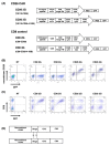
Figure 1
Figure 1
Transduction and expression of CD26 CAR. Gene transduction of CD26 2/3G and control CD8 2/3G into PBMCs was confirmed by GFP marker fluorescence three days following retroviral infection. (A) Schematic diagram of CD26 CAR and CD8 control. (B) Expression of YS110 scFv on CD26 2/3G-transfected PBMCs was confirmed by binding to Fc-tagged recombinant CD26, which was detected by the anti-Fc antibody conjugated with PE. (C) Expression of CD8 on control CD8 2/3G-transfected PBMCs was confirmed with the anti-CD8 antibody conjugated with PE. (D) Construct schema for rh-CD26 FC-chimera. Data are representative of three independent experiments (n = 3).
Figure 2
Figure 2
Expansion of CD26 CAR-T-cells. (A) Cell numbers of CD26 2/3G CAR-T-cells and CD8 2/3G control cells were determined at day 3, 6, 9 and 12 following retroviral infection. (B) CD26 expression on CD26 2/3G CAR-T-cells and CD8 2/3G control cells was analyzed at day 3, 6, 9, and 12 by flow cytometry. Data are representative of three independent experiments (n = 1).
Figure 3
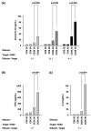
Figure 3
Activation of CD26 CAR-T-cells. Effector cells (CD26 2/3G CAR-T-cells and CD8 2/3G control cells) were cultured with HSB2 cells as target cells at an effector:target ratio of 1:1, 2:1, and 4:1. (A) Secretion of granzyme B was evaluated by ELISA assay. (B,C) Secretion of TNFα (B) and IL-8 (C) were evaluated by flow cytometry. Data are representative of three independent experiments (n = 3).
Figure 4
Figure 4
In vitro anti-tumor activity of CD26 CAR-T-cells. (A) Target cells (HSB2 cells) were co-cultured with effector cells (CD26 2/3G CAR-T-cells or CD8 2/3G control cells) at an effector:target ratio of 0:1, 1:1, 5:1, and 10:1. (B) Target cells (H9 and KARPAS299 cells) were co-cultured with effector cells (CD26 3G CAR-T-cells or CD8 3G control cells) at an effector:target ratio of 1:1, 5:1, and 10:1. Anti-tumor activity of effector cells was evaluated by measurement of luciferase activity from the target cells. Data are representative of three independent experiments (n = 3).
Figure 5
Figure 5
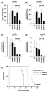
In vivo anti-tumor activity of CD26 CAR-T-cells. (A,B) KARPAS299 cells transduced with luciferase gene were transplanted subcutaneously into NSG mice at day 0. CD26 2/3G CAR-T-cells or CD8 2/3G control cells were then injected intravenously twice at day 6 and day 13. Tumor volume (mm3) (A) and luminescence (photon/sec) (B) at day 20 are presented (n = 6). (CE) HSB2 cells transduced with the luciferase gene were transplanted intravenously into NSG mice at day 0 as a murine model of systemic dissemination. CD26 2/3G CAR-T-cells or CD8 2/3G control cells were then injected intravenously once at day 1. Luminescence (photon/sec) at day 7 (C) and day 21 (D) are presented (n = 6). For the same model of systemic dissemination, CD26 2/3G CAR-T-cells were injected intravenously once at day 1 and survival of mice was monitored (n = 5) (E).
Figure 6
Figure 6
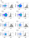
In vitro anti-tumor activity of CD26 CAR-T-cells against patient lymphoma samples. CD26-expressing lymphoma cells from patients with PTCL (A) (n = 1) or AITL (B) (n = 1) were co-cultured with CD26 2/3G CAR-T-cells or CD8 2/3G control cells. CD26 2/3G CAR-T-cells or CD8 2/3G control cells were pre-stained with CTOG and were distinguished from lymphoma cells by flow cytometry as the Alexa Fluor 488-positive cells. CD26 expression was evaluated in the Alexa Fluor 488-negative lymphoma cells. Data are representative of three independent experiments (n = 3).

References

    1. Ohnuma K., Hosono O., Dang N.H., Morimoto C. Dipeptidyl Peptidase in Autoimmune Pathophysiology. Adv. Clin. Chem. 2011;53:51–84. - PubMed
    1. Thompson M., Ohnuma K., Abe M., Morimoto C., Dang N. CD26/Dipeptidyl Peptidase IV as a Novel Therapeutic Target for Cancer and Immune Disorders. Mini-Rev. Med. Chem. 2007;7:253–273. doi: 10.2174/138955707780059853. - DOI - PubMed
    1. Cordero O.J., Rafael-Vidal C., Varela-Calviño R., Calviño-Sampedro C., Malvar-Fernández B., García S., Viñuela J.E., Pego-Reigosa J.M. Distinctive CD26 Expression on CD4 T-Cell Subsets. Biomolecules. 2021;11:1446. doi: 10.3390/biom11101446. - DOI - PMC - PubMed
    1. Ohnuma K., Hatano R., Komiya E., Otsuka H., Itoh T., Iwao N., Kaneko Y., Yamada T., Dang N.H., Morimoto C. A Novel Role for CD26/Dipeptidyl Peptidase IV as a Therapeutic Target. Front. Biosci. 2018;23:1754–1779. doi: 10.2741/4671. - DOI - PubMed
    1. Havre P.A., Abe M., Urasaki Y., Ohnuma K., Morimoto C., Dang N.H. The Role of CD26/Dipeptidyl Peptidase IV in Cancer. Front. Biosci. 2008;13:1345–1351. doi: 10.2741/2787. - DOI - PubMed

Publication types