. 2023 Aug 19;12(16):2097.
doi: 10.3390/cells12162097.

Catch of the Day: New Serum Amyloid A (SAA) Antibody Is a Valuable Tool to Study Fish Health in Salmonids

Affiliations

Catch of the Day: New Serum Amyloid A (SAA) Antibody Is a Valuable Tool to Study Fish Health in Salmonids

Ralfs Buks et al. Cells. .

Abstract

Serum amyloid A (SAA) proteins belong to a family of acute-phase reactants, playing an integral role in defending the organism from pathological damage. Despite a wealth of data on the regulation of SAA transcripts in teleosts, there is only limited information on these proteins' abundance in fish. The aim of this study is to characterise SAA protein levels in salmonids using a newly developed antibody specific to salmonid SAA. The salmonid SAA antibody detected SAA and accurately discriminated between stimulated and control specimens from rainbow trout macrophage cell line (RTS-11) in vitro, as well as rainbow trout challenged with Aeromonas salmonicida- or flagellin-stimulated Atlantic salmon in vivo. The presence of SAA protein was analysed in RTS-11 cell line supernatants, liver, and spleen samples using ELISA, immunoblotting, and immunohistochemistry. This study is the first to characterise SAA protein levels in salmonids in vivo and in vitro. The newly developed salmonid SAA antibody was able to discriminate between stimulated and unstimulated specimens, showing that it can be used to study the acute-phase response in salmonids with the potential to be further developed into assays to monitor and evaluate health in wild and farmed fish.

Keywords: RTS-11 cell line; SAA antibody; SAA recombinant protein; fish health; research tools; salmonids; serum amyloid A (SAA).


Conflict of interest statement

R.B. is a knowledge transfer partnership (KTP) associate funded by Innovate UK and VAL. A.A. (Ayham Alnabulsi), A.A. (Abdo Alnabulsi), and A.W. are employees of VAL.

Figures

Figure 1
Figure 1
AlphaFold-predicted 3D structure of the SAA-5 protein (UniProt B9EPA2). AlphaFold produces a per-residue confidence score (pLDDT) between 0 and 100. Model Confidence is categorised as very high (pLDDT > 90), confident (90 > pLDDT > 70), low (70 > pLDDT > 50), or very low (pLDDT < 50). Black dotted rectangle denotes predicted signal peptide of the SAA-5 (MKLLLAGLVLTLVVGAQA). Green dotted rectangle highlights the peptide sequence (RYRPNGLPRNY) used as the immunogen for the SAA antibody development.
Figure 2
Figure 2
Anti-SAA A10 antibody detection of recombinant Atlantic salmon SAA-5 protein via Western blotting and ELISA. (A) A total of 40 ng of Atlantic salmon rSAA-5 was size separated in a 4–12% (w/v) Bis-Tris SDS-PAGE under reducing conditions and immunostained with anti-SAA A10 monoclonal antibody. (B) The 96-well plate was coated with Atlantic salmon rSAA-5 (0.2–10 ng) and the optical density from the anti-SAA-5-biotin-linked antibody was measured via an indirect ELISA. Data are fitted to a one-phase exponential association and each datapoint is presented as mean (n = 3) with SD.
Figure 3
Figure 3
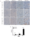
Localisation and analysis of SAA in Atlantic salmon liver using IHC. Fish were stimulated with a flagellin or PBS (control). Liver samples were collected 6, 24, or 48 h post-stimulation, and IHC was performed using anti-SAA A10 antibody. (A) Representative images of control (100× magnification, (a,e,i)) and flagellin-stimulated fish (100× (b,f,j), 200× (c,g,k) and 400× magnification (d,h,l)) are shown. Scale bars represent 100 μm. Black arrows represent granulated cytoplasmic SAA, while white arrows denote homogenous SAA staining. (B) Area (%) covered by SAA staining measured at 200× magnification. Mann–Whitney test, mean with SD, n = 4, * p < 0.05.
Figure 4
Figure 4
Localisation and analysis of SAA in Atlantic salmon spleen using IHC. Fish were stimulated with a flagellin or PBS (control). Liver samples were collected 6, 24, or 48 h post-stimulation and IHC was performed using the anti-SAA A10 antibody. (A) Representative images of control (100× magnification, (a,e,i)) and flagellin-stimulated fish (100× (b,f,j), 200× (c,g,k) and 400× magnification (d,h,l)) are shown. Scale bars represent 100 μm. Black arrows represent granulated cytoplasmic SAA, while white arrows denote homogenous SAA staining. (B) Area (%) covered by SAA staining measured at 200× magnification. Mann–Whitney test, mean with SD, n = 4. ns—not significant, * p < 0.05.
Figure 5
Figure 5
Recombinant rainbow trout SAA and endogenous SAA immunoblot detection by the new salmonid SAA antibody. (A) Recombinant rainbow trout SAA protein (40 ng); (B) 20 μL of IL-1β-stimulated RTS-11 cell line supernatant were size separated in a 4–12% (w/v) Bis-Tris SDS-PAGE under reducing conditions and immunostained with the anti-salmonid SAA antibody.
Figure 6
Figure 6
SAA protein is synthesised early following IL-1β stimulation in the RTS-11 cell line. (A) SDS-PAGE immunoblots of SAA in RTS-11 cell supernatants 6, 12, 24, 48, and 72 h post-stimulation with IL-1β or PBS (control). Rainbow trout rSAA (10 ng) was used as a positive control. (B) Densitometry plot comparing SAA protein abundance from the SDS-PAGE immunoblot (A): grey (control), black (IL-1β) stimulation. (C) Optical density (OD) of SAA protein levels in supernatants from RTS-11 control (grey) and IL-1β (black)-stimulated cells for 4, 6, 8, 10, 12, and 24 h measured using indirect ELISA. Background signal (OD = 0.048) from the RTS-11 culture media without cells is noted by the black dashed line. The Y-axis is divided into two segments visualising the SAA protein level differences 4 and 6 h post-stimulation. (D) Representative standard curve of OD values from 0.2, 0.5, 1, 5, and 10 ng rainbow trout rSAA protein. n = 3, mean with SD. (E) Estimated SAA extracellular protein levels in 100 μL RTS-11-stimulated media based on mean OD values (C) and the standard curve (D). RTS-11 cells were stimulated in three independent experiments. Each sample was measured in duplicates by ELISA. Data are presented as the means with SDs, Mann–Whitney test. ns—not significant, **** p < 0.0001.
Figure 7
Figure 7
Localisation and analysis of SAA in rainbow trout liver using IHC. Fish were challenged with A. salmonicida or PBS (control). Liver samples were collected 48 h post-challenge, and IHC was performed using anti-SAA A10 antibody. (A) Representative images of control (100× magnification (a)) and A. salmonicida-challenged fish (100× (b), 200× (c) and 400× magnification (d)) are shown. Scale bars represent 100 μm. Black arrows represent granulated cytoplasmic SAA, while white arrows denote homogenous SAA staining. (B) Area (%) covered by SAA staining measured at 200× magnification. Mann–Whitney test, mean with SD, n = 4. * p < 0.05.

References

    1. Sack G.H., Jr. Serum Amyloid A (SAA) Proteins. Subcell. Biochem. 2020;94:421–436. doi: 10.1007/978-3-030-41769-7_17. - DOI - PubMed
    1. Ericsson L.H., Eriksen N., Walsh K.A., Benditt E.P. Primary structure of duck amyloid protein A. The form deposited in tissues may be identical to its serum precursor. FEBS Lett. 1987;218:11–16. doi: 10.1016/0014-5793(87)81008-6. - DOI - PubMed
    1. Jensen L.E., Hiney M.P., Shields D.C., Uhlar C.M., Lindsay A.J., Whitehead A.S. Acute phase proteins in salmonids: Evolutionary analyses and acute phase response. J. Immunol. 1997;158:384–392. doi: 10.4049/jimmunol.158.1.384. - DOI - PubMed
    1. Jorgensen J.B., Lunde H., Jensen L., Whitehead A.S., Robertsen B. Serum amyloid A transcription in Atlantic salmon (Salmo salar L.) hepatocytes is enhanced by stimulation with macrophage factors, recombinant human IL-1 beta, IL-6 and TNF alpha or bacterial lipopolysaccharide. Dev. Comp. Immunol. 2000;24:553–563. doi: 10.1016/S0145-305X(00)00022-7. - DOI - PubMed
    1. Wei J., Guo M., Ji H., Qin Q. Molecular cloning, characterization of one key molecule of teleost innate immunity from orange-spotted grouper (Epinephelus coioides): Serum amyloid A. Fish Shellfish Immunol. 2013;34:296–304. doi: 10.1016/j.fsi.2012.11.014. - DOI - PubMed

Publication types

LinkOut - more resources