. 2023 Jul 28;12(8):1063.
doi: 10.3390/biology12081063.

3D Culture and Interferon-γ Priming Modulates Characteristics of Mesenchymal Stromal/Stem Cells by Modifying the Expression of Both Intracellular and Exosomal microRNAs

Affiliations

3D Culture and Interferon-γ Priming Modulates Characteristics of Mesenchymal Stromal/Stem Cells by Modifying the Expression of Both Intracellular and Exosomal microRNAs

Matteo Bulati et al. Biology (Basel). .

Abstract

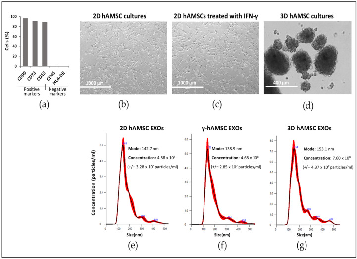
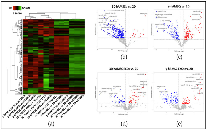
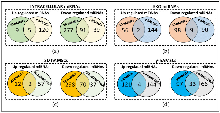
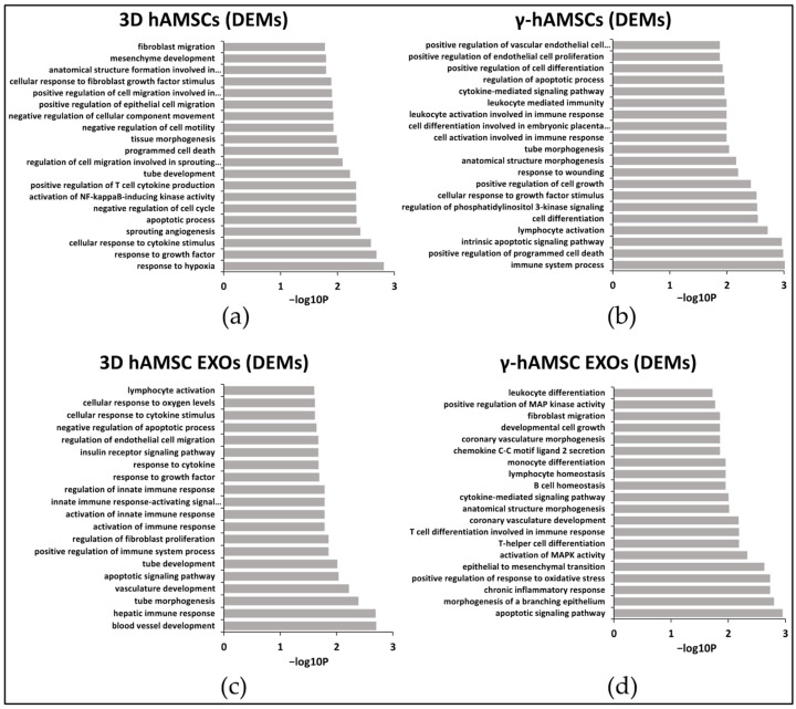
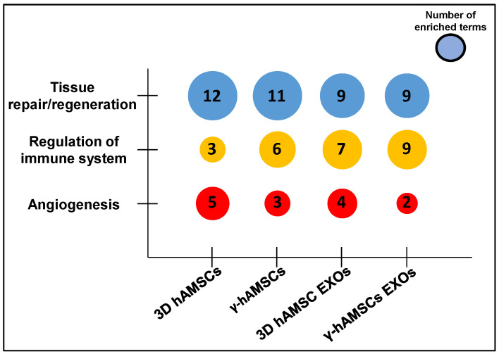
Mesenchymal stromal/stem cells (MSCs) have emerged as a therapeutic tool in regenerative medicine. Recent studies have shown that exosome (EXO)-derived microRNAs (miRNAs) play a crucial role in mediating MSC functions. Additionally, intracellular miRNAs have been found to regulate MSC therapeutic capacities. However, the molecular mechanisms underlying miRNA-mediated MSC effects are not fully understood. We used 3D culture and IFN-γ to prime/enhance the MSC therapeutic effects in terms of functional miRNAs. After priming, our analysis revealed stable variations in intracellular miRNA among the MSC biological replicates. Conversely, a significant variability of miRNA was observed among EXOs released from biological replicates of the priming treatment. For each priming, we observed distinct miRNA expression profiles between the MSCs and their EXOs. Moreover, in both types of priming, gene ontology (GO) analysis of deregulated miRNAs highlighted their involvement in tissue repair/regeneration pathways. In particular, the 3D culture enhanced angiogenic properties in both MSCs and EXOs, while IFN-γ treatment enriched miRNAs associated with immunomodulatory pathways. These findings suggest that 3D culture and IFN-γ treatment are promising strategies for enhancing the therapeutic potential of MSCs by modulating miRNA expression. Additionally, the identified miRNAs may contribute to understanding the molecular mechanisms underlying the miRNA-mediated therapeutic effects of MSCs.

Keywords: IFN-γ priming; MSC priming; MSC spheroids; MSC therapeutic properties; exosomes; mesenchymal stromal/stem cells; microRNAs; regenerative medicine.


Conflict of interest statement

The authors declare no conflict of interest. The funders had no role in the design of the study; in the collection, analysis, and interpretation of data; in the writing of the manuscript; or in the decision to publish the results.

Figures

Figure 1
Figure 1
Human amnion-derived mesenchymal stem cells (hAMSCs) cultured as both a monolayer (2D culture) and spheroids (3D cultures). (a) Cytofluorimetric results of the positive and negative surface markers of hAMSCs. (b) Representative differential interference contrast (DIC) images of hAMSCs grown in a monolayer (2D cultures). (c) Representative DIC images of hAMSCs grown in a monolayer and treated with IFN-γ. (d) Representative DIC images of hAMSCs grown as spheroids (3D cultures). (e) Size and concentration (dilution 1:500) of exosomes isolated from the hAMSC 2D cultures. (f) Size and concentration (dilution 1:500) of exosomes isolated from the hAMSC 2D cultures treated with IFN-γ. (g) Size and concentration (dilution 1:500) of exosomes isolated from the hAMSC 3D cultures. The red lines in (eg) describes the relationship between particle number distribution (left y-axis) and particle size (x-axis).
Figure 2
Figure 2
Cluster analysis to compare the fold changes (FC) in miRNA expression in primed hAMSCs and their EXOs. (a) Hierarchical clustering of z score transformed FC of miRNA in each treatment group compared to 2D untreated samples (control). (b) Volcano plot analysis (fold change > 1.5 and p < 0.05) of miRNA expression in the 3D culture of hAMSCs vs. control (2D culture). (c) Volcano plot analysis (fold change > 1.5 and p < 0.05) of miRNA expression in hAMSCs treated with IFN-γ vs. control (2D). (d) Volcano plot analysis (fold change > 1.5 and p < 0.05) of miRNA expression in EXOs derived from the 3D hAMSCs vs. control (2D). (e) Volcano plot analysis (fold change > 1.5 and p < 0.05) of miRNA expression in EXOs derived from hAMSCs treated with IFN-γ vs. control (2D). For all the volcano plots in the figure, red dots represent the up-regulated miRNAs in the experimental samples, while the blu dots represent the miRNAs up-regulated in the experimental controls.
Figure 3
Figure 3
Venn diagram showing differentially expressed miRNAs between (a) 3D hAMSCs and hAMSCs treated with IFN-γ (γ-hAMSCs); (b) 3D hAMSC EXOs and γ-hAMSC EXOs; (c) 3D hAMSCs and 3D hAMSC EXOs; (d) γ-hAMSCs and γ-hAMSC EXOs.
Figure 4
Figure 4
GO analysis of deregulated miRNAs (DEMs) in (a) hAMSCs grown as 3D spheroids (3D hAMSCs); (b) hAMSCs treated with IFN-γ (γ-hAMSCs); (c) exosomes (EXOs) derived from the 3D culture of hAMSCs; (d) EXOs derived from IFN-γ treated hAMSCs. Partial list of biological process enrichment analysis.
Figure 5
Figure 5
Bubble plot visualizing categories for GO-enriched terms of the DEMs in hAMSCs and their EXOs primed with either 3D culture or IFN-γ. Partial list of the 20 more significant enriched terms.

References

    1. Han Y., Li X., Zhang Y., Han Y., Chang F., Ding J. Mesenchymal Stem Cells for Regenerative Medicine. Cells. 2019;8:886. doi: 10.3390/cells8080886. - DOI - PMC - PubMed
    1. Margiana R., Markov A., Zekiy A.O., Hamza M.U., Al-Dabbagh K.A., Al-Zubaidi S.H., Hameed N.M., Ahmad I., Sivaraman R., Kzar H.H., et al. Clinical application of mesenchymal stem cell in regenerative medicine: A narrative review. Stem Cell Res. Ther. 2022;13:366. doi: 10.1186/s13287-022-03054-0. - DOI - PMC - PubMed
    1. Miceli V., Bulati M., Gallo A., Iannolo G., Busa R., Conaldi P.G., Zito G. Role of Mesenchymal Stem/Stromal Cells in Modulating Ischemia/Reperfusion Injury: Current State of the Art and Future Perspectives. Biomedicines. 2023;11:689. doi: 10.3390/biomedicines11030689. - DOI - PMC - PubMed
    1. Chang C., Yan J., Yao Z., Zhang C., Li X., Mao H.Q. Effects of Mesenchymal Stem Cell-Derived Paracrine Signals and Their Delivery Strategies. Adv. Healthc. Mater. 2021;10:e2001689. doi: 10.1002/adhm.202001689. - DOI - PMC - PubMed
    1. Miceli V., Zito G., Bulati M., Gallo A., Busa R., Iannolo G., Conaldi P.G. Different priming strategies improve distinct therapeutic capabilities of mesenchymal stromal/stem cells: Potential implications for their clinical use. World J. Stem Cells. 2023;15:400–420. doi: 10.4252/wjsc.v15.i5.400. - DOI - PMC - PubMed

LinkOut - more resources