. 2023 Aug 19;12(8):1147.
doi: 10.3390/biology12081147.

Effect of Mesenchymal Stem Cells Overexpressing BMP-9 Primed with Hypoxia on BMP Targets, Osteoblast Differentiation and Bone Repair

Affiliations

Effect of Mesenchymal Stem Cells Overexpressing BMP-9 Primed with Hypoxia on BMP Targets, Osteoblast Differentiation and Bone Repair

Jessica Emanuella Rocha Moura Paz et al. Biology (Basel). .

Abstract

Bone formation is driven by many signaling molecules including bone morphogenetic protein 9 (BMP-9) and hypoxia-inducible factor 1-alpha (HIF-1α). We demonstrated that cell therapy using mesenchymal stem cells (MSCs) overexpressing BMP-9 (MSCs+BMP-9) enhances bone formation in calvarial defects. Here, the effect of hypoxia on BMP components and targets of MSCs+BMP-9 and of these hypoxia-primed cells on osteoblast differentiation and bone repair was evaluated. Hypoxia was induced with cobalt chloride (CoCl2) in MSCs+BMP-9, and the expression of BMP components and targets was evaluated. The paracrine effects of hypoxia-primed MSCs+BMP-9 on cell viability and migration and osteoblast differentiation were evaluated using conditioned medium. The bone formation induced by hypoxia-primed MSCs+BMP-9 directly injected into rat calvarial defects was also evaluated. The results demonstrated that hypoxia regulated BMP components and targets without affecting BMP-9 amount and that the conditioned medium generated under hypoxia favored cell migration and osteoblast differentiation. Hypoxia-primed MSCs+BMP-9 did not increase bone repair compared with control MSCs+BMP-9. Thus, despite the lack of effect of hypoxia on bone formation, the enhancement of cell migration and osteoblast differentiation opens windows for further investigations on approaches to modulate the BMP-9-HIF-1α circuit in the context of cell-based therapies to induce bone regeneration.

Keywords: BMP-9; HIF-1α; bone; cell therapy; hypoxia; stem cells.


Conflict of interest statement

The authors declare no conflict of interest.

Figures

Figure 1
Figure 1
Hypoxia induction and duration of its effect on MSCs+BMP-9. Viability of MSCs+BMP-9 cultured in either absence (control) or presence of CoCl2 at 100, 150 and 200 μM for the last 3 (A), 5 (B) and 7 days (C) of culture and at 10, 20, 40 and 50 μM in the last 3 (D), 5 (E) and 7 days (F) of culture, measured on day 8. Protein expression of HIF-1α in MSCs+BMP-9 cultured in either absence (control) or presence of CoCl2 at 40 and 50 μM for the last 3 and 5 days, detected on day 8 (G). Gene expression of Glut1 (H) and Vegfa (I) in MSCs+BMP-9 cultured for 8 days, with the last 3 days in either absence (control) or presence of CoCl2 at 40 μM, detected 0, 4, 8 and 12 h after CoCl2 removal. The data are presented as mean ± standard deviation, and the * indicates statistically significant differences (p ≤ 0.05).
Figure 2
Figure 2
Effect of hypoxia on the expression of components and targets of the BMP signaling pathway in MSCs+BMP-9. Protein expression of BMP-9 (A), and gene expression of Bmp-2 (B), Bmp-4 (C), Bmpr1a (D), Smad1 (E), Smad5 (F), Hey1 (G) and Runx2 (H) in MSCs+BMP-9 cultured under either control or hypoxia condition, on day 8. The data are presented as mean ± standard deviation, and the * indicates statistically significant differences (p ≤ 0.05).
Figure 3
Figure 3
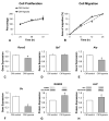
Effect of CM of MSCs+BMP-9 primed with hypoxia on cell proliferation, migration and osteoblast differentiation of MSCs. Cell proliferation at 24, 48 and 72 h (A) and migration at 0, 6, 12 and 24 h (B) of MSCs cultured in CM of MSCs+BMP-9 cultured under either control (CM control) or hypoxia condition (CM hypoxia). Gene expression of Runx2 (C), Sp7 (D), Alp (E) and Oc (F) and protein expression of RUNX2 (G) and ALP (H) of MSCs cultured either in CM control or CM hypoxia, on day 7. The data are presented as mean ± standard deviation, and the * indicates statistically significant differences (p ≤ 0.05).
Figure 4
Figure 4
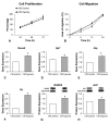
Effect of CM of MSCs+BMP-9 primed with hypoxia on cell proliferation, migration and osteoblast differentiation of MC3T3-E1 cells. Cell proliferation at 24, 48 and 72 h (A) and migration at 0, 6, 12 and 24 h (B) of MC3T3-E1 cells cultured in CM of MSCs+BMP-9 cultured under either control (CM control) or hypoxia condition (CM hypoxia). Gene expression of Runx2 (C), Sp7 (D), Alp (E) and Oc (F) and protein expression of RUNX2 (G) and ALP (H) of MC3T3-E1 cells cultured either in CM control or CM hypoxia, on day 7. The data are presented as mean ± standard deviation, and the * indicates statistically significant differences (p ≤ 0.05).
Figure 5
Figure 5
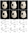
Effect of MSCs+BMP-9 primed with hypoxia on bone repair of rat calvarial defects. Analysis of the bone tissue by µCT. Three-dimensional reconstructions of rat calvarial defects treated with either control MSCs+BMP-9 (Control, (AC)) or MSCs+BMP-9 primed with hypoxia (Hypoxia, (DF)) locally injected 2 weeks post-defect creation (day 0) and evaluated at 14 and 28 days post-treatment. Morphometric parameters bone volume (BV, (G)), percentage of bone volume (BV/TV, (H)), bone surface (BS, (I)), trabecular thickness (Tb.Th, (J)) trabecular number (Tb.N, (K)) and bone mineral density (BMD, (L)) evaluated in the region of interest, within the 5 mm diameter of the calvarial defect. The data are presented as mean ± standard deviation (n = 11 for control and n = 12 for hypoxia). Scale bar: (AF) = 3 mm.
Figure 6
Figure 6
Effect of MSCs+BMP-9 primed with hypoxia on bone repair of rat calvarial defects. Histological analysis of the bone tissue. Micrographs of rat calvarial defects treated with either control MSCs+BMP-9 (Control, (AC)) or MSCs+BMP-9 primed with hypoxia (Hypoxia, (DF)) locally injected 2 weeks post-defect creation and evaluated at 28 days post-treatment. The square in (A) is presented in (B), and the square in (D) is presented in (E). Scale bar: (A,D) = 1.25 mm; (B,E) = 200 μm; (C,F) = 50 μm. bv: blood vessel; cl: cement line; ct: connective tissue; ib: immature bone; lb: lamellar bone; ob: osteoblast; ot: osteocyte.

References

    1. Dominici M., Le Blanc K., Mueller I., Slaper-Cortenbach I., Marini F.C., Krause D.S., Deans R.J., Keating A., Prockop D.J., Horwitz E.M. Minimal criteria for defining multipotente mesenchymal stromal cells. The International Society for Cellular Therapy position statement. Cytotherapy. 2006;8:315–317. doi: 10.1080/14653240600855905. - DOI - PubMed
    1. Freitas G.P., Lopes H.B., Souza A.T.P., Oliveira P.G.F.P., Almeida A.L.G., Coelho P.G., Ferreira F.U., Covas D.T., Beloti M.M., Rosa A.L. Effect of cell therapy with osteoblasts differentiated from bone marrow or adipose tissue stromal cells on bone repair. Regen. Med. 2019;14:1107–1119. doi: 10.2217/rme-2019-0036. - DOI - PubMed
    1. Teotia A.K., Qayoom I., Singh P., Mishra A., Jaiman D., Seppälä J., Lidgren L., Kumar A. Exosome-functionalized ceramic bone substitute promotes critical-sized bone defect repair in rats. ACS Appl. Bio Mater. 2021;4:3716–3726. doi: 10.1021/acsabm.1c00311. - DOI - PubMed
    1. Venkataiah V.S., Yahata Y., Kitagawa A., Inagaki M., Kakiuchi Y., Nakano M., Suzuki S., Handa K., Saito M. Clinical applications of cell-scaffold constructs for bone regeneration therapy. Cells. 2021;10:2687. doi: 10.3390/cells10102687. - DOI - PMC - PubMed
    1. Chan N.T., Lee M.-S., Wang Y., Galipeau J., Li W.-J., Xu W. CTR9 drives osteochondral lineage differentiation of human mesenchymal stem cells via epigenetic regulation of BMP-2 signaling. Sci. Adv. 2022;8:eadc9222. doi: 10.1126/sciadv.adc9222. - DOI - PMC - PubMed

LinkOut - more resources