. 2023 Aug 11;15(16):4066.
doi: 10.3390/cancers15164066.

BRD4 Inhibition as a Strategy to Prolong the Response to Standard of Care in Estrogen Receptor-Positive Breast Cancer

Affiliations

BRD4 Inhibition as a Strategy to Prolong the Response to Standard of Care in Estrogen Receptor-Positive Breast Cancer

Ahmed M Elshazly et al. Cancers (Basel). .

Abstract

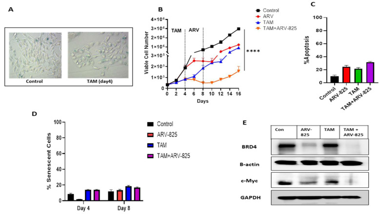
Breast cancer is the most commonly occurring malignancy in women and the second most common cause of cancer-related deaths. ER+ breast cancer constitutes approximately 70% of all breast cancer cases. The standard of care for ER+ breast cancer involves estrogen antagonists such as tamoxifen or fulvestrant in combination with CDK4/6 inhibitors such as palbociclib. However, these treatments are often not curative, with disease recurrence and metastasis being responsible for patient mortality. Overexpression of the epigenetic regulator, BRD4, has been shown to be a negative prognostic indicator in breast cancer, and BET family inhibitors such as ARV-825 and ABBV-744 have garnered interest for their potential to improve and prolong the response to current therapeutic strategies. The current work examined the potential of utilizing ARV-825 and ABBV-744 to increase the effectiveness of tamoxifen or fulvestrant plus palbociclib. ARV-825 was effective in both p53 wild-type (WT) breast tumor cells and in cells lacking functional p53 either alone or in combination with tamoxifen, while the effectiveness of ABBV-744 was limited to fulvestrant plus palbociclib in p53 WT cells. These differential effects may be related to the capacity to suppress c-Myc, a downstream target of BRD4.

Keywords: ABBV-744; ARV-825; BRD4; c-Myc; p53; senescence.


Conflict of interest statement

The authors declare no conflict of interest.

Figures

Figure 1
Figure 1
ARV-825 suppresses proliferative recovery of TAM-treated MCF-7 cells. (A) MCF-7 cells were treated with 5 µM TAM for 4 days. Cells were fixed on day 4, stained with x-gal staining solution, and imaged using a bright field microscope. All images were generated under the same magnification (20X). (B) Cells were treated with TAM (5 µM) for 4 days followed by ARV-825 (50 nM) addition for 4 days. Cell viability was monitored over a period of 16 days through trypan blue exclusion. (C) Cells were treated with TAM (5 µM) for 4 days followed by ARV-825 (50 nM) for 4 days. Apoptosis was evaluated at day 4 of ARV-825 treatment via flow cytometry using an Annexin V-FITC Apoptosis Detection Kit. (D) Percentage of SA-β-gal positive was quantified using C12FDG at the indicated time points. (E) Western blotting for BRD4 and c-Myc at day 4 of ARV-825 treatment. All images are representative fields or blots from at least two/three independent experiments. The uncropped blots are shown in the Supplementary Materials. **** p ≤ 0.001 indicates statistical significance of each condition compared to the control as determined using two-way ANOVA with Sidak’s post hoc test.
Figure 2
Figure 2
ARV-825 induces significant growth suppression in combination with TAM in MCF-7 p53−/− cells. (A) Western blot for p53, and p21 after 24 h of doxorubicin treatment. (B) MCF-7 p53−/− cells were treated with TAM (5 µM) for 4 days followed by ARV-825 (50 nM) addition for 4 days. Viable cell number was monitored over a period of 16 days through trypan blue exclusion. (C) Apoptosis was evaluated via flow cytometry using an Annexin V-FITC Apoptosis Detection Kit. (D) Percentage of SA-β-gal positive was quantified using C12FDG at the indicated time points. (E) Western blotting for BRD4 and c-Myc at day 4 of ARV-825 treatment. All images are representative fields or blots from at least two/three independent experiments. The uncropped blots are shown in the Supplementary Materials.
Figure 3
Figure 3
ABBV-744 extends the growth inhibitory response initiated by fulvestrant plus palbociclib in MCF-7 cells. (A) Cells were treated with fulvestrant (100 nM) plus palbociclib (1 µM) for 6 days followed by ABBV-744 (100 nM) addition starting from day 6 to 10. Cell viability was monitored over a period of 18 days through trypan blue exclusion. (B) Cells were treated with fulvestrant (100 nM) plus palbociclib (1 µM) for 6 days followed by ABBV-744 (100 nM) from day 6 to 10. Apoptosis was evaluated at day 4 of ABBV-744 treatment via flow cytometry using an Annexin V-FITC Apoptosis Detection Kit. (C) Cells were treated with fulvestrant (100 nM) plus palbociclib (1 µM) for 6 days and fixed on Day 6, stained with x-gal staining solution, and imaged using a bright field microscope. Both images were generated under the same magnification (20X). (D) Quantification of SA-β-gal using C12FDG at the indicated time points. (E) Western blotting for BRD4, c-Myc, and p53 at day 4 of ABBV-744 treatment. (F) MCF-7 p53 WT cells were subjected to immunoprecipitation for BRD4 and p53 using BRD4 and p53 antibodies. The arrows indicate the location of BRD4 and p53 bands at 152 kDa and 53 kDa, respectively. All images are representative fields or blots from at least two/three independent experiments. The uncropped blots are shown in the Supplementary Materials. **** p ≤ 0.001 indicates statistical significance of each condition compared to fulvestrant plus palbociclib as determined using two-way ANOVA with Sidak’s post hoc test.
Figure 4
Figure 4
Absence of ABBV-744 effects in MCF-7 p53−/− cells. (A) Both MCF-7 p53 WT and p53−/− cells were treated with different concentrations of ABBV-744 (25 nM, 50 nM, 75 nM, and 100 nM) for 4 days; cell viability was determined after 96, 120, and 144 h using the MTT assay. (B) MCF-7 p53−/− cells were treated with fulvestrant (100 nM) plus palbociclib (1 µM) for 6 days followed by ABBV-744 (100 nM) addition starting from day 6 to 10. Cell viability was monitored over a period of 14 days through trypan blue exclusion. (C) Western blotting for BRD4 and c-Myc at day 4 of ABBV-744 treatment. All images are representative fields or blots from at least two/three independent experiments. The uncropped blots are shown in the Supplementary Materials. NS indicates non-statistical significance of each condition compared to fulvestrant plus palbociclib as determined using two-way ANOVA with Sidak’s post hoc test.
Figure 5
Figure 5
ABBV-744 did not extend growth arrest when combined with fulvestrant plus palbociclib in T47D cells. (A) Cells were treated with ABBV-744 (25 nM, 50 nM, 75 nM, and 100 nM) for 4 days. Percent cell viability was measured by the MTS viability assay. (B) Cell viability was monitored over a period of 10 days through trypan blue exclusion. (C) Apoptosis was evaluated via flow cytometry using an APC Annexin V Apoptosis Detection Kit. (D) Cells were treated with fulvestrant (100 nM) plus palbociclib (1 µM) for 6 days followed by ABBV-744 (100 nM) addition for 4 days. Cell viability was monitored over a period of 14 days through trypan blue exclusion. (E) Apoptosis was evaluated via flow cytometry using an Annexin V-FITC Apoptosis Detection Kit. (F) Western blotting for BRD4, and c-Myc at day 4 of ABBV-744 treatment. All images are representative fields or blots from at least two/three independent experiments. The uncropped blots are shown in the Supplementary Materials. NS indicates non-statistical significance of each condition compared to control and fulvestrant plus palbociclib as determined using two-way ANOVA with Sidak’s post hoc test.
Figure 6
Figure 6
ABBV-744 did not suppress the recovery of TAM-treated cells. (A) Cells were treated with TAM (5 µM) for 4 days followed by ABBV-744 (100 nM) addition starting from day 4 to day 8. Cell viability was monitored over a period of 16 days through trypan blue exclusion. (B) Apoptosis was evaluated via flow cytometry using an Annexin V-FITC Apoptosis Detection Kit. (C) Western blotting for BRD4, c-Myc, and p53 at day 4 of ABBV-744 treatment. All images are representative fields or blots from at least two/three independent experiments. The uncropped blots are shown in the Supplementary Materials. NS (non-significant) indicates non-statistical significance of TAM plus ABBV-744 compared to TAM alone as determined using two-way ANOVA with Sidak’s post hoc test.

Similar articles

Cited by

References

    1. Siegel R.L., Miller K.D., Fuchs H.E., Jemal A. Cancer Statistics, 2021. CA Cancer J. Clin. 2021;71:7–33. doi: 10.3322/caac.21654. - DOI - PubMed
    1. Elshazly A.M., Gewirtz D.A. An overview of resistance to Human epidermal growth factor receptor 2 (Her2) targeted therapies in breast cancer. Cancer Drug Resist. 2022;5:472–486. doi: 10.20517/cdr.2022.09. - DOI - PMC - PubMed
    1. Kohler B.A., Sherman R.L., Howlader N., Jemal A., Ryerson A.B., Henry K.A., Boscoe F.P., Cronin K.A., Lake A., Noone A.M., et al. Annual Report to the Nation on the Status of Cancer, 1975–2011, Featuring Incidence of Breast Cancer Subtypes by Race/Ethnicity, Poverty, and State. J. Natl. Cancer Inst. 2015;107:djv048. doi: 10.1093/jnci/djv048. - DOI - PMC - PubMed
    1. Howlader N., Altekruse S.F., Li C.I., Chen V.W., Clarke C.A., Ries L.A., Cronin K.A. US incidence of breast cancer subtypes defined by joint hormone receptor and HER2 status. J. Natl. Cancer Inst. 2014;106:dju055. doi: 10.1093/jnci/dju055. - DOI - PMC - PubMed
    1. Finnegan R.M., Elshazly A.M., Schoenlein P.V., Gewirtz D.A. Therapeutic Potential for Targeting Autophagy in ER+ Breast Cancer. Cancers. 2022;14:4289. doi: 10.3390/cancers14174289. - DOI - PMC - PubMed