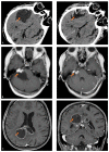
Proteomic Analysis on Sequential Samples of Cystic Fluid Obtained from Human Brain Tumors
- PMID: 37627098
- PMCID: PMC10452907
- DOI: 10.3390/cancers15164070
Proteomic Analysis on Sequential Samples of Cystic Fluid Obtained from Human Brain Tumors
Abstract
Cystic formation in human primary brain tumors is a relatively rare event whose incidence varies widely according to the histotype of the tumor. Composition of the cystic fluid has mostly been characterized in samples collected at the time of tumor resection and no indications of the evolution of cystic content are available. We characterized the evolution of the proteome of cystic fluid using a bottom-up proteomic approach on sequential samples obtained from secretory meningioma (SM), cystic schwannoma (CS) and cystic high-grade glioma (CG). We identified 1008 different proteins; 74 of these proteins were found at least once in the cystic fluid of all tumors. The most abundant proteins common to all tumors studied derived from plasma, with the exception of prostaglandin D2 synthase, which is a marker of cerebrospinal fluid origin. Overall, the protein composition of cystic fluid obtained at different times from the same tumor remained stable. After the identification of differentially expressed proteins (DEPs) and the protein-protein interaction network analysis, we identified the presence of tumor-specific pathways that may help to characterize tumor-host interactions. Our results suggest that plasma proteins leaking from local blood-brain barrier disruption are important contributors to cyst fluid formation, but cerebrospinal fluid (CSF) and the tumor itself also contribute to the cystic fluid proteome and, in some cases, as with immunoglobulin G, shows tumor-specific variations that cannot be simply explained by differences in vessel permeability or blood contamination.
Keywords: cystic fluid; cystic schwannoma (CS); glioblastoma (CG); proteomics; secretory meningioma (SM); tumor cyst.
Conflict of interest statement
The authors declare no conflict of interest.
Figures


Similar articles
-
Diagnostic biomarkers from proteomic characterization of cerebrospinal fluid in patients with brain malignancies.J Neurochem. 2021 Jul;158(2):522-538. doi: 10.1111/jnc.15350. Epub 2021 Apr 9. J Neurochem. 2021. PMID: 33735443
-
Proteomic analysis of cerebrospinal fluid extracellular vesicles: a comprehensive dataset.J Proteomics. 2014 Jun 25;106:191-204. doi: 10.1016/j.jprot.2014.04.028. Epub 2014 Apr 24. J Proteomics. 2014. PMID: 24769233
-
Glioblastoma microenvironment contains multiple hormonal and non-hormonal growth-stimulating factors.Fluids Barriers CNS. 2022 Jun 4;19(1):45. doi: 10.1186/s12987-022-00333-z. Fluids Barriers CNS. 2022. PMID: 35659255 Free PMC article.
-
Utilization of Cerebrospinal Fluid Proteome Analysis in the Diagnosis of Meningioma: A Systematic Review.Cureus. 2021 Dec 26;13(12):e20707. doi: 10.7759/cureus.20707. eCollection 2021 Dec. Cureus. 2021. PMID: 34966627 Free PMC article. Review.
-
Intraparenchymal cystic chordoid meningioma: a case report and review of the literature.Neuropathology. 2011 Dec;31(6):648-53. doi: 10.1111/j.1440-1789.2011.01214.x. Epub 2011 Apr 11. Neuropathology. 2011. PMID: 21481008 Review.
Cited by
-
Deciphering the causal relationship between plasma and cerebrospinal fluid metabolites and glioblastoma multiforme: a Mendelian Randomization study.Aging (Albany NY). 2024 May 10;16(9):8306-8319. doi: 10.18632/aging.205818. Epub 2024 May 10. Aging (Albany NY). 2024. PMID: 38742944 Free PMC article.
-
Computational Tools and Methods for the Study of Systemic Amyloidosis at the Clinical and Molecular Level.Methods Mol Biol. 2025;2884:369-387. doi: 10.1007/978-1-0716-4298-6_22. Methods Mol Biol. 2025. PMID: 39716014
-
Differential proteomic profiles of exosomes in pediatric and adult adamantinomatous craniopharyngioma cyst fluid.Mol Biol Rep. 2024 Nov 7;51(1):1126. doi: 10.1007/s11033-024-10073-y. Mol Biol Rep. 2024. PMID: 39505756
-
Glioblastoma: Overview of Proteomic Investigations and Biobank Approaches for the Development of a Multidisciplinary Translational Network.Cancers (Basel). 2025 Jun 26;17(13):2151. doi: 10.3390/cancers17132151. Cancers (Basel). 2025. PMID: 40647449 Free PMC article. Review.
References
-
- Curtin L., Whitmire P., Rickertsen C.R., Mazza G.L., Canoll P., Johnston S.K., Mrugala M.M., Swanson K.R., Hu L.S. Assessment of Prognostic Value of Cystic Features in Glioblastoma Relative to Sex and Treatment With Standard-of-Care. Front. Oncol. 2020;10:580750. doi: 10.3389/fonc.2020.580750. - DOI - PMC - PubMed
Grants and funding
- codice ric. 08016019/Policlinico San Matteo Fondazione
- GJC2021 (n. 2022-0578)/Fondazione Cariplo
- PON ELIXIR CNRBiOMICS (PIR01_00017)/Ministry of Education, Universities and Research
- Elixir Implementation Study Proteomics 2021-23, ELIXIRxNextGenIT [PNRR, Prot. IR0000010]/Ministero Università e Ricerca (MUR)
- (RF2019-12370396)/Ministero della Salute
LinkOut - more resources
Full Text Sources

