Review
. 2023 Aug 14;15(16):4100.
doi: 10.3390/cancers15164100.

Emergence of Lipid Droplets in the Mechanisms of Carcinogenesis and Therapeutic Responses

Affiliations
Review

Emergence of Lipid Droplets in the Mechanisms of Carcinogenesis and Therapeutic Responses

Dominique Delmas et al. Cancers (Basel). .

Abstract

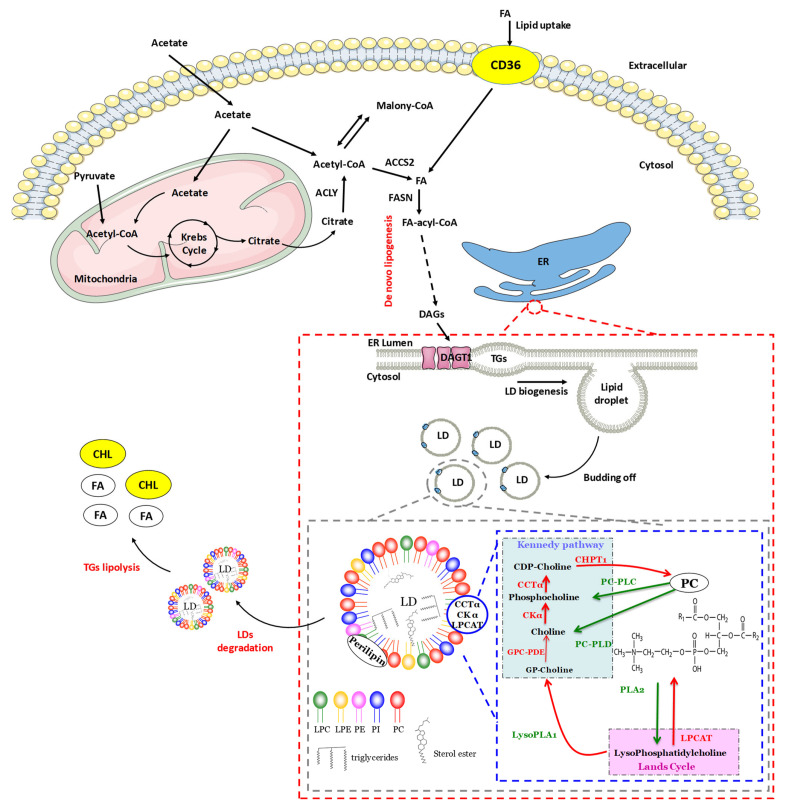
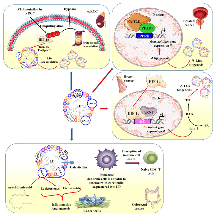
Cancer shares common risk factors with cardiovascular diseases such as dyslipidemia, obesity and inflammation. In both cases, dysregulations of lipid metabolism occur, and lipid vesicles emerge as important factors that can influence carcinogenesis. In this review, the role of different lipids known to be involved in cancer and its response to treatments is detailed. In particular, lipid droplets (LDs), initially described for their role in lipid storage, exert multiple functions, from the physiological prevention of LD coalescence and regulation of endoplasmic reticulum homeostasis to pathological involvement in tumor progression and aggressiveness. Analysis of LDs highlights the importance of phosphatidylcholine metabolism and the diversity of lipid synthesis enzymes. In many cancers, the phosphatidylcholine pathways are disrupted, modifying the expression of genes coding for metabolic enzymes. Tumor microenvironment conditions, such as hypoxia, different types of stress or inflammatory conditions, are also important determinants of LD behavior in cancer cells. Therefore, LDs represent therapeutic targets in cancer, and many lipid mediators have emerged as potential biomarkers for cancer onset, progression, and/or resistance.

Keywords: biomarkers; cancers; chemoresistance; lipid droplets; lipid metabolism.


Conflict of interest statement

The authors declare no conflict of interest.

Figures

Figure 1
Figure 1
The formation of lipid droplets (LDs) results from the budding of the endoplasmic reticulum (ER) from the outer leaflet. Newly formed LDs can fuse together to increase their volume and surface area. These LDs are made up of very heterogeneous PLs where PC is predominant on the surface of LDs and thus prevents their coalescence, which is essential for the growth of LDs. Some of the enzymes of the Kennedy pathway and Land’s cycle seem to actively participate in the synthesis of PC at the level of LDs. In mammals, it can be synthesized de novo via two synthetic pathways [7] the main of which is the cytidine diphosphate-choline (CDP-choline) pathway, more commonly known as the Kennedy pathway. Choline is first phosphorylated to phosphocholine by choline kinase alpha (CKα) and then phosphocholine cytidylyltransferase alpha (CTTα), the limiting enzyme of this pathway, catalyzes the reaction between phosphocholine and cytidine triphosphate (CTP) to form CDP-choline which is then converted into PC by 1,2-diacylglycerol choline phosphotransferase (CHPT1) [8]. The second pathway for de novo synthesis of PC, known as the phosphatidylethanolamine N-methyltransferase (PEMT) pathway, occurs only in the liver and consists of a series of three PEMT-catalyzed PE methylations [9]. Finally, PC can also be synthesized via the remodeling of PLs during the Lands cycle by reacylation of LPC catalyzed by lysophosphatidylcholine acyltransferases 1, 2, and 4 (LPCAT1, LPCAT2 and LPCAT4) [10]. Ether from sterols is synthesized by coupling sterols with an FA using isoforms 1 and 2 of the enzyme acetyl-coenzyme A acetyltransferase (ACAT). The surface of LDs is composed of amphipathic and polar lipids forming a monolayer distinguishable from other cell membranes by their composition.
Figure 2
Figure 2
Involvement of LDs in different types of cancers where the accumulation of LDs is associated with the aggressiveness of cancers. In renal cancers, the VHL mutation favors the ubiquitination of HIF, thus allowing the increase in the expression of perilipin 2, which plays an essential role in the biogenesis of LDs. Moreover, the phenomenon of hypoxia comes to counter the proteosomal degradation of ubiquitylated HIF, thus reinforcing the action of the latter on the quantity of perilipin 2. Consequently, this results in the accumulation of LDs. In prostate cancer, the interaction of PPARγ with KMT2D promotes the activation of gene transcription such as fasn, acly, and acc, which makes it possible to increase lipogenesis and in fact the biogenesis of LDs that will accumulate. In the same way in breast cancer, HIF-1α activates the transcription of the lipin-1 gene coding for the lipin-1 protein, whose enzymatic activity catalyzes the conversion of phosphatidic acid (PA) to DAG and consequently to LD biogenesis and its accumulation. In colon cancer, various mechanisms are involved through the enzymes present in LDs, such as COX-2 and LOX, which participate in inflammatory processes and tumor angiogenesis. Furthermore, the sequestration of calreticulin into LDs induces a failure in dendritic cell maturation, which leads to limited recruitment and activation of naïve CD8+ T cells and leads to a disruption of immune cell death.

References

    1. Gross D.A., Silver D.L. Cytosolic lipid droplets: From mechanisms of fat storage to disease. Crit. Rev. Biochem. Mol. Biol. 2014;49:304–326. doi: 10.3109/10409238.2014.931337. - DOI - PubMed
    1. Buchanan F.G., McReynolds M., Couvillon A., Kam Y., Holla V.R., Dubois R.N., Exton J.H. Requirement of phospholipase D1 activity in H-RasV12-induced transformation. Proc. Natl. Acad. Sci. USA. 2005;102:1638–1642. doi: 10.1073/pnas.0406698102. - DOI - PMC - PubMed
    1. Fei W., Zhong L., Ta M.T., Shui G., Wenk M.R., Yang H. The size and phospholipid composition of lipid droplets can influence their proteome. Biochem. Biophys. Res. Commun. 2011;415:455–462. doi: 10.1016/j.bbrc.2011.10.091. - DOI - PubMed
    1. Brasaemle D.L. Thematic review series: Adipocyte biology. The perilipin family of structural lipid droplet proteins: Stabilization of lipid droplets and control of lipolysis. J. Lipid Res. 2007;48:2547–2559. doi: 10.1194/jlr.R700014-JLR200. - DOI - PubMed
    1. Ducharme N.A., Bickel P.E. Lipid droplets in lipogenesis and lipolysis. Endocrinology. 2008;149:942–949. doi: 10.1210/en.2007-1713. - DOI - PubMed